Endocrinol Metab 2013;28:226-230
http://dx.doi.org/10.3803/EnM.2013.28.3.226 pISSN 2093-596X · eISSN 2093-5978
Case Report
Graves’ Disease that Developed Shortly after Surgery for Thyroid Cancer
Hea Min Yu1, Soon Hyun Park2, Jae Min Lee1, Kang Seo Park1
1Department of Internal Medicine, Eulji University School of Medicine; 2Soe’s Internal Medicine Clinic, Daejeon, Korea
Graves’ disease is an autoimmune disorder that may present with various clinical manifestations of hyperthyroidism. Patients with Graves’ disease have a greater number of thyroid nodules and a higher incidence of thyroid cancer compared with patients with nor- mal thyroid activity. However, cases in which patients are diagnosed with recurrence of Graves’ disease shortly after partial thyroid- ectomy for thyroid cancer are very rare. Here we report a case of hyperthyroid Graves’ disease that occurred after partial thyroidec- tomy for papillary thyroid cancer. In this case, the patient developed hyperthyroidism 9 months after right hemithyroidectomy, and antithyroglobulin autoantibody and thyroid stimulating hormone receptor stimulating autoantibody were positive. Therefore, we di- agnosed Graves’ disease on the basis of the laboratory test results and thyroid ultrasonography findings. The patient was treated with and maintained on antithyroid drugs. The mechanism of the recurrence of Graves’ disease in this patient is still unclear. The mechanism may have been the improper response of the immune system after partial thyroidectomy. To precisely determine the mechanisms in Graves’ disease after partial thyroidectomy, further studies based on a greater number of cases are needed.
Keywords: Graves’ disease; Partial thyroidectomy; Thyroid cancer, papillary
INTRODUCTION
Graves’ disease is an autoimmune disorder that may present with various clinical manifestations. The mechanism of develop- ment is thought to arise from thyroid stimulating hormone (TSH) receptor stimulating autoantibodies (TSHR Ab) which cause hyperthyroidism, and is accompanied by a diffuse and hy- pervascular goiter [1]. As a result, patients with Graves’ disease have more thyroid nodules and a higher incidence of thyroid cancer compared with patients who have normal thyroid activity [2,3]. The incidence of thyroid cancer in Graves’ disease patients has been reported to range from 0% to 9.8%, and papillary thy- roid cancer is known to account for the majority of cases [4,5].
However cases of patients who are diagnosed with Graves’ dis- ease within a short period of time after partial thyroidectomy for thyroid cancer are very rare. Ten cases have been reported in foreign countries, but no cases have been reported in Korea [6- 9]. Here we report a case of hyperthyroid Graves’ disease that occurred after partial thyroidectomy for papillary thyroid cancer, together with the literature review results.
CASE REPORT
Patient: A 41-year-old female.
Main phenomena: Palpitations, heat intolerance, perspiration.
Current medical history: In November 2010, 0.5×0.6 cm
Received: 5 October 2012, Accepted: 12 November 2012 Corresponding author: Kang Seo Park
Division of Endocrinology and Metabolism, Department of Internal Medicine, Research Institute of Clinical Medicine, Eulji University Hospital,
Eulji University School of Medicine, 95 Dunsanseo-ro, Seo-gu, Daejeon 302-799, Korea
Tel: +82-42-611-3000, Fax: +82-42-259-1111, E-mail: [email protected]
Copyright © 2013 Korean Endocrine Society
This is an Open Access article distributed under the terms of the Creative Com- mons Attribution Non-Commercial License (http://creativecommons.org/
licenses/by-nc/3.0/) which permits unrestricted non-commercial use, distribu- tion, and reproduction in any medium, provided the original work is properly cited.
hypoechoic nodules were found in the right thyroid of a female patient. Fine needle aspiration biopsy was performed. Through the pathology examination, she was diagnosed with papillary thyroid cancer and had robot thyroidectomy on the right thy- roid in January 2011. During the postsurgery recovery period, she was found to have clinical features of Graves’ disease, and the surgery department sent her to our department for investi- gation and treatment.
Past history: She had been taking antituberculous drugs since September as she had been treated for tuberculosis.
Social history: She drank two to three times a week (one bot- tle of Soju each time), but never smoked.
Family history: Nothing noteworthy was seen.
Scientific view: Her initial blood pressure was 133/78 mm Hg, pulse 95 beats per minute, respirations 20 breaths per min- ute, and body temperature 36.4°C. She had a clear level of consciousness without acute signs of disease, but She looked rather sick. The thyroid was not swollen and was painless, and there were no palpable nodules. Also, retraction of the palpe- bral fissures, proptosis, hyperemia of the conjunctivae, and edema were not observed.
Lab opinion: Peripheral blood was examined and the blood results were white blood cells 7,220/mm3, hemoglobin 13.6 g/
dL, hematocrit 41.2%, and platelets 353,000/mm3, while the blood sedimentation rate was 7 mm/hr; therefore, all of the test results were normal. According to the serum biochemical examination, blood urea nitrogen was 13 mg/dL, serum creati- nine 0.6 mg/dL, calcium 8.8 mg/dL, phosphorus 4.0 mg/dL, gross protein 7.0 g/dL, albumin 4.0 g/dL, aspartate amino-
transferase 25 IU/L, alanine aminotransferase 34 IU/L, and gross bilirubin 0.4 mg/dL; therefore, all of the biochemical re- sults were normal. Meanwhile, the alkaline phosphatase den- sity was elevated (111 IU/L). According to the serum electro- lyte examination, sodium density was 144 mEq/L and potassi- um 4.4 mEq/L.
Endocrine examination opinion: Before the operation, triio- dothyronine (T3) was 80 ng/dL (normal range, 65 to 150), free thyroxine (free T4) 1.07 ng/dL (normal range, 0.89 to 1.76), and TSH 1.80 μIU/mL (normal range, 0.35 to 5.50), all of which were in the normal range, but 9 months after the opera- tion, the patient was diagnosed with thyrotoxicosis with T3 363 ng/dL, free T4 2.33 ng/dL, and TSH 0.02 μIU/mL. Mean- while, there was an increase in thyroglobulin (Tg) 16.6 ng/mL (normally, 1.4 to 78.0), antithyroperoxidase antibody 39.6 IU/
mL (normally, 0 to 34), antithyroglobulin antibody (anti-Tg Ab) 335.8 IU/mL (normally, 0 to 60.0), and TSHR Ab 5.44 IU/L (normally, 0 to 1.75).
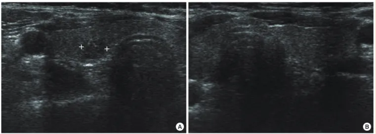
Image examination opinion: In the thyroid sonogram before the operation, 0.5×0.6-cm sized hypoechoic nodules were found in the right lobe of the thyroid which was a normal size, and there was no sign of increased blood flow (Fig. 1). On thy- roid ultrasound after the operation, we could not find any tumor or nodule in the remaining thyroid, but the left thyroid was a little increased in size, the overall phosphorus shade reduced, and the blood flow increased (Fig. 2). However, there was no sign of enlarged cervical lymph nodes in the sonogram either before or after the operation.
Clinical course: The patient’s thyroid activity examination
Fig. 1. Thyroid ultrasound before surgery. (A) Thyroid ultrasound shows an approximately 0.5×0.6-cm sized hypoechoic nodule in the right thyroid. (B) Ultrasound of the left thyroid shows normal findings.
A B
was normal 1 year before she was diagnosed with thyroid can- cer and remained normal just before the thyroid cancer opera- tion. On the preoperative thyroid ultrasound, nothing was note- worthy except for hypoechoic nodules in the right thyroid.
However, she was diagnosed with thyrotoxicosis during an examination performed 3 months after the right thyroidecto- my. When she was transferred to the surgery department for the operation, her T3 was 226 ng/dL, free T4 2.39 ng/dL, and TSH 0.03 μIU/mL. The surgery department decided to reduce the amount of her levothyroxine from 100 to 50 μg (Fig. 3).
Since her condition had not improved at all, she stopped taking the drug, but her condition worsened 3 months later (9 months after the operation) and a blood test was performed, and even-
tually she was sent to the endocrinology department.
When we considered the clinical features, blood examina- tion results, and ultrasound result, we came to the conclusion that the patient had Graves’ disease resulting from the thyroid cancer operation, and we began observing the development of her disease while treating her with carbimazole 20 mg/day and propranolol 30 mg/day. After a month, follow-up tests were performed and we found that her results had greatly improved, with T3 at 177 ng/dL, free T4 1.21 ng/dL, and TSH 0.02 μIU/
mL. After 6 months of drug treatment, all the values stabilized:
T3 was 102 ng/dL, Free T4 1.14 ng/dL, TSH 2.02 μIU/mL, and TSHR Ab, and she currently comes to the hospital for checkups while taking carbimazole 10 mg/day.
Fig. 2. Thyroid ultrasound after right partial thyroidectomy. (A) Thyroid ultrasound shows an enlarged left thyroid gland and hypoecho- genicity in the transverse section. (B) In the longitudinal section, the aforementioned findings were also observed.
Fig. 3. Histological examination of the surgical specimen. Papillary thyroid cancer cells were seen (A, H&E stain, ×100; B, H&E stain,
×300). A papillary growth pattern is evident.
A
A
B
B
DISCUSSION
Graves’ disease is an organ-specific autoimmune disease that causes hyperthyroidism via the production of TSH receptor autoantibodies which stimulate the thyroid. Graves’ disease is known to have a genetic-environmental etiology. Due to its ability to stimulate the growth of thyroid nodules, it may be accompanied by a colloid goiter, autoimmune lymphocytic disease, thyroid osteoarthritic changes, and hyperplastic ade- nomatous tissue [1,10]. It is common to detect Graves’ disease in patients with thyroid cancer clinically, but cases of Graves’
disease recurrence after partial thyroidectomy for thyroid cancer are very rare. Cases of Graves’ disease occurring after thyroid operation for thyroid nodules or cancer were first described in three case reports by Tamai et al. [8] in 1982, and three more cases were reported by Misaki et al. [11] in 1997 [7]. When we looked into the seven case reports that have appeared since 1997, in four of those cases, antithyroid microsomal antibody and anti-Tg Ab were measured before the operation, and in three of those cases, antithyroid antibody tested positive. Ac- cording to our analysis, all of the above seven cases took a considerable time (from 2 to 7 years) for Graves’ disease to be diagnosed following thyroidectomy. In one case the disease was diagnosed 4 weeks after the operation, but the patient in that case had a thyroid that had normal function without toxic symptoms before the operation. However, antithyroid micro- somal antibodies and thyroid stimulating antibodies were al- ready elevated and a goiter was discovered on thyroid sono- gram before the operation. When we looked at the time of oc- currence and examination results of the cases that have been reported so far, the findings did not support the theory that thyroidectomy has a direct relationship to Graves’ disease.
For the patient in the present case, although thyroid autoan- tibody was not measured before the operation, and there was no suspicion of thyroid autoimmune disease judging from the sonogram, blood examination, or clinical features, the patient also developed Graves’ disease in a comparatively short peri- od of time after thyroidectomy, and this case could provide good evidence that the thyroid operation itself can induce Graves’ disease.
The mechanism of this phenomenon is not obvious, but some hypotheses have been presented. The first hypothesis proposes that the operation destroys thyroid cells, generating TSH re- ceptor elevation, which causes the disease. In short, when thy- roid epithelial cells are damaged during an operation, TSH re- ceptor, Tg antigen, and microsomal antigen are secreted from
the thyroid and they then stimulate the helper T cells to gener- ate these autoantibodies, finally resulting in an increase in thy- roid activity [12,13]. In contrast, the second hypothesis pro- poses that it is not the excessive antigens that cause the auto- immune thyroiditis, but rather an abnormality of the antigen presenting cells that keep the activation of suppressor cells or regulatory cells under control, which then attack the immune system, causing postoperative Graves’ disease [14]. This theo- ry may be supported by cases in which antibody levels were stabilized or reduced either before or after surgery in patients who already had Graves’ disease [15,16]. The third hypothesis proposes that the stress from general anesthesia and surgery affects the patient physically and mentally, causing neuroen- docrine fluctuations which induce immunological homeostasis [17,18]. Since the first report by Dr. Parry in 1825, there have been many reports indicating that stress is associated with Graves’ disease, but the relevant mechanism has not yet been elucidated [19]. The final hypothesis is that postoperative bac- terial or viral infection increases the number of CD5+ B cells and these CD5+ B cells stimulate the TSH receptor antibodies to provoke Graves’ disease [6,20]. However, the patient in this case was not infected after the operation, and it was also un- clear whether the patients in the preceding cases were infected after surgery.
In conclusion, we report our experience of the recurrence of Graves’ disease possibly caused by immunological malfunc- tioning after partial thyroid resection. Further research is need- ed to establish the pathological mechanism of this disease by studying similar cases, although the condition is rare. In addi- tion, we recommend keeping a close watch on thyroid activity both before and after partial resection for thyroid cancer or tu- mor.
CONFLICTS OF INTEREST
No potential conflict of interest relevant to this article was re- ported.
REFERENCES
1. Laurberg P, Wallin G, Tallstedt L, Abraham-Nordling M, Lundell G, Torring O. TSH-receptor autoimmunity in Graves’ disease after therapy with anti-thyroid drugs, sur- gery, or radioiodine: a 5-year prospective randomized study. Eur J Endocrinol 2008;158:69-75.
2. Carnell NE, Valente WA. Thyroid nodules in Graves’ dis-
ease: classification, characterization, and response to treat- ment. Thyroid 1998;8:647-52.
3. Chung JO, Cho DH, Chung DJ, Chung MY. Ultrasono- graphic features of papillary thyroid carcinoma in patients with Graves’ disease. Korean J Intern Med 2010;25:71-6.
4. Dobyns BM, Sheline GE, Workman JB, Tompkins EA, McConahey WM, Becker DV. Malignant and benign neo- plasms of the thyroid in patients treated for hyperthyroid- ism: a report of the cooperative thyrotoxicosis therapy fol- low-up study. J Clin Endocrinol Metab 1974;38:976-98.
5. Pacini F, Elisei R, Di Coscio GC, Anelli S, Macchia E, Concetti R, Miccoli P, Arganini M, Pinchera A. Thyroid carcinoma in thyrotoxic patients treated by surgery. J En- docrinol Invest 1988;11:107-12.
6. Umena S, Takano T, Iijima T, Hidaka Y, Yagoro A, Takai S, Amino N. A case of repeated painless thyroiditis followed by Graves’ disease. Endocr J 1995;42:821-6.
7. Kasuga Y, Kobayashi S, Fujimori M, Shingu K, Hama Y, Ito K, Amano J. Development of Graves’ disease after sur- gical treatment for thyroid nodules: report of four cases.
Endocr J 1997;44:567-70.
8. Tamai H, Hirota Y, Matsuzuka F, Kuma K, Nagataki S. Three cases developed Graves’ disease after surgical treatment for thyroid carcinomas. Horumon To Rinsho 1982;30:124-5.
9. Nagai K, Tamai H, Matsubayashi S, Takaichi Y, Matsuzu- ka F, Kobayashi A, Hirai K, Kuma K, Nagataki S. A case developed Graves’ disease after thyroid nodule. Horumon To Rinsho 1987;35:107-9.
10. Collins J, Gough S. Autoimmunity in thyroid disease. Eur J Nucl Med Mol Imaging 2002;29 Suppl 2:S417-24.
11. Misaki T, Iwata M, Kasagi K, Iida Y, Akamizu T, Kosugi S, Konishi J. Hyperthyroid Graves’ disease after hemithy- roidectomy for papillary carcinoma: report of three cases.
Endocr J 2000;47:191-5.
12. Cho BY, Oh SK, Shong YK, Kim SE, Yoo HY, Lee HK, Koh CS, Min HK. Changes in thyrotropin receptor anti-
body after subtotal thyroidectomy in Graves’ disease: com- parison with the degree of lymphocytic infiltration in the thyroid. Autoimmunity 1990;8:143-7.
13. De Bruin TW, Patwardhan NA, Brown RS, Braverman LE.
Graves’ disease: changes in TSH receptor and anti-micro- somal antibodies after thyroidectomy. Clin Exp Immunol 1988;72:481-5.
14. Volpe R. Autoimmune diseases of the endocrine system.
Boca Raton: CRC Press; 1990. Chapter 4, Immunology of the thyroid; p. 73-239.
15. Teng CS, Yeung RT, Khoo RK, Alagaratnam TT. A pro- spective study of the changes in thyrotropin binding inhibi- tory immunoglobulins in Graves’ disease treated by subto- tal thyroidectomy or radioactive iodine. J Clin Endocrinol Metab 1980;50:1005-10.
16. Nakamura S, Ishiyama-Takuno M, Kosaka J, Kondo K, Horiya Y, Hara S, Shima H, Shiraki T. A case of Graves’
disease and papillary thyroid carcinoma with predominant production of thyroid-stimulation-blocking antibodies (TSBAb) persisted after total thyroidectomy. Endocrinol Jpn 1992;39:133-9.
17. Kiecolt-Glaser JK, Page GG, Marucha PT, MacCallum RC, Glaser R. Psychological influences on surgical recov- ery: perspectives from psychoneuroimmunology. Am Psy- chol 1998;53:1209-18.
18. Bauer M. Anesthesia and perioperative immune function.
Remarks on the work of M. Bauer, H. Rensing and T.
Ziegenfuss (Anaesthesist 1998,47:538-556). Anaesthesist 1998;47:939.
19. Chung JH. Genetics of Graves’ disease. J Korean Soc En- docrinol 2003;18:5-11.
20. Iwatani Y, Amino N, Kaneda T, Ichihara K, Tamaki H, Tachi J, Matsuzuka F, Fukata S, Kuma K, Miyai K. Marked in- crease of CD5 + B cells in hyperthyroid Graves’ disease.
Clin Exp Immunol 1989;78:196-200.