Case Report
ISSN 1738-5997 (Print) • ISSN 2092-9935 (Online) Electrolyte Blood Press 16:11-14, 2018 https://doi.org/10.5049/EBP.2018.16.1.11Copyright © 2018 The Korean Society of Electrolyte Metabolism
Esophageal Artery Pseudoaneurysm and Takayasu Arteritis in a Patient with Autosomal Dominant Polycystic Kidney Disease
Hyunsuk Kim1*, Yeonsil Yu2*, Kwang Eon Shim1, Jin Eop Kim1, Junga Koh3, Jong-Woo Yoon1, Curie Ahn4, Yun Kyu Oh5
1Department of Internal Medicine, Hallym University Medical Center, Chuncheon; 2Department of Internal Medicine, J Hospital, Seongnam;
3Department of Internal Medicine, Gangneung Dongin Hospital, Gangneung; 4Department of Internal Medicine, Seoul National University Hospital, Seoul; 5Department of Internal Medicine, Seoul National University Boramae Medical Center, Seoul, Korea
Received: January 20, 2018 Accepted: April 12, 2018
Corresponding Author: Yun Kyu Oh, MD, PhD, Department of Internal Medicine, Seoul National University Boramae Medical Center, 5Gil 20, Boramae-Road, Dongjak-Gu, Seoul 07061, Korea Tel: +82-2-870-2219
Fax: +82-2-870-3863 E-mail: [email protected]
*Hyunsuk Kim and Yeonsil Yu contributed equally to this work.
A 47-year-old female previously diagnosed with ADPKD visited the hospital due to sudden pain in her upper abdomen and back. Esophagogastroduode- noscopy, contrast-enhanced abdominal computed tomography (CT), and CT angiography identified an esophageal artery pseudoaneurysm and hematoma in the esophagus. Urgent angiography and embolization were performed.
After the procedure, CT angiography and positron emission tomography were performed due to differences in blood pressure between the arms. The pati- ent was also found to have Takayasu arteritis and subsequently received outpatient follow-up care. The possible mechanisms that cause vascular abnormalities in ADPKD patients include damaged vascular integrity due to abnormal polycystin expression caused by PKD mutations and connec- tive tissue abnormalities. Further research is needed to confirm these mecha- nisms, and ADPKD patients should be assessed for vascular abnormalities.
Key Words: Autosomal dominant polycystic kidney disease (ADPKD), Aneurysms, Takayasu arteritis
This is an Open Access article distributed under the terms of the Creative Commons Attribution Non-Commercial License(http://creativecommons.org/licenses/by-nc/4.0/) which permits unrestricted non-commercial use, distribution, and reproduction in any medium, provided the original work is properly cited.
Introduction
Autosomal dominant polycystic kidney disease (ADPKD) is the most common inherited renal disease and is charac- terized by numerous cysts in both kidneys1). In addition to affecting the kidneys, ADPKD is associated with various extrarenal vascular abnormalities including cerebral aneu- rysms, coronary artery aneurysms and dissection, cervico- cephalic artery dissection, and aortic aneurysms and dissec- tion2,3). Visceral artery aneurysms are rare; although gas- troduodenal artery pseudoaneurysm and splenic artery aneurysm have been reported, no previous case of an eso- phageal artery pseudoaneurysm has been reported4).
Takayasu arteritis is a chronic granulomatous, large- vessel vasculitis primarily involving the aorta, its main branches, and the pulmonary arteries. Most arterial lesions in Takayasu arteritis are stenotic, whereas aneurysms can be found at various sites, including the visceral abdominal arteries5,6).
We present the case of an ADPKD patient who was diagnosed with an esophageal artery pseudoaneurysm and Takayasu arteritis.
Case Report
A 47-year-old female visited the emergency room due to sudden pain in her upper abdomen and back. The
12 HS Kim•Esophageal Artery Pseudoaneurysm in ADPKD
Copyright © 2018 The Korean Society of Electrolyte Metabolism Fig. 1. Contrast-enhanced abdominopelvic computed tomogra-
phy, showing numerous cysts in the liver and both kidneys.
Fig. 2. Esophagogastroduodenoscopy. External compression is no- ted at the lower body, antrum, and duodenal bulb (arrowheads).
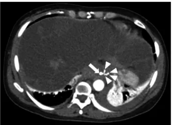
Fig. 3. Computed tomography angiography, showing multifocal pseudoaneurysms (arrowheads) of the esophageal artery from the left gastric artery (arrow), with a suspected hematoma along the esophageal wall.
Fig. 4. Angiography (arteriography of the superior mesenteric ar- tery). A 9-mm pseudoaneurysm (circle) was found in the esopha- geal artery and it was confirmed that it communicated with the distal left gastric artery (arrow). The distal portion of the esopha- geal artery was embolized and the esophageal artery was no longer seen (dashed circle) on angiography after the procedure.
patient had been diagnosed with ADPKD and severe poly- cystic liver disease 13 years prior (Fig. 1) and had received follow-up care with no symptoms. Additionally, as a com- plication of ADPKD, an arachnoid cyst in the left middle cranial fossa was found on brain magnetic resonance ima- ging.
The patient’s blood pressure at the time of hospitali- zation was 140/100 mm Hg, her heart rate was 62 beats per minute, her respiratory rate was 18 breaths per mi- nute, and her temperature was 36.3℃. The laboratory findings showed a white blood cell count of 12,770/μL, a hemoglobin count of 13.5 g/dL, a platelet count of 184,000/
μL, a blood urea nitrogen level of 15 mg/dL, a creatinine level of 0.85 mg/dL, and a C-reactive protein level of 0.35 mg/dL. Esophagogastroduodenoscopy showed exter- nal compression in the lower gastric body, but no hemor- rhage of the gastroesophageal mucosa (Fig. 2).
Computed tomography (CT) angiography showed an esophageal artery pseudoaneurysm and a hematoma in
the esophagus (Fig. 3). Urgent angiography and emboliza- tion of the esophageal artery pseudoaneurysm were per- formed (Fig. 4). CT angiography was performed due to a blood pressure difference of 30 mmHg (right arm, 124/
77 mmHg; left arm, 156/74 mmHg) and the presence of a left subclavian bruit. This examination revealed that the patient also had Takayasu arteritis (Fig. 5). Autoantibody tests, including antineutrophilic cytoplasmic antibody, anti- cyclic citrullinated peptide antibody, and rheumatoid fac- tor, were all negative. Positron emission tomography was performed to assess inflammation, but did not show any concerning signs of active vasculitis. The patient was trans- ferred to outpatient management with low-dose aspirin
Electrolyte Blood Press 16:11-14, 2018 • https://doi.org/10.5049/Ebp.2018.16.1.11 13
Copyright © 2018 The Korean Society of Electrolyte Metabolism Fig. 5. Computed tomography angiography with maximum inten-
sity projection reconstruction, showing 50%-70% luminal narro- wing of the proximal right axillary artery (arrow) and near-total occlusion of the left common carotid artery from the os to the bulb (arowheads).
(100 mg once per day), and has not shown any signs of complications.
Discussion
Both ADPKD and Takayasu arteritis can cause vascular complications, but involvement of the visceral arteries is uncommon4,7). There have been no reports of the simulta- neous diagnosis of ADPKD and Takayasu arteritis, and this is also the first reported case of esophageal artery pseu- doaneurysm in a patient with both ADPKD and Takayasu arteritis.
ADPKD is a hereditary disorder, and is accompanied by various systemic manifestations1,8). Among these, vas- cular abnormalities, such as cerebral or coronary aneurysm and aortic aneurysm and dissection, are relatively well- known, although the mechanisms are not entirely under- stood2). The possible mechanisms that cause vascular ab- normalities in ADPKD patients include damaged vascular integrity due to abnormal polycystin expression caused by PKD mutations and connective tissue abnormalities.
ADPKD is known to be caused by mutations in the PKD1 and PKD2 genes, which code for the polycystin 1 and polycystin 2 proteins, respectively9). These muta- tions cause the growth of numerous renal and extrarenal cysts in ADPKD patients8). Studies have suggested that
the vascular abnormalities found in ADPKD have to do with the polycystin 1 and polycystin 2 present in the vas- cular smooth muscles and endothelium and their involve- ment in the mechanism of vascular remodeling and early aneurysm development2,3,10).
Researchers have long suspected that ADPKD is a type of connective tissue disorder. The histopathological fin- dings of ADPKD patients are generally suggestive of con- nective tissue disorders. Based on vessel aneurysmal chan- ges in the renal tissue, as well as excessive collagen matrix and edema seen in an ADPKD patient who underwent ne- phrectomy, it was suggested that weak, excessive collagen is the common pathogenic factor that is responsible for various ADPKD symptoms11). Moreover, in an autopsy of an individual with vertebrobasilar dolichoectasia, changes were observed in the integrity of the extracellular matrix, with degeneration and multiple gaps in the internal elastic lamina, thinning of the media, and smooth muscle atrophy;
these observations were considered to provide grounds for seeing ADPKD as a connective tissue disorder12).
Takayasu arteritis is a systemic inflammatory vasculitis of unknown etiology. The pathogenesis of Takayasu arte- ritis is poorly understood, but T cell-mediated mechanisms are thought to be the most important13). The inflammatory process within the vessel can result in stenosis, occlusion, dilatation, or aneurysm formation in the involved portions of the arteries5). Although many studies have investigated Takayasu arteritis, its diagnosis is still challenging. This patient fulfilled 3 of 6 American College of Rheumatology criteria for the diagnosis of Takayasu arteritis14): differ- ence in blood pressure between arms, the presence of a left subclavian bruit, and arteriographic abnormalities.
However, to meet the modified Ishikawa diagnostic cri- teria, esophageal artery pseudoaneurysm should be in- cluded5).
The differential diagnosis of esophageal artery pseudoa- neurysm includes atherosclerosis, connective tissue disor- ders (e.g., Marfan, Ehlers-Danlos, or fibromuscular dys- plasia), vasculitis (e.g., polyarteritis nodosa or Takayasu arteritis), and polycystic kidney disease15). In this case, the esophageal artery pseudoaneurysm may have been caused by either ADPKD or Takayasu arteritis. We reviewed the patient’s images before the onset of symptoms and found
14 HS Kim•Esophageal Artery Pseudoaneurysm in ADPKD
Copyright © 2018 The Korean Society of Electrolyte Metabolism celiac axis stenosis, which can be a manifestation of Taka-
yasu arteritis.
Since this was the first report of ADPKD and Takayasu arteritis in a single patient, it was not easy to identify a possible link between the 2 diseases. Baek et al.16) propo- sed the possibility of a T cell-mediated autoimmune re- sponse to an abnormal extracellular matrix protein result- ing from the mutation implicated in Marfan syndrome, with regard to the pathogenesis of a case of Marfan syn- drome accompanied by Takayasu arteritis. In this report, we suggest that Takayasu arteritis may be caused by a T cell-mediated autoimmune response to abnormal polycys- tin proteins, similar to the proposal by Baek et al.
We presented a novel and interesting case of esopha- geal artery pseudoaneurysm in a patient with ADPKD, proving that ADPKD is a type of connective tissue dis- order. Vascular abnormalities are a major complication of ADPKD, but if rare vascular complications occur in ADPKD patients, it is important to reassess the possibility of other forms of vasculitis or another connective tissue disorder. Further study is required to confirm the relevant mechanisms.
Declarations Notes
The authors have no financial conflicts of interest.
Acknowledgments
This research was supported by a grant from the Natio- nal Research Foundation, Republic of Korea (grant num- ber: NRF-2016R1D1A1B03934173).
References
1. Gabow PA: Autosomal dominant polycystic kidney dis- ease. N Engl J Med 329:332-342, 1993
2. Perrone RD, Malek AM, Watnick T: Vascular complica- tions in autosomal dominant polycystic kidney disease.
Nat Rev Nephrol 11:589-598, 2015
3. Ecder T, Schrier RW: Cardiovascular abnormalities in autosomal-dominant polycystic kidney disease. Nat Rev Nephrol 5:221-228, 2009
4. Uğur M, Ersoy E, Oztepe B, Kinay E, Sezer S. A Rare Etiology of Gastrointestinal Bleeding: Polycystic Kidney Disease. Gavin J Urol Renal Dis 2016:13-15, 2016 5. de Souza AWS, de Carvalho JF. Diagnostic and classifica-
tion criteria of Takayasu arteritis. Journal of autoimmunity 48:79-83, 2014
6. Matsumura K, Hirano T, Takeda K, Matsuda A, Naka- gawa T, Yamaguchi N, et al. Incidence of aneurysms in Takayasu’s arteritis. Angiology 42:308-315, 1991 7. Matsumoto T, Ishizuka M, Iso Y, Kita J, Kubota K. Mini-
Laparotomy for Superior Mesenteric Artery Aneurysm Due to Takayasu’s Arteritis. Int Surg 100:765-769, 2015 8. Sung P-H, Yang Y-H, Chiang H-J, Chiang JY, Chen C-J,
Liu C-T, et al. Risk of aortic aneurysm and dissection in patients with autosomal-dominant polycystic kidney disease: a nationwide population-based cohort study. On- cotarget 8:57594, 2017
9. Igarashi P, Somlo S. Genetics and pathogenesis of poly- cystic kidney disease. J Am Soc Nephrol 13:2384-2398, 2002
10. Graf S, Schischma A, Eberhardt KE, Istel R, Stiasny B, Schulze BD. Intracranial aneurysms and dolichoectasia in autosomal dominant polycystic kidney disease. Nephrol Dial Transplant 17:819-23, 2002
11. Anwar Ul Haque AM. Adult polycystic kidney disease:
a disorder of connective tissue? International journal of clinical and experimental pathology 1:84, 2008 12. Nakagawa S, Furuichi K, Sagara A, Shinozaki Y, Kitajima
S, Toyama T, et al. An autopsy case of vertebrobasilar dolichoectasia under hemodialysis due to autosomal do- minant polycystic kidney disease. CEN Case Rep 5:51-55, 2016
13. Serra R, Butrico L, Fugetto F, Chibireva MD, Malva A, De Caridi G, et al. Updates in Pathophysiology, Diagno- sis and Management of Takayasu Arteritis. Ann Vasc Surg 35:210-225, 2016
14. Arend WP, Michel BA, Bloch DA, Hunder GG, Cala- brese LH, Edworthy SM, et al. The American College of Rheumatology 1990 criteria for the classification of Takayasu arteritis. Arthritis Rheum 33:1129-1134, 15. Van Rijn M, ten Raa S, Hendriks J, Verhagen H. Visceral
aneurysms: Old paradigms, new insights? Best Practice
& Research Clinical Gastroenterology 31:97-104, 2017 16. Baek HJ, Shin KC, Lee YJ, Kang SW, Lee EB, Song YW.
Takayasu’s arteritis concurrent with Marfan syndrome--a case report. Angiology 51:435-439, 2000