182 Ann Dermatol
Received May 22, 2008, Accepted for publication October 9, 2008 Reprint request to: Seok-Kweon Yun, M.D., Department of Dermatology, Chonbuk National University Medical School, 634-18, Geumam 2-dong, Deokjin-gu, Jeonju 561-712, Korea. Tel: 82- 63-250-1894, Fax: 82-63-250-1970, E-mail: dermayun@chonbuk.
ac.kr
Ann Dermatol Vol. 21, No. 2, 2009
CASE REPORT
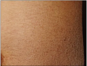
Fig. 1. Poorly defined irregular and polygonal scales on the abdomen.
Mycosis Fungoides as an Ichthyosiform Eruption
Kyung-Hwa Nam, M.D., Jin Park, M.D., Jin-Seok Hong, M.D., Si-Gyun Roh, M.D.
1, Dae-Shick Kim, M.D.
2, Seok-Kweon Yun, M.D., Ph.D.
Departments of Dermatology and 1Plastic Surgery, Chonbuk National University Medical School, Jeonju, 2Department of Pathology, Samsung Medical Center, Sungkyunkwan University School of Medicine, Seoul, Korea
Ichthyosiform eruption as a specific manifestation of mycosis fungoides is very rare and only a few such cases have currently been reported in the medical literature. A 63- year-old Korean man presented with a 4-year history of a pruritic ichthyotic eruption. There was no personal or family history of ichthyosis or atopy. The ichthyosiform skin chan- ges involved the abdomen, arms, thighs and shins. The face, palms and soles were spared. There was no peripheral lymphadenopathy or organomegaly. The typical lesions of mycosis fungoides were not present. The results of the routine investigations were normal or negative. A skin bio- psy specimen revealed the findings of early mycosis fun- goides. He was successfully treated with photochemo- therapy. (Ann Dermatol 21(2) 182∼184, 2009)
-Keywords-
Ichthyosis, Mycosis fungoides
INTRODUCTION
Mycosis fungoides (MF) is the most common type of cutaneous T-cell lymphoma (CTCL), and MF is a mali- gnant lymphoma that’s characterized by the expansion of a clone of the CD4+ (or helper) memory T cells that frequently lacks other normal T-cell antigens (CD7). The skin rash in MF patients usually consists of patches, plaques or tumors that may have a long natural history;
however many atypical variants have also been reported1. The MF patients with malignancy sometimes have ichth-
yosis. Ichthyosiform eruption as a specific manifestation of MF is very rare and this represents 1.8% of all MF cases.
Only a few cases of ichthyosiform eruption as a specific manifestation of MF have currently been reported in the English literature1-8. We report here on the case of a 63- year-old Korean man who presented with ichthyosiform eruption and a histological pattern of MF.
CASE REPORT
A 63-year-old Korean man had a 4-year history of a pruritic eruption. There was no personal or family history of ichthyosis or atopy. The ichthyosiform skin changes were widespread, but they were mainly located on the abdomen, arms, thighs and shins (Fig. 1). The face, palms, soles and most flexure areas were spared. Yet it was interesting that we did find a finely scaled eruption on the axilla. There was no peripheral lymphadenopathy or organomegaly. The typical lesions of MF were not present.
The results of the routine investigations were normal or
Mycosis Fungoides as an Ichthyosiform Eruption
Vol. 21, No. 2, 2009 183 Fig. 2. The biopsy specimen shows a compact, thick orthoke-
ratosis, a thinned epidermis with a slightly decreased granular layer and an upper dermal mononuclear cell infiltrate with Pautrier’s microabscess in the epidermis (H&E, ×400).
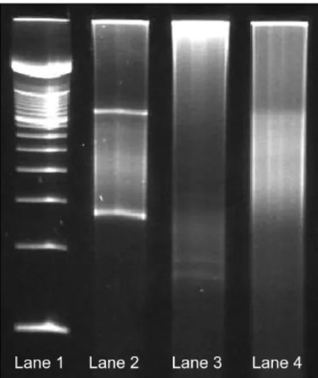
Fig. 3. PCR product run on SSCP gel. Lane 1: molecular weight marker. Lane 2 and Lane 3 show the Vγ1-8 and Vγ11 PCR products from the case, respectively. A well-defined monoclonal pattern was produced with the Vγ1-8 primer. The polyclonal control produced the pattern of smears in lane 4.
negative, including the complete blood count, the di- fferential leukocyte count, the erythrocyte sedimentation rate and the blood chemistry studies. Antinuclear anti- bodies were not detected. The serologic studies were negative for HIV. No Sézary cells were present in the peripheral blood. Chest x-ray examination, gastroscopy and proctoscopy revealed no abnormalities. A skin biopsy specimen from the ichthyosiform lesion on the abdomen revealed parakeratosis and focally compact orthokeratosis of the epidermis with an underlying thinned granular layer. In the superficial dermis, there was a lichenoid infiltrate that mainly consisted of lymphocytes and his- tiocytes and it also showed epidermotrophism and micro- abscess (Fig. 2). The lymphocyte nuclei were hyperchro- matic and cerebriform. Immunostaining confirmed the T cell helper pattern of the dermal and epidermal lym- phocytes (CD2+, CD3+ and CD4+, but CD20- and CD30-). To assess the clonality in the paraffin-embedded samples, polymerase chain reaction single-strand confor- mational polymorphism (PCR-SSCP) was performed as previously described9 to detect T-cell receptor γ (TCR-γ) gene rearrangements. On the results, a monoclonal T-cell clone was detected in the ichthyosiform skin lesion (Fig.
3). Genomic DNA was obtained from the 5-μm sections of the formalin-fixed paraffin-embedded tissues by de- waxing with xylene, proteinase K digestion and phenol/
chloroform extraction. The DNA was precipitated in etha- nol, dried and dissolved in distilled water. Vγ1-8, Vγ9, V γ10, Vγ11 and Jγ1/Jγ2 consensus primers were used and the PCR products were analyzed by SSCP combined with 20% polyacrylamide gel electrophoresis. A chronic nonspecific dermatitis sample was used as a polyclonal
control.
The diagnosis of Mycosis fungoides stage Ib (T2N0M0) was established. The patient was treated with twice weekly PUVA photochemotherapy for 3 months, and the ichthyosiform eruption regressed.
DISCUSSION
In general, MF is a slowly progressing chronic disorder. It usually begins as flat patches, which may or may not be histologically diagnosed as MF. Many variations of MF have been described, such as the follicular, granuloma- tous, hypo- or hyperpigmented and unilesional varients1. Moreover, ichthyosis-like eruption may very rarely be the first sign of MF. Acquired ichthyosis is recognized as one of the cutaneous manifestations of malignancies9. A few cases have recently been reported in which the ichthyosi- form eruption proved to be a specific manifestation of MF2-8.
Acquired ichthyosis may develop in patients of any age and who have several systemic diseases. It has occurred in association with Hodgkin’s lymphoma, non-Hodgkin’s lymphoma, MF, multiple myeloma and carcinomatosis.
Ichthyosiform MF differs from acquired ichthyosis asso- ciated with cutaneous lymphoma10. In the latter condi- tion, the patient presents with both the cutaneous specific lesions of lymphoma and ichthyosis. However, a skin biopsy of the ichthyotic lesions does not show any pathologic aspect of lymphoma, but rather, it shows only epidermal hyperplasia. In the former condition, on the
KH Nam, et al
184 Ann Dermatol
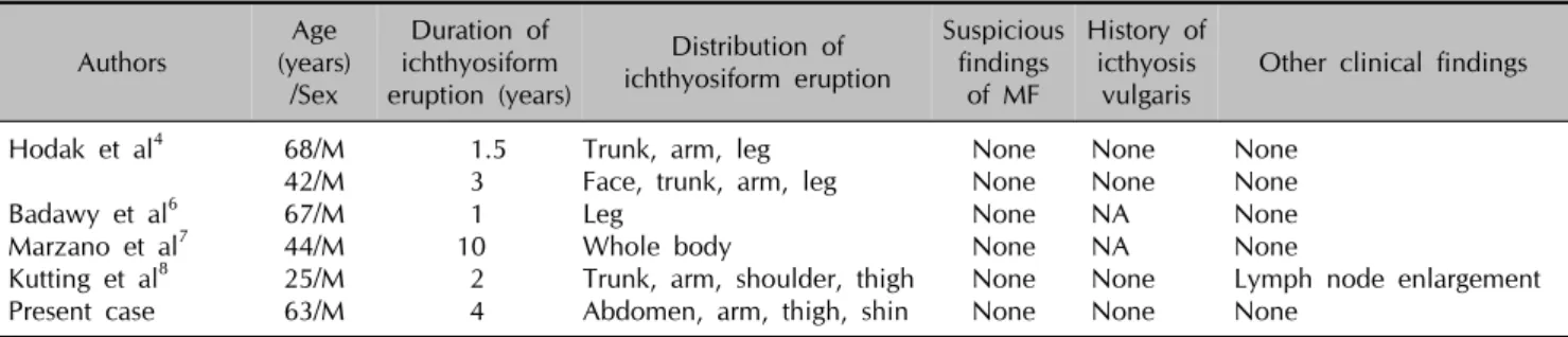
Table 1. Summary of the reported cases that present ichthyosiform eruption as the sole manifestation of mycosis fungoides Authors
Age (years)
/Sex
Duration of ichthyosiform eruption (years)
Distribution of ichthyosiform eruption
Suspicious findings
of MF
History of icthyosis
vulgaris Other clinical findings Hodak et al4
Badawy et al6 Marzano et al7 Kutting et al8 Present case
68/M 42/M 67/M 44/M 25/M 63/M
1.5 3 1 10 2 4
Trunk, arm, leg Face, trunk, arm, leg Leg
Whole body
Trunk, arm, shoulder, thigh Abdomen, arm, thigh, shin
None None None None None None
None None NA NA None None
None None None None
Lymph node enlargement None
NA: information not available
other hand, ichthyosis can be the only clinical mani- festation of MF, and the histologic findings are consistent with both ichthyosis and MF as well. According to the clinical findings, ichthyosiform MF can be divided into 3 types: 1) ichthyosiform eruption as the sole manifestation of the disease, 2) ichthyosiform eruption in conjunction with additional atypical findings of MF and 3) ichthyo- siform eruption in combination with the classic types of MF. To date, 5 cases4,6-8 that showed ichthyosiform erup- tion as the sole manifestation of MF have been reported (Table 1). Our patient also had the sole manifestation of ichthyosiform MF. Clinical remission in such cases is obtained with nonaggressive therapies such as topical treatments, PUVA therapy and ultraviolet light B (UVB)6. Acquired ichthyosis can either indicate the presence of a severe underlying disease or it reveals an atypical form of cutaneous T cell lymphoma. Thus, the patients with ac- quired ichthyosis should be carefully evaluated by per- forming a through biopsy.
REFERENCES
1. Kazakov DV, Burg G, Kempf W. Clinicopathological spec- trum of mycosis fungoides. J Eur Acad Dermatol Venereol 2004;18:397-415.
2. Bianchi L, Papoutsaki M, Orlandi A, Citarella L, Chimenti S.
Ichthyosiform mycosis fungoides: a neoplastic acquired ich- thyosis. Acta Derm Venereol 2007;87:82-83.
3. Sato M, Sohara M, Kitamura Y, Hatamochi A, Yamazaki S.
Ichthyosiform mycosis fungoides: report of a case associated with IgA nephropathy. Dermatology 2005;210:324-328.
4. Hodak E, Amitay I, Feinmesser M, Aviram A, David M.
Ichthyosiform mycosis fungoides: an atypical variant of cu- taneous T-cell lymphoma. J Am Acad Dermatol 2004;50:
368-374.
5. Eisman S, O'Toole EA, Jones A, Whittaker SJ. Granulomatous mycosis fungoides presenting as an acquired ichthyosis. Clin Exp Dermatol 2003;28:174-176.
6. Badawy E, D'Incan M, El Majjaoui S, Franck F, Fabricio L, Dereure O, et al. Ichthyosiform mycosis fungoides. Eur J Dermatol 2002;12:594-596.
7. Marzano AV, Borghi A, Facchetti M, Alessi E. Ichthyosiform mycosis fungoides. Dermatology 2002;204:124-129.
8. Kutting B, Metze D, Luger TA, Bonsmann G. Mycosis fun- goides presenting as an acquired ichthyosis. J Am Acad Dermatol 1996;34:887-889.
9. Signoretti S, Murphy M, Cangi MG, Puddu P, Kadin ME, Loda M. Detection of clonal T-cell receptor gamma gene rearrangements in paraffin-embedded tissue by polymerase chain reaction and nonradioactive single-strand conforma- tional polymorphism analysis. Am J Pathol 1999;154:67-75.
10. Schwartz RA, Williams ML. Acquired ichthyosis: a marker for internal disease. Am Fam Physician 1984;29:181-184.