http://dx.doi.org/10.4174/astr.2014.86.6.289 Annals of Surgical Treatment and Research
Higher incidence of gastroesophageal reflux disease after gastric wedge resections of gastric submucosal tumors located close to the gastroesophageal junction
Seung Yeon Ko, Jeong Sun Lee, Jin-Jo Kim, Seung-Man Park
Division of Gastrointestinal Surgery, Department of Surgery, Incheon St. Mary’s Hospital, The Catholic University of Korea College of Medicine, Incheon, Korea
INTRODUCTION
It is difficult to make a preoperative pathologically confirmed diagnosis of a gastrointestinal stromal tumor (GIST) despite the advancement in endoscopic ultrasonography (EUS) and EUS-guided biopsy techniques [1-5]. Therefore, many patients with a gastric submucosal tumor (SMT) undergo gastric wedge resection without a confirmed diagnosis. Gastric wedge
resection of an SMT located close to the gastroesophageal junction (GEJ) is technically challenging because there is a substantial risk of stenosis of the GEJ or injury to the lower esophageal sphincter (LES). Much concern has been expressed about these issues in the literature [6-17]. However, to the best of our knowledge, there are no reports on the incidence of stenosis of the GEJ or the incidence of gastroeophageal reflux disease (GERD) caused by injury to the LES after surgery. We
Received November 25, 2013, Revised January 6, 2014, Accepted February 12, 2014
Corresponding Author: Jin-Jo Kim
Division of Gastrointestinal Surgery, Department of Surgery, Incheon St.
Mary’s Hospital, The Catholic University of Korea College of Medicine, 56 Dongsu-ro, Bupyeong-gu, Incheon 403-720, Korea
Tel: +82-32-280-5609, Fax: +82-32-280-5988 E-mail: [email protected]
Copyright ⓒ 2014, the Korean Surgical Society
cc Annals of Surgical Treatment and Research is an Open Access Journal. All articles are distributed under the terms of the Creative Commons Attribution Non- Commercial License (http://creativecommons.org/licenses/by-nc/3.0/) which permits unrestricted non-commercial use, distribution, and reproduction in any medium, provided the original work is properly cited.
Purpose: We hypothesized that gastroesophageal reflux disease (GERD) would be more prevalent after a gastric wedge resection of a submucosal tumor (SMT) located close to the gastroesophageal junction (GEJ) than after a gastric wedge resection of an SMT at other locations because of the damage to the lower esophageal sphincter during surgery.
Methods: Fifty-eight patients with gastric SMT who underwent open or laparoscopic gastric wedge resection between January 2000 and August 2012 at the Department of Surgery, Incheon St. Mary's Hospital were enrolled into this study.
The patients were divided into 2 groups according to the location of the tumor, upper or lateral border of the tumor within 5 cm of the GEJ (GEJ ≤ 5 cm group) and upper or lateral border of the tumor greater than 5 cm distal to the GEJ (GEJ >
5 cm group). The surgical records, clinicopathologic findings, postoperative GERD symptoms, postoperative use of acid suppressive medications and preoperative and postoperative endoscopic findings were retrospectively reviewed and compared between the 2 groups.
Results: There was no difference in the frequency of the preoperative GERD symptoms between the 2 groups, whereas postoperative GERD symptoms and postoperative use of acid suppressive medications were more frequent in the GEJ ≤ 5 cm group (P = 0.045 and P = 0.031). However, there were no differences in the follow-up endoscopic findings in terms of reflux esophagitis and Hill’s grade between the 2 groups.
Conclusion: The incidence of GERD was higher after gastric wedge resection of SMTs located close to the GEJ. Hence, adequate care should be taken during the follow-up of these patients.
[Ann Surg Treat Res 2014;86(6):289-294]
Key Words: Gastric wedge resection, Esophagogastric Junction, Gastroesophageal reflux
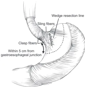
hypothesized that a wedge resection of an SMT whose upper or lateral border is located within 5 cm of the GEJ, but does not involve the GEJ, may cause injury to the LES, especially to the sling fibers (Fig. 1), and may result in GERD at a later stage. The objective of this study is to determine the incidence of GERD after gastric wedge resections of SMTs located close to the GEJ and compare it with the incidence of GERD after gastric wedge resection of SMTs at other locations in the stomach.
METHODS
From January 2000 to August 2012, 69 patients received surgical treatment for a gastric SMT at the Department of Surgery, Incheon St. Mary's Hospital. Among them, 9 patients underwent formal gastrectomies (2 patients underwent total gastrectomy, and 7 patients underwent distal gastrectomy) because of the location and/or the size of the tumor. Among the 2 patients who underwent total gastrectomy, 1 patient initially underwent laparoscopic enucleation for a 2-cm SMT at the GEJ.
However, the final pathologic report revealed a GIST and hence, she underwent a laparoscopic proximal gastrectomy. After the proximal gastrectomy, the patient suffered from severe reflux esophagitis and she finally underwent a laparoscopic completion total gastrectomy at 2 years and 6 months after the proximal gastrectomy. In another 2 patients, gastric SMTs were located at the GEJ. These patients underwent a laparoscopic wedge resection and a prophylactic antireflux surgery, and one
of the 2 cases has been reported elsewhere [18]. The remaining 58 patients who underwent open or laparoscopic gastric wedge resection were enrolled in this study. This study was approved by the Institutional Review Board of the Department of Surgery, Incheon St. Mary's Hospital (OC13RISI0002). Fifty-eight patients were divided into 2 groups according to the location of the tumor. The GEJ ≤ 5 cm group included the patients in whom the upper or lateral border of the tumor was located within 5 cm of the GEJ but it did not involve the GEJ, and the GEJ > 5 cm group included the patients in whom the upper or lateral border of the tumor was located more than 5-cm distal to the GEJ. The distance between the GEJ and the tumor was directly measured with endoscope during the preoperative endoscopy.
Surgical records, clinicopathologic findings, postoperative GERD symptoms, postoperative use of acid suppressive medications and preoperative and postoperative endoscopic findings were retrospectively reviewed and compared between the 2 groups.
Presence of postoperative GERD symptoms during the follow-up period was assessed with a careful review of the medical records when the patient complained of typical GERD symptoms, such as heartburn and/or acid regurgitation, and when the patient complained of epigastric pain and the endoscopic findings showed any evidence of reflux esophagitis; this symptom was also regarded as a GERD symptom. Postoperative use of acid suppressive medications, such as proton pump inhibitors (PPI), H2 receptor antagonists and antacids, in each patient was investigated and the use of such medications for more than 30-day was recorded.
Preoperative endoscopic findings and the last postoperative follow-up endoscopic findings in each patient were reviewed simultaneously by 2 expert endoscopists (J.S.L. and S.M.P.). The degree of reflux esophagitis was graded by the Los Angeles (LA) classification [19] and the endoscopic morphology of the GEJ was classified by Hill’s grade [20]. When there was disagreement in the findings between the 2 endoscopists, they discussed the findings and arrived at consensus. Endoscopically, when there was absence of reflux esophagitis or the LA classification grade was M (minimal change), it was considered that the patient did not have reflux esophagitis, and when the LA classification grade was more than A, it was considered that the patient had reflux esophagitis.
If the final pathology result in a patient indicated a GIST, then the patient was regularly followed up. If the final pathology result indicated a benign SMT, then the patient was followed up based on the symptoms.
All continuous variables are expressed as a mean ± standard deviation. A chi-square test was used to compare the categorical variables, and a Student t-test was used to compare the continuous variables. P-value less than 0.05 was considered statistically significant.
Fig. 1. Illustration for the hypothesis of the development of gastroesophageal reflux disease after a gastric wedge resection for a submucosal tumor located close to the gastroesophageal junction. If the upper or lateral border of the tumor is located within 5 cm from the gastroesophageal junction, there is a substantial risk of damage to the lower esophageal sphincter, especially to the sling fibers after wedge resection for this tumor.
RESULTS
There were 25 men and 33 women and the mean age of the patients was 59 years (range, 31–86 years). The clinical characteristics of each group are shown in Table 1. The incidence of preoperative GERD symptoms and preoperative endoscopic findings were not different between the 2 groups.
The pathologic analysis of 58 SMTs revealed GIST in 48 cases and other benign SMTs in 10 cases (3 leiomyoma, 3 heterotopic pancreas, 1 Schwanoma, 1 glomus tumor, 1 gastric cyst, and 1 inflammatory myofibroblastic tumor). All of the resection margins were not involved by the tumor. A postoperative complication was noted in one case of the GEJ > 5 cm group.
An intraabdominal hematoma developed in this patient and he recovered well after percutaneous drainage and transfusions.
There were no cases of stenosis of the GEJ in the GEJ ≤ 5 cm group. There was no postoperative mortality in either group.
Among the 48 GIST patients, there was no postoperative recurrence during the mean 30 (±23) months of postoperative follow-up (Table 2).
The mean follow-up periods in the GEJ ≤ 5 cm group and the GEJ > 5 cm group were 26.3 (±22.1) months and 21.3 (±20.7) months, respectively. During the follow-up, many patients complained of various upper gastrointestinal symptoms (Table 3), and the patients in the GEJ ≤ 5 cm group had a tendency for developing more upper gastrointestinal symptoms than the patients in the GEJ > 5 cm group, but there was no statistically significant difference (P = 0.072). Eight patients (23.5%) complained of GERD symptoms in the GEJ ≤ 5 cm group and
a greater number of patients complained of GERD symptoms in the GEJ ≤ 5 cm group than in the GEJ > 5 cm group (P = 0.045). The postoperative use of acid suppressive medications for more than 30 days was more frequently observed in the GEJ ≤ 5 cm group (P = 0.031). The mean duration of GERD symptoms of 8 patients in the GEJ ≤ 5 cm group was 21.8 (±18.1) months, and the duration of GERD symptoms of 1 patient in the GEJ > 5 cm group was 18 months. Among 8 patients with GERD symptoms in the GEJ ≤ 5 cm group, symptoms improved by PPI medication for not more than 6 months in 3 patients and symptoms improved by PPI medication for not more than
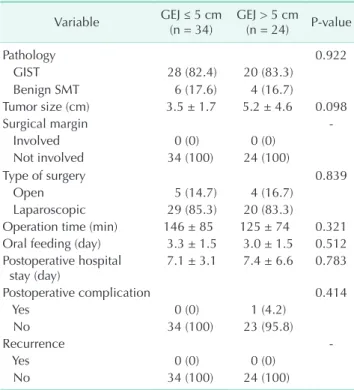
Table 2. Postoperative outcomes after gastric wedge resections
Variable GEJ ≤ 5 cm
(n = 34) GEJ > 5 cm (n = 24) P-value
Pathology 0.922
GIST 28 (82.4) 20 (83.3)
Benign SMT 6 (17.6) 4 (16.7)
Tumor size (cm) 3.5 ± 1.7 5.2 ± 4.6 0.098
Surgical margin -
Involved 0 (0) 0 (0)
Not involved 34 (100) 24 (100)
Type of surgery 0.839
Open 5 (14.7) 4 (16.7)
Laparoscopic 29 (85.3) 20 (83.3)
Operation time (min) 146 ± 85 125 ± 74 0.321 Oral feeding (day) 3.3 ± 1.5 3.0 ± 1.5 0.512 Postoperative hospital
stay (day) 7.1 ± 3.1 7.4 ± 6.6 0.783
Postoperative complication 0.414
Yes 0 (0) 1 (4.2)
No 34 (100) 23 (95.8)
Recurrence -
Yes 0 (0) 0 (0)
No 34 (100) 24 (100)
Values are presented as number (%) or mean ± standard deviation.
GEJ, gastroesophageal junction; GIST, gastrointestinal stromal tumor; SMT, submucosal tumor.
Table 1. Clinical characteristics of the patients with gastric submucosal tumor
Characteristic GEJ ≤ 5 cm
(n = 34) GEJ > 5 cm (n = 24) P-value
Gender 0.373
Male 13 (38.2) 12 (50.0)
Female 21 (61.8) 12 (50.0)
Age (yr) 57 ± 13 61 ± 12 0.231
Preoperative GERD symptoms 0.564
Yes 1 (2.9) 2 (8.3)
No 33 (97.1) 22 (91.7)
Preoperative EGD finding
Reflux esophagitis 0.640
Yes 2 (5.9) 3 (12.5)
No 32 (94.1) 21 (87.5)
Hill’s grade 0.564
≤2 33 (97.1) 22 (91.7)
>2 1 (2.9) 2 (8.3)
Values are presented as number (%) or mean ± standard deviation.
GEJ, gastroesophageal junction; GERD, gastroesophageal reflux disease; EGD, esophagogastroduodenoscopy.
Table 3. Postoperative upper gastrointestinal complaints after gastric wedge resections
Variable GEJ ≤ 5 cm (n = 34)a) GEJ > 5 cm (n = 24)
Indigestion 4 2
Epigastric pain 5 3
Heartburn 4 0
Regurgitation 4 1
Gas bloat 2 0
Dysphagia 1 0
Nausea 0 1
GEJ, gastroesophageal junction.
a)There were more than one symptoms in some patients.
24 months in another 3 patients. However, the remaining 2 patients became chronic PPI users. One of the 2 chronic PPI users who underwent antireflux surgery at 4 years after developing symptoms recovered.
The postoperative follow-up endoscopies were not performed in 10 patients of the GEJ ≤ 5 cm group and in 9 patients of the GEJ > 5 cm group because of benign pathology, old age, etc.
There were no differences in the endoscopic findings in terms of reflux esophagitis and Hill’s grade between the 2 groups (Table 4).
DISCUSSION
The treatment for a gastric GIST is complete surgical resection with a microscopically negative margin [17]. Although it appears to be simple, sometimes it is very difficult to perform such a simple task, especially when the tumor is located close to the GEJ or the pylorus. Some investigators have suggested performing an enucleation or an enucleation-like resection when the tumor is located at or very close to the GEJ [8,13,21].
However, this approach can be dangerous because there is high risk of a microscopically positive margin resulting in a negative impact on the patient’s survival [22]. Uchikoshi et al.
[8] reported a case of recurrence at 2 years after enucleation of a GIST located near the GEJ. There were no cases of recurrent
disease and all of the tumors were excised with negative surgical margins in our study. We think that this was possible because of our surgical policy of performing an open or a laparoscopic complete wedge resection of a gastric SMT mea suring more than 2 cm in size, whenever possible. We performed a formal gastrectomy rather than an enucleation when a complete wedge resection was not possible. There was one exception to our surgical policy as previously mentioned.
The patient is recovering well after 6 years of the initial operation (laparoscopic enucleation) without any evidence of recurrence and after 3 years and 6 months of the laparoscopic completion total gastrectomy without any symptoms of reflux esophagitis.
If the upper or lateral border of an SMT involves the GEJ and the wedge resection line for the tumor crosses the GEJ, there is an apparent resultant damage to the LES. However, it is not certain whether this will result in GERD at a later stage because there is a lack of data about this subject in the literature. Then what will happen in cases in which the lateral border of the tumor is located close to the GEJ but it does not involve the GEJ?
As shown in Fig. 1, LES is composed of 2 muscular components;
clasp and sling fibers. If the clasp and sling fibers are damaged at the same time, as in the wedge resection of an SMT located at the GEJ, there will be an apparent weakening of the LES and resultant development of GERD. What will happen if the damage is limited to some parts of the sling fibers only, as in the wedge resection of a SMT located near the GEJ but does not reach the GEJ? The objective of the current study was to find an answer to this question.
The incidence of GERD after gastric wedge resection of an SMT located close to the GEJ in our study was 23.5%. It was higher than the incidence of GERD in patients who underwent gastric wedge resections of gastric SMTs at other locations in the stomach and the incidence of GERD among the general population in Korea [23]. Moreover, the postoperative use of acid suppressive medications for more than one month was more frequently observed in the GEJ ≤ 5 cm group. This finding supports the higher incidence of GERD in the GEJ
≤ 5 cm group. Many patients complained of various upper gastrointestinal symptoms after gastric wedge resection as shown in Table 3. However, most of the symptoms except for the symptoms of GERD did not persist for more than one month. Hence, we set the cutoff for the duration of medication at one month.
The LA classification and Hill’s grade were used as indicators of GERD in endoscopic finding because Hill’s grade correlates well with the existence and severity of GERD [24]. We failed to demonstrate increased frequency of GERD in the GEJ ≤ 5 cm group with endoscopic findings. However, we think that this was because of a lack of endoscopic follow-up in some of the patients in both groups for several reasons. Therefore, a Table 4. Findings of postoperative follow-up after gastric
wedge resections
Variable GEJ ≤ 5 cm
(n = 34) GEJ > 5 cm (n = 24) P-value Postoperative upper
gastrointestinal symptoms 0.072
Yes 18 (52.9) 7 (29.2)
No 10 (47.1) 17 (70.8)
Postoperative GERD symptoms 0.045
Yes 8 (23.5) 1 (4.2)
No 26 (76.5) 23 (95.8)
Postoperative use of acid
suppressive medication 0.031
Yes 11 (32.4) 2 (8.3)
No 23 (67.6) 22 (91.7)
Postoperative EGD findingsa)
Reflux esophagitis 1.000
Yes 2 (8.3) 1 (7.7)
No 22 (91.7) 12 (92.3)
Hill’s grade 0.690
≤2 17 (70.8) 10 (76.9)
>2 7 (29.2) 3 (23.1)
Values are presented as number (%).
GEJ, gastroesophageal junction; GERD, gastroesophageal reflux disease; EGD, esophagogastroduodenoscopy.
a)Postoperative follow-up endoscopies were not performed in 10 patients of the GEJ ≤ 5 cm group and in 9 patients of the GEJ > 5 cm group because of benign pathology, old age, etc.
prospective study in a large number of patients is needed.
In conclusion, the incidence of GERD was 23.5% after wedge resection of a gastric SMT located close to the GEJ. The incidence of GERD after wedge resection of a gastric SMT located close to the GEJ was higher than the incidence of GERD after wedge resection of an SMT at other locations in the stomach. Therefore, adequate care should be taken during the
follow-up of these patients.
CONFLICTS OF INTEREST
No potential conflict of interest relevant to this article was reported.
1. Buscaglia JM, Nagula S, Jayaraman V, Robbins DH, Vadada D, Gross SA, et al.
Diagnostic yield and safety of jumbo biopsy forceps in patients with sub- epithelial lesions of the upper and lower GI tract. Gastrointest Endosc 2012;75:1147- 52.
2. Papanikolaou IS, Triantafyllou K, Kou- rikou A, Rosch T. Endoscopic ultra sono- graphy for gastric submucosal le sions.
World J Gastrointest Endosc 2011;3: 86-94.
3. Mekky MA, Yamao K, Sawaki A, Mizuno N, Hara K, Nafeh MA, et al. Diagnostic utility of EUS-guided FNA in patients with gastric submucosal tumors. Gastrointest Endosc 2010;71:913-9.
4. Karaca C, Turner BG, Cizginer S, Forcione D, Brugge W. Accuracy of EUS in the evaluation of small gastric subepithelial lesions. Gastrointest Endosc 2010;71:722-7.
5. Polkowski M, Gerke W, Jarosz D, Nasie- rowska-Guttmejer A, Rutkowski P, No- wecki ZI, et al. Diagnostic yield and safety of endoscopic ultrasound-guided trucut [corrected] biopsy in patients with gastric submucosal tumors: a prospective study.
Endoscopy 2009;41:329-34.
6. Tagaya N, Mikami H, Kogure H, Kubota K, Hosoya Y, Nagai H. Laparoscopic intra gastric stapled resection of gastric submucosal tumors located near the eso phagogastric junction. Surg Endosc 2002;16:177-9.
7. Morinaga N, Sano A, Katayama K, Suzuki K, Kamisaka K, Asao T, et al. Laparoscopic transgastric tumor-everting resection of the gastric submucosal tumor located near the esophagogastric junction. Surg Laparosc Endosc Percutan Tech 2004;
14:344-8.
8. Uchikoshi F, Ito T, Nishida T, Kitagawa T, Endo S, Matsuda H. Laparoscopic intragastric resection of gastric stromal tumor located at the esophago-cardiac junction. Surg Laparosc Endosc Percutan Tech 2004;14:1-4.
9. Granger SR, Rollins MD, Mulvihill SJ, Glasgow RE. Lessons learned from laparo- scopic treatment of gastric and gastro- esophageal junction stromal cell tumors.
Surg Endosc 2006;20:1299-304.
10. Song KY, Kim SN, Park CH. Tailored- approach of laparoscopic wedge resection for treatment of submucosal tumor near the esophagogastric junction. Surg Endosc 2007;21:2272-6.
11. Ke ZW, Chen DL, Cai JL, Zheng CZ.
Extraluminal laparoscopic wedge-resection of submucosal tumors on the posterior wall of the gastric fundus close to the esophagocardiac junction. J Laparoendosc Adv Surg Tech A 2009;19:741-4.
12. Ke CW, Cai JL, Chen DL, Zheng CZ.
Extraluminal laparoscopic wedge resec- tion of gastric submucosal tumors: a retrospective review of 84 cases. Surg Endosc 2010;24:1962-8.
13. Shim JH, Lee HH, Yoo HM, Jeon HM, Park CH, Kim JG, et al. Intragastric ap- proach for submucosal tumors located near the Z-line: a hybrid laparoscopic and endoscopic technique. J Surg Oncol 2011;104:312-5.
14. Hara J, Nakajima K, Takahashi T, Yama- saki M, Miyata H, Kurokawa Y, et al.
Laparoscopic intragastric surgery revi- sited: its role for submucosal tumors adjacent to the esophagogastric junction.
Surg Laparosc Endosc Percutan Tech 2012;22:251-4.
15. Sakamoto Y, Sakaguchi Y, Akimoto H, Chinen Y, Kojo M, Sugiyama M, et al.
Safe laparoscopic resection of a gastric gastrointestinal stromal tumor close to the esophagogastric junction. Surg Today 2012;42:708-11.
16. Kim HS, Kim MG, Kim BS, Lee IS, Lee S, Yook JH, et al. Laparoscopic sur gery for submucosal tumor near the esopha- gogastric junction. J Laparoendosc Adv Surg Tech A 2013;23:225-30.
17. Kong SH, Yang HK. Surgical treatment of gastric gastrointestinal stromal tumor. J Gastric Cancer 2013;13:3-18.
18. Lee JS, Kim JJ, Park SM. Laparoscopic gastric wedge resection and prophylactic antireflux surgery for a submucosal tumor of gastroesophageal junction. J Gastric Cancer 2011;11:131-4.
19. Lundell LR, Dent J, Bennett JR, Blum AL, Armstrong D, Galmiche JP, et al.
Endoscopic assessment of oesophagitis:
clinical and functional correlates and further validation of the Los Angeles classification. Gut 1999;45:172-80.
20. Hill LD, Kozarek RA, Kraemer SJ, Aye RW, Mercer CD, Low DE, et al. The gastroesophageal flap valve: in vitro and in vivo observations. Gastrointest Endosc 1996;44:541-7.
21. Coccolini F, Catena F, Ansaloni L, Lazza- reschi D, Pinna AD. Esophagogastric junc tion gastrointestinal stromal tumor:
resection vs enucleation. World J Gastro- enterol 2010;16:4374-6.
22. Langer C, Gunawan B, Schuler P, Huber W, Fuzesi L, Becker H. Prognostic factors
REFERENCES
influencing surgical management and outcome of gastrointestinal stromal tumours. Br J Surg 2003;90:332-9.
23. Kim JJ. Upper gastrointestinal cancer and
reflux disease. J Gastric Cancer 2013;13:79- 85.
24. Kayaoglu HA. Correlation of the gastro- esophageal flap valve grade with the
surgery rate in patients with gastro- esophageal reflux disease. Surg Endosc 2013;27:801-7.