J Korean Surg Soc 2012;83:307-315 http://dx.doi.org/10.4174/jkss.2012.83.5.307
ORIGINAL ARTICLE
Journal of the Korean Surgical Society
JKSS
pISSN 2233-7903ㆍeISSN 2093-0488
Received May 29, 2012, Revised August 10, 2012, Accepted August 23, 2012 Correspondence to: Young-Wook Kim
Division of Vascular Surgery, Department of Surgery, Samsung Medical Center, Sungkyunkwan University School of Medicine, 81 Irwon-ro, Gangnam-gu, Seoul 135-710, Korea
Tel: +82-2-3410-3461, Fax: +82-2-3410-0040, E-mail: [email protected]
cc Journal of the Korean Surgical Society is an Open Access Journal. All articles are distributed under the terms of the Creative Commons Attribution Non-Commercial License (http://creativecommons.org/licenses/by-nc/3.0/) which permits unrestricted non-commercial use, distribution, and reproduction in any medium, provided the original work is properly cited.
Treatment of failing vein grafts in patients who underwent lower extremity arterial bypass
Keun-Myoung Park, Yang Jin Park, Shin-Seok Yang, Dong-Ik Kim, Young-Wook Kim
Division of Vascular Surgery, Department of Surgery, Samsung Medical Center, Sungkyunkwan University School of Medicine, Seoul, Korea
Purpose: We attempted to determine risk factors for the development of failing vein graft and optimal treatment in patients with infrainguinal vein grafts. Methods: We retrospectively reviewed a database of patients who underwent infrainguinal bypass using autogenous vein grafts due to chronic atherosclerotic arterial occlusive disease of lower extremity (LE) at a sin- gle institute between September 2003 and December 2011. After reviewing demographic, clinical, and angiographic features of the patients with failing grafts, we analyzed those variables to determine risk factors for the development of failing grafts.
To determine an optimal treatment for the failing vein grafts, we compared results of open surgical repair (OSR), endovas- cular treatment (EVT) and conservative treatment. Results: Two hundred and fifty-eight LE arterial bypasses using autoge- nous vein grafts in 242 patients were included in this study. During the follow-up period of 39 ± 25 months (range, 1 to 89 months), we found 166 (64%) patent grafts with no restenosis, 41 (15.9%) failing grafts, 39 (15.1%) graft occlusions, and 12 (4.7%) grafts lost in follow-up. In risk factor analysis for the development of a failing graft, no independent risk factors were identified. After 50 treatments of the 41 failing grafts (24 OSR, 18 EVT, 8 conservative management), graft occlusion was sig- nificantly more common in conservative treatment group and severe (>75%) restenosis was significantly more common fol- lowing EVT than OSR (P = 0.001). Reintervention-free graft patency was also superior in the OSR group to that of the EVT group (87% vs. 42%, P = 0.015). Conclusion: OSR of failing grafts has better outcomes than EVT or conservative management in treating failing grafts.
Key Words: Lower extremity, Bypass, Failing graft, Stenosis, Graft occlusion
INTRODUCTION
While the frequency of lower extremity (LE) bypass has recently declined due to widespread use of endovascular procedures, leg bypass with autogenous vein graft still re- mains the gold standard in treatment of long or multi-lev- el, advanced atherosclerotic occlusive lesions of LE arte-
ries. After leg bypass using vein graft, vein graft patency is threatened by subsequent development of stenosis of the vein graft itself or at the anastomotic site in up to 20% of the cases, which is usually due to intimal hyperplasia or progression of native arterial disease [1-3].
The original use of the term “failing graft” was in- troduced by Veith et al. [4] in 1984. A failing graft is defined
Fig. 1. A failing vein graft near the proximal anastomosis of femoro-distal bypass. (A) Duplex ultrasonography showed high (730 cm/sec) peak systolic velocity at the stenotic lesion; (B) tight stenosis (arrow) on digital subtraction angiography; and (C) operative photo of stenotic segment of vein graft close to the proximal anastomosis.
as a vein graft that is patent but hemodynamically failing due to stenosis and exposed to risk for graft occlusion.
Underlying causes of failing grafts are stenotic or occlu- sive lesions in the inflow or outflow arteries, anastomotic sites, or within the vein graft itself. Among these, intrinsic vein graft lesions were known to be the most common cause (60 to 80%) of failing graft, which usually develop in the first 3 to 5 years following vein graft implantation [5].
The emergence of duplex ultrasonography (DUS) has al- lowed for vein graft surveillance following LE arterial by- pass, increasing the likelihood of detecting failing grafts before occurrence of the graft occlusions.
Previous studies have reported that 10 to 50% of failing grafts will occlude within 12 months if not properly treat- ed [6,7]. Therefore, various endovascular or open surgical procedures have been utilized for the treatment of failing grafts, but, there is no consensus on which method is most effective to prolong the patency of the vein graft. In this study, we attempted to determine risk factors for the de- velopment of failing graft after infrainguinal vein grafts implantation and an optimal treatment of the failing graft by comparing the treatment outcomes of failing grafts be- tween open surgical repair (OSR) versus endovascular treatment (EVT) versus conservative treatment.
METHODS
We retrospectively reviewed a database of patients who underwent infrainguinal bypass of chronic LE arterial oc- clusive disease using autogenous vein grafts at a single in- stitution between September 2003 and December 2011. LE bypasses performed on patients with nonatherosclerotic disease or vascular trauma were excluded from this study.
Postoperatively, all patients were prescribed an anti- platelet medication (aspirin, n = 42 limbs; aspirin and clo- pidogrel, n = 216 limbs) and a lipid-lowering medication (e.g., statin) for patients with hyperlipidemia unless contraindicated. Vein graft surveillance was performed using DUS (iU22, Philips, Amsterdam, The Netherlands) at 1 month after bypass surgery and every 6 months thereafter. On DUS, findings of increased peak systolic ve- locity (PSV >300 cm/sec), velocity ratio (≥4) at the sten- otic site, or diminished PSV (<45 cm/sec) distal to the stenotic lesion were used as diagnostic criteria for failing grafts regardless of whether there was symptomatic re- currence (Fig. 1A). Digital subtraction or computed to- mography (CT) angiography was performed to confirm the location and severity of the stenotic lesion before treatment.
To determine risk factors for developing a failing graft after LE vein bypass, we conducted a multivariate analysis using demographic, clinical, and procedural variables.
Fig. 2. Treatment of a failing graft. (A) Failing graft (arrow) close to the proximal anastomosis; (B) after balloon angioplasty (arrow) of the stenotic lesion; (C) vein patch angioplasty; and (D) proximal graft extension with a new vein graft.
Treatment modality of failing grafts was determined by the length of the stenotic lesion; short (≤2 cm) stenotic le- sions were treated with percutaneous transluminal angio- plasty (PTA) (Fig. 2A, B) while longer (>2 cm) stenotic le- sions were treated with vein patch angioplasty or graft ex- tension using a new autogenous vein graft.
For vein patch angioplasty, we exposed the stenotic seg- ment of the vein graft by meticulous dissection with No. 15 blade. To facilitate identification of the stenotic lesion by surgical dissection, vein graft and the stenotic lesion was marked on the overlying skin using DUS before the operation. After administration of intravenous heparin, the proximal and distal of the vein graft stenosis were controlled. Longitudinal incision was made along the an- terior wall of the stenotic segment of the vein graft which sometimes crossed the anastomotic line. Patch closure of the stenotic segment was completed with 6-0 polyprolene continuous suture (Fig. 2C) of the autogenous vein patch.
Proximal or distal extension of the vein graft was also per- formed using a new segment of great saphenous vein by- passing the stenotic lesion (Fig. 2D).
Following the intervention, we continued to use anti- platelet agent and periodic vein graft surveillance using DUS. Patients who refused EVT or OSR were con- servatively treated with prescribing antiplatelet and or lipid lowering agent and performed periodic follow-up
examinations of DUS.
We compared primary or primary assisted patency and reintervention-free patency of the original vein grafts among EVT, OSR, and conservative treatment groups.
Graft patency rates were calculated using the Kaplan- Meier method, and compared them among groups using log rank tests. Reintervention-free patency was calculated from the time of vein graft revision. To determine factors related to treatment failure, multivariate analysis was con- ducted using logistic regression and the Cox proportional hazards model.
RESULTS
Among 395 LE bypasses performed in 341 patients be- tween September 2003 and December 2011, vein grafts were used in 274 limbs (69%) in 242 patients. In this study, we included 258 bypasses, excluding 16 bypasses (in 16 patients) performed for LE arterial occlusion due to non-atherosclerotic causes.
On postoperative follow-up examination of the vein grafts (mean, 39 ± 25 months; range, 1 to 89 months) with DUS, we found 166 (64%) patent grafts with no restenosis, 41 (15.9%) failing grafts, 39 (15.1%) graft occlusions, 12 (4.7%) grafts lost to follow-up and 24 (9.5%) patients were
Characteristic Failing graft (n = 41)
Patent graft with no restenosis
(n = 166)
P-value
Age (yr) 67.7 ± 9.6
(42–82)
68.2 ± 11.5 (39–89)
NS
Male 36 (88) 146 (88) 0.539
Indications for leg bypass 0.598
Claudication 24 (58) 101 (61)
Critical limb ischemia 17 (41) 55 (39)
Type of vein graft 0.219
Reversed saphenous vein 39 (95) 164 (99)
In situ vein 2 (5) 2 (1)
Length of vein graft 0.178
Short vein grafta) 14 (34) 73 (44) Long vein graftb) 27 (66) 95 (57)
Inflow procedure 10 (24) 31 (18) 0.356 Aorto-femoral bypass 4 (10) 3 (2)
Femoro-femoral bypass 2 (4) 9 (5) Iliac stent or angioplasty 4 (10) 19 (11) Co-morbidity or risk factor
Hypertension 32 (78) 120 (72) 0.281
Diabetes mellitus 21 (51) 80 (48) 0.438 Hyperlipidemia 11 (27) 48 (29) 0.367 Ischemic heart disease 17 (42) 59 (36) 0.456 Current smoking 26 (63) 97 (58) 0.377 Cerebral ischemic disease 8 (20) 34 (20) 0.588 Chronic renal failure 3 (7) 14 (9) 0.134 Values are presented as mean ± SD (range) or number (%).
NS, not significant.
a)Vein graft not crossing the knee joint such as femoro-above-knee popliteal bypass or below knee popliteal-distal bypass. b)Vein graft crossing the knee joint such as femoro-below-knee popliteal or below-knee popliteal or femoro-distal bypass.
Table 1. Comparison of demographic, clinical features and vein graft characteristics between the groups of failing graft and patent graft with no restenosis
Characteristic Value
Location of the stenotic lesion Inflow artery
Vein graft close to the proximal anastomosis Mid-graft
Vein graft close to the distal anastomosis Outflow artery
Combined
4 (10) 27 (64) 3 (7) 3 (7) 1 (2) 3 (7) Time to the detection of failing graft (mo)
<6 mo after bypass surgery ≥6 mo after bypass surgery
Length of stenotic lesion on angiography (cm) ≤2
>2
14.3 ± 15.7 (1–84) 11 (27) 30 (73)
27 (66) 14 (34)
Ankle brachial index 0.83 ± 0.02
Clinical presentation
No recurrent ischemic symptom Claudication
Rest pain
Non-healing ulcer or gangrene
14 (34) 24 (59) 1 (2) 2 (5) Duplex US findings
Peak systolic velocity (cm/sec) >300
<45 Velocity ratio
466.19 ± 198.6 36 (88) 5 (12) 6.71 ± 2.81 Values are presented as number (%), mean ± SD (range) or mean ± SD.
US, ultrasonography.
Table 2. Characteristics of failing grafts (n = 41) dead.
Demographic and clinical features of the patients and characteristics of vein grafts were compared between groups with failing graft (n = 41 limbs in 37 patients) and patent graft with no restenosis (n = 166 limbs in 157 pa- tients) (Table 1). There was no significant difference in demographic or clinical features of the patients and char- acteristics of the vein graft between the 2 groups.
The most common site of the stenotic lesions in the fail- ing grafts was found near the proximal anastomosis (64%) as shown in Fig. 2B. Mean time to the detection of a failing graft was 14.3 ± 15.7 months (range, 1 to 84 months) and
most frequently detected later than 6 months after vein graft implantations. Failing grafts clinically presented with recurrent ischemic symptoms in 27 (66%) grafts while 14 (34%) were asymptomatic and detected by peri- odic surveillance using DUS. Two-thirds of stenotic le- sions were found to be short (≤2 cm) on angiography.
Table 2 shows characteristics of the failing grafts.
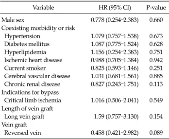
On a risk factor analysis for the development of a failing graft, we were unable to find independent risk factors (Table 3).
In 50 treatments for 41 failing grafts, 24 were treated with OSR (13 graft extensions with new vein grafts; 11 vein patch angioplasties), 18 were treated with EVT (13 balloon angioplasties and 5 stentings), and 8 were managed conservatively. Among 24 OSR group, 5 patients were pre- viously treated with EVT which resulted in an occurrence of restenosis.
During the mean follow-up period of 12.4 ± 16.0 months
Variable HR (95% CI) P-value
Male sex 0.778 (0.254–2.383) 0.660
Coexisting morbidity or risk Hypertension
Diabetes mellitus Hyperlipidemia Ischemic heart disease Current smoker
Cerebral vascular disease Chronic renal disease
1.079 (0.757–1.538) 1.087 (0.775–1.524) 1.156 (0.254–2.383) 0.988 (0.705–1.384) 0.825 (0.593–1.146) 1.031 (0.681–1.561) 0.827 (0.243–1.751)
0.673 0.628 0.751 0.942 0.251 0.885 0.113 Indications for bypass
Critical limb ischemia 1.016 (0.506–2.041) 0.549 Length of vein graft
Long vein graft 1.59 (0.757–3.130) 0.154 Vein graft
Reversed vein 0.458 (0.421–2.982) 0.089 HR, hazard ratio; CI, confidence interval.
a)Cox’s proportional hazard model.
Table 3. Risk factor analysisa) for the development of a failing graft after lower extremity vein graft
Variable
Treatment
P-value Endovascular
(n = 18)
Open surgical (n = 24)
Conservative (n = 8)
Mean age (yr) 63.4 69.5 70.8 NSb)
Male sex 16 (88) 21 (87) 7 (88) 0.990a)
Coexisting morbidity/ risk Hypertension
Diabetes mellitus Hyperlipidemia Ischemic heart disease Current smoking Cerebral vascular disease Chronic renal disease
13 (72) 10 (56) 3 (23) 8 (44) 8 (44) 1 (6) 0 (0)
18 (75) 13 (54) 9 (38) 9 (38) 15 (63) 5 (21) 2 (4)
7 (88) 4 (50) 1 (13) 5 (63) 7 (88) 2 (25) 2 (25)
0.693a) 0.966a) 0.200a) 0.467a) 0.111a) 0.307a) 0.068a)
Long vein graft 10 (56) 17 (70) 4 (50) 0.449a)
Lesion location
Close to prox. anastomosis 11 (61) 15 (63) 7 (88) 0.358a)
Time to reintervention after vein graft implantation (mo) 17.1 ± 14.3 15.4 ± 18.9 8.4 ± 5.5 NSb)
Length of stenotic lesion (cm) 0.83 ± 0.51 1.24 ± 1.23 0.91 ± 0.25 NSb)
Mean duration of follow-up (mo) 9.5 ± 12.8 19.1 ± 17.7 4.9 ± 10.5 NSb)
Treatment results Graft occlusion
Restenosis >75% or required intervention
2 (12) 10 (56)
1 (4) 2 (8)
2 (25) -
0.001a)
Values are presented as number (%) or mean ± SD.
Endovascular treatment included 13 balloon angioplasties and 5 stentings; open surgical repair included 13 graft extensions and 11 patch angioplasties using an autogenous vein; conservative treatment included antiplatelet medication with or without lipid lowering medication.
NS, not significant.
a)Chi-square test. b)Student’s t-test.
Table 4. Comparison of patient data and treatment results (range, 1 to 56 months) after treatment (n = 50) for 41 limbs with failing grafts, we have experienced 5 (10%) graft oc-
clusion, 12 (24%) severe restenosis, and 1 (2%) major limb amputation. Graft occlusion developed in 4% in OSR group, 12% in EVT group and 25% in conservative treat- ment group. We have observed that graft occlusion was most common in conservative treatment group and occur- rence of severe restenosis was significantly more common in EVT group than in OSR (56% vs. 8%, P = 0.001) (Table 4).
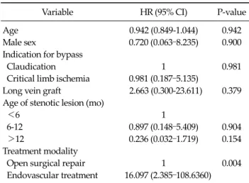
On a multivariate analysis using Cox’s proportional haz- ard model to determine risk factors for treatment failure in patients with failing vein grafts, we found that EVT was an independent risk factor for the development of severe restenosis or graft occlusion (P = 0.004; hazard ratio, 16.097) (Table 5).
Primary assisted patency rates in OSR and EVT group were not significantly different at 1, 3 and 5 years (Fig. 3A) but, 1 year reintervention-free patency rate was sig- nificantly higher in OSR group than in EVT group (87% vs.
42%, P = 0.015, log rank test) (Fig. 3B).
Variable HR (95% CI) P-value
Age 0.942 (0.849-1.044) 0.942
Male sex 0.720 (0.063–8.235) 0.900
Indication for bypass
Claudication 1 0.981
Critical limb ischemia 0.981 (0.187–5.135)
Long vein graft 2.663 (0.300-23.611) 0.379 Age of stenotic lesion (mo)
<6 1
6-12 0.897 (0.148–5.409) 0.904
>12 0.236 (0.032–1.719) 0.154
Treatment modality
Open surgical repair 1 0.004
Endovascular treatment 16.097 (2.385–108.6360) HR, hazard ratio; CI, confidence interval.
a)Cox’s proportional hazard model. b)Treatment failure: graft occlusion, restenosis >75% or reintervention.
Table 5. Multivariate analysisa) for treatment failureb) in patients with failing vein graft
Fig. 3. Primary assisted (A) and reintervention-free patency rates (B) in surgical and endovascular treatment groups.
DISCUSSION
In contrast to term “failed” graft, a “failing” graft de- notes a patent autologous vein graft that is at risk of graft occlusion due to hemodynamically significant narrowing of the inflow artery, outflow artery, or the vein graft itself [4]. In current practice of vascular surgery, early detection of the failing grafts and optimal treatment of the target le- sion has known to be important to prolong patency of the original vein grafts. As many as 80% of patients who pres- ent with recurrent limb ischemia after LE bypass have graft occlusions, and treatment option for the failed graft is usually limited to redo bypass or EVT which demands
more challenging technique and usually results in poorer results than that of the primary bypass surgery [8,9].
Unlike the treatment of the failed graft, the graft patency (primary assisted patency) of the failing vein graft can be prolonged by a less invasive surgical or endovascular procedure.
Many patients with failing grafts are known to experi- ence recurrent ischemic symptoms during the follow-up period, but 10 to 30% of the failing grafts are asymptomatic at the time of detection [7]. In the present study, two thirds of failing grafts presented with recurrent leg ischemic symptom while one-third of failing grafts were detected without recurrent ischemic symptoms. Like other pre- vious reports [5,10], we also experienced an importance of the postoperative DUS surveillance of the vein grafts for early detection of failing grafts after LE bypass surgery with the vein graft.
Suggested DUS-based diagnostic criteria of failing grafts include high velocity criteria such as PSV > 300 cm/sec, PSV ratio > 3.5-4 at the stenotic segment, and low velocity criterion such as PSV < 45 cm/sec distal to the stenotic lesion [11,12]. The diagnostic criteria for failing grafts may differ from institution to institution. CT or con- ventional angiography is usually recommended to re- confirm the location and severity of the stenotic lesion be- fore treatment. We also used digital subtraction or CT an- giography after detection of failing graft with DUS.
Regarding the time interval between vein graft im- plantation and detection of a failing graft, many studies have reported that failing grafts are often detected be-
tween 3 months and 18 months after bypass surgery [10,13]. Schneider et al. [14] reported that 50% of failing grafts were detected within 1 year after bypass. Based on this observation, they recommended more frequent DUS examinations of the vein graft within the first 1 or 2 years after LE bypass grafting. In our study, 73% of the failing grafts were detected later than 6 months and 80% of failing grafts were detected within 2 years after bypass surgery.
The location of vein graft stenosis may be related to the type of vein graft (reversed vein graft vs. in situ vein), di- ameter of the vein graft, location of venous valves, or intra- operative vein graft injury by clamping or cannulation.
Mills et al. [13] reported that the proximal and the distal anastomosis was similar in frequency of stenotic lesions.
According to Berkowitz et al. [15] and Schneider et al. [14], 40% of stenotic lesions were located within 4 cm of the proximal anastomosis due to the location of venous valves or intraoperative vein graft injury from clamping or manipulation. However, Avino et al. [2] and Berceli et al.
[10] reported that stenotic lesions were most commonly seen midgraft due to the diameter of the vein graft and the location of the vein valve. After performing LE bypasses with reversed vein grafts in majority of the patients, we ob- served that 64% of stenotic lesion was located near the proximal anastomosis which was followed by inflow ar- tery, mid graft and vein graft close to the distal anasto- mosis.
A failing graft will typically progress to graft occlusion.
Mofidi et al. [7] reported that 80% of failing grafts became occluded within 2 years when left untreated. It has been reported, however, that revision of the stenotic segment can significantly improve graft patency. According to pre- vious reports, a successful revision of a failing graft can prolong the primary patency by 15% [11,16,17].
The optimal treatment for failing grafts has been a source of debate for decades. Though Mills et al. [11] rec- ommended anticoagulation therapy for the treatment of failing grafts, many other authors recommended an ear- lier treatment of the target lesion with endovascular or surgical intervention [6,18]. Various procedures have been used to treat failing grafts including EVT (e.g., PTA, cut- ting balloon angioplasty, cryoplasty or stenting) and OSR (e.g., vein patch angioplasty, proximal or distal vein graft
extension). Avino et al. [2] recommended the use of PTA for patients with short (<2 cm) stenosis, vein graft diame- ter >3.5 mm, and age of the vein graft >3 months after implantation.
Several studies have reported higher failure rates of PTA compared to OSR [19-21]. Additionally, conflicting evidence has been reported regarding improved results in treatment of failing grafts with cutting balloon angio- plasty [14,22]. Cutting balloon angioplasty was known to be associated with a higher technical success, low short-term patency and higher complication rates so that some authors do not recommend cutting balloon angio- plasty for the treatment of the failing graft [23]. Recently, cryoplasty and drug-eluting stents (DES) were also in- troduced as a treatment for failing vein grafts. Vikram et al. [24] reported a 50% reintervention-free patency rate at 12 months following cryoplasty. As seen in coronary ap- plications, experimental data for DES in infrainguinal fail- ing grafts has recently begun to be released. [25]
On our retrospective study comparing outcomes of OSR, EVT, and conservative management for treatment of failing vein grafts, we found that significantly better re- intervention-free patency rate after OSR compared to EVT.
OSR is reported to provide long-term primary assisted pa- tency in patients with a failing graft [1,14]. We report a 90%
primary assisted patency rate at 5 years in OSR group.
And we found higher rates of graft restenosis (>75%) or occlusion in EVT and conservative treatment groups than in OSR group. On a multivariate risk factor analysis for the treatment failure of the failing graft, EVT showed sig- nificantly higher risk of restenosis despite they were se- lectively performed for patients with a short stenotic le- sion [5].
Surgical options in treatment of failing grafts depend on the location and length of the stenotic lesion as well as the availability of an available autogenous vein. Vein patch angioplasty or vein graft extension (proximal or distal) have been reported with excellent medium and long term patency rates [5]. Focal short stenotic lesions are often caused by intimal hyperplasia or sclerotic valve leaflets, which is a good candidate of vein patch angioplasty.
Stenotic lesions near the anastomotic site or athero- sclerotic lesions involving the distal runoff artery are suit-
able candidates for graft extension with a new vein graft [26].
The present study has several limitations including ret- rospective study design, different indication for each treatment group, use of balloon angioplasty in most cases for EVT and small number in each treatment group. This study was conducted during a period in which treatment protocols, techniques, and device availability was sub- stantially changing.
To conclude our observation, we have experienced that infrainguinal failing vein can be detected in asymptomatic patients by postoperative DUS surveillance. Though there were above described limitations in this study, we found that successful OSR resulted in better outcomes than EVT in treating failing grafts.
CONFLICTS OF INTEREST
No potential conflict of interest relevant to this article was reported.
REFERENCES
1. Nguyen LL, Conte MS, Menard MT, Gravereaux EC, Chew DK, Donaldson MC, et al. Infrainguinal vein bypass graft revision: factors affecting long-term outcome. J Vasc Surg 2004;40:916-23.
2. Avino AJ, Bandyk DF, Gonsalves AJ, Johnson BL, Black TJ, Zwiebel BR, et al. Surgical and endovascular intervention for infrainguinal vein graft stenosis. J Vasc Surg 1999;29:60-70.
3. Landry GJ, Moneta GL, Taylor LM Jr, Edwards JM, Yeager RA, Porter JM. Long-term outcome of revised low- er-extremity bypass grafts. J Vasc Surg 2002;35:56-62.
4. Veith FJ, Weiser RK, Gupta SK, Ascer E, Scher LA, Samson RH, et al. Diagnosis and management of failing lower ex- tremity arterial reconstructions prior to graft occlusion. J Cardiovasc Surg (Torino) 1984;25:381-4.
5. Berceli SA. Revision of vein bypass grafts: factors affecting durability of interventions. Semin Vasc Surg 2009;22:261-6.
6. Carter A, Murphy MO, Halka AT, Turner NJ, Kirton JP, Murray D, et al. The natural history of stenoses within lower limb arterial bypass grafts using a graft surveillance program. Ann Vasc Surg 2007;21:695-703.
7. Mofidi R, Kelman J, Berry O, Bennett S, Murie JA, Dawson AR. Significance of the early postoperative duplex result in
infrainguinal vein bypass surveillance. Eur J Vasc Endo- vasc Surg 2007;34:327-32.
8. Baldwin ZK, Pearce BJ, Curi MA, Desai TR, McKinsey JF, Bassiouny HS, et al. Limb salvage after infrainguinal by- pass graft failure. J Vasc Surg 2004;39:951-7.
9. Eagleton MJ, Erez O, Srivastava SD, Henke PK, Upchurch GR Jr, Stanley JC, et al. Outcome of surgical and endolumi- nal intervention for infrainguinal bypass anastomotic strictures. Vasc Endovascular Surg 2006;40:11-22.
10. Berceli SA, Hevelone ND, Lipsitz SR, Bandyk DF, Clowes AW, Moneta GL, et al. Surgical and endovascular revision of infrainguinal vein bypass grafts: analysis of midterm outcomes from the PREVENT III trial. J Vasc Surg 2007;46:1173-9.
11. Mills JL Sr, Wixon CL, James DC, Devine J, Westerband A, Hughes JD. The natural history of intermediate and critical vein graft stenosis: recommendations for continued sur- veillance or repair. J Vasc Surg 2001;33:273-8.
12. Westerband A, Mills JL, Kistler S, Berman SS, Hunter GC, Marek JM. Prospective validation of threshold criteria for intervention in infrainguinal vein grafts undergoing du- plex surveillance. Ann Vasc Surg 1997;11:44-8.
13. Mills JL, Fujitani RM, Taylor SM. The characteristics and anatomic distribution of lesions that cause reversed vein graft failure: a five-year prospective study. J Vasc Surg 1993;17:195-204.
14. Schneider PA, Caps MT, Nelken N. Infrainguinal vein graft stenosis: cutting balloon angioplasty as the first-line treat- ment of choice. J Vasc Surg 2008;47:960-6.
15. Berkowitz HD, Fox AD, Deaton DH. Reversed vein graft stenosis: early diagnosis and management. J Vasc Surg 1992;15:130-41.
16. Davies AH, Hawdon AJ, Sydes MR, Thompson SG; VGST Participants. Is duplex surveillance of value after leg vein bypass grafting? Principal results of the Vein Graft Surveillance Randomised Trial (VGST). Circulation 2005;
112:1985-91.
17. Lundell A, Lindblad B, Bergqvist D, Hansen F. Femoropo- pliteal-crural graft patency is improved by an intensive surveillance program: a prospective randomized study. J Vasc Surg 1995;21:26-33.
18. Bui TD, Mills JL Sr, Ihnat DM, Gruessner AC, Goshima KR, Hughes JD. The natural history of duplex-detected stenosis after femoropopliteal endovascular therapy suggests ques- tionable clinical utility of routine duplex surveillance. J Vasc Surg 2012;55:346-52.
19. Conte MS, Bandyk DF, Clowes AW, Moneta GL, Seely L, Lorenz TJ, et al. Results of PREVENT III: a multicenter, randomized trial of edifoligide for the prevention of vein graft failure in lower extremity bypass surgery. J Vasc Surg 2006;43:742-51.
20. Alexander JQ, Katz SG. The efficacy of percutaneous trans- luminal angioplasty in the treatment of infrainguinal vein bypass graft stenosis. Arch Surg 2003;138:510-3.
21. Perler BA, Osterman FA, Mitchell SE, Burdick JF, Williams GM. Balloon dilatation versus surgical revision of infra-in- guinal autogenous vein graft stenoses: long-term fol-
low-up. J Cardiovasc Surg (Torino) 1990;31:656-61.
22. Kasirajan K, Schneider PA. Early outcome of "cutting" bal- loon angioplasty for infrainguinal vein graft stenosis. J Vasc Surg 2004;39:702-8.
23. Garvin R, Reifsnyder T. Cutting balloon angioplasty of au- togenous infrainguinal bypasses: short-term safety and efficacy. J Vasc Surg 2007;46:724-30.
24. Vikram R, Ross RA, Bhat R, Griffiths GD, Stonebridge PA, Houston JG, et al. Cutting balloon angioplasty versus
standard balloon angioplasty for failing infra-inguinal vein grafts: comparative study of short- and mid-term primary patency rates. Cardiovasc Intervent Radiol 2007;30:607-10.
25. Yeo KK, Malik U, Laird JR. Outcomes following treatment of femoropopliteal in-stent restenosis: a single center experience. Catheter Cardiovasc Interv 2011;78:604-8.
26. Caps MT, Cantwell-Gab K, Bergelin RO, Strandness DE Jr.
Vein graft lesions: time of onset and rate of progression. J Vasc Surg 1995;22:466-74.