Vol. 25, No. 1, 2013 107
Letter to the Editor
Received January 20, 2012, Revised April 24, 2012, Accepted for publication April 25, 2012
Corresponding author: Jun Young Lee, Department of Dermatology, Seoul St. Mary’s Hospital, College of Medicine, The Catholic University of Korea, 222 Banpo-daero, Seocho-gu, Seoul 137-701, Korea. Tel: 82-2-2258-1386, Fax: 82-2-594-3255, E-mail: [email protected]
This is an Open Access article distributed under the terms of the Creative Commons Attribution Non-Commercial License (http://
creativecommons.org/licenses/by-nc/3.0) which permits unrestricted non-commercial use, distribution, and reproduction in any medium, provided the original work is properly cited.
6. Godar DE. UVA1 radiation triggers two different final apop- totic pathways. J Invest Dermatol 1999;112:3-12.
7. Yamauchi R, Morita A, Yasuda Y, Grether-Beck S, Klotz LO, Tsuji T, et al. Different susceptibility of malignant versus nonmalignant human T cells toward ultraviolet A-1 radia- tion-induced apoptosis. J Invest Dermatol 2004;122:477- 483.
8. Von Kobyletzki G, Heine O, Stephan H, Pieck C, Stücker M, Hoffmann K, et al. UVA1 irradiation induces deoxyribonu- clease dependent apoptosis in cutaneous T-cell lymphoma in vivo. Photodermatol Photoimmunol Photomed
2000;16:271- 277.
9. Tuchinda C, Kerr HA, Taylor CR, Jacobe H, Bergamo BM, Elmets C, et al. UVA1 phototherapy for cutaneous diseases:
an experience of 92 cases in the United States. Photoderma- tol Photoimmunol Photomed 2006;22:247-253.
10. Besaratinia A, Synold TW, Chen HH, Chang C, Xi B, Riggs AD, et al. DNA lesions induced by UV A1 and B radiation in human cells: comparative analyses in the overall genome and in the p53 tumor suppressor gene. Proc Natl Acad Sci U S A 2005;102:10058-10063.
http://dx.doi.org/10.5021/ad.2013.25.1.107
Cutaneous Metastasis of Hepatocellular Carcinoma Following Skin Injury after Transcatheter Arterial Chemoembolization
Eujin Cho, Hei Sung Kim, Young Min Park, Hyung Ok Kim, Jun Young Lee
Department of Dermatology, College of Medicine, The Catholic University of Korea, Seoul, Korea
Dear Editor:
Transcatheter arterial chemoembolization (TACE) is widely used as an approach for patients with hepato- cellular carcinoma (HCC). Cutaneous complications related to TACE such as erythema, necrosis, and scarring may rarely occur1. This report describes the first known case of cutaneous metastasis of HCC following skin injury after TACE.
A 39-year old man had been diagnosed with HCC in January 2010. On the initial computed tomography (CT) image, a 7.8 cm sized tumor was noted. The patient was treated with TACE from February 2010 via the femoral artery. On a follow-up CT in May 2010, the tumor size decreased, however, daughter nodules were noted. The 7th TACE was performed via the internal mammary artery (IMA) due to the presence of collateral pathways. In November 2010, an 8th course of TACE of the IMA was
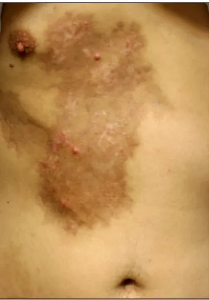
performed. However, an erythematous to violaceous patch developed shortly after the infusion of chemo- therapeutic agents and resulted in pigmentation and induration. In January 2011, the patient was referred to the dermatology clinic with two erythematous papules on the right chest that developed at the end of December 2010 (Fig. 1). The papules had developed within the pigmented induration, which was associated with skin injury after the TACE in November 2010.
Histopathologic examination of the erythematous papule showed asymmetrical, lobulated nests invading the dermis (Fig. 2A). The large cells had a polyhedral shape, eosino- philic cytoplasm, large central nuclei and prominent nucleoli (Fig. 2B). The specimen was negative for AFP (Fig. 2C). CD31 stain did not show evidence of hemato- genous spread of tumor nests (Fig. 2D).
As the papules occurred on the scar and a series of
108 Ann Dermatol Letter to the Editor
Fig. 1. Two, asymptomatic, erythematous papules and underlying pigmentation and wood plate-like induration on the right chest.
Fig. 2. (A) Asymmetrical, lobulated cords and nests invading the dermis (H&E, ×40). (B) The large epithelial cells of polyhedral shape showed granular, eosinophilic cytoplasm, large, vesicular, central nuclei and prominent nucleoli (H&E, ×400). (C) The specimen was negative for α-fetoprotein (AFP) (AFP, ×200). (D) CD31 stain did not show any evidence of hematogenous spread (CD31, ×200).
examinations showed no other metastasis, the lesions were thought to be associated with the skin injury after TACE. The biopsy specimen was negative for AFP.
However, positive staining for AFP has been reported to be present in 2 to 61% of HCC2. Expression of AFP can be weak, especially in small biopsies.
Tumor seeding of HCC after interventional procedures is a rare complication. Cutaneous tumor seeding of HCC along the tract of the needle used in the diagnosis has been reported3. The frequency of needle tract implan- tation after radiofrequency ablation ranges from 0 to 4.4%.3 Iatrogenic cutaneous tumor seeding by ultrasound- guided percutaneous ethanol injection has also been rarely reported4.
TACE via the IMA may be performed when collateral pathways are evident. Repetitive TACEs or large tumor sizes may be associated with the presence of collateral pathways. In our case, TACE-induced injury may have caused inflammatory changes or adhesion of the chest
Vol. 25, No. 1, 2013 109
Letter to the Editor
Received February 14, 2012, Revised April 21, 2012, Accepted for publication May 4, 2012
Corresponding author: Beom Joon Kim, Department of Dermatology, Chung-Ang University Hospital, 102 Heukseok-ro, Dongjak-gu, Seoul 156-755, Korea.
Tel: 82-2-6299-1525, Fax: 82-2-823-1049, E-mail: [email protected]
This is an Open Access article distributed under the terms of the Creative Commons Attribution Non-Commercial License (http://
creativecommons.org/licenses/by-nc/3.0) which permits unrestricted non-commercial use, distribution, and reproduction in any medium, provided the original work is properly cited.
wall that helped the progression of the residual viable tumor along the liver surface to the skin. Adhesion between the HCC and other tissues including vessels such as the peritoneum and/or omentum after TACE has been shown in a laparoscopic study5.
We have presented a case of cutaneous metastasis from HCC following skin injury after TACE that was thought to be relevant to the cutaneous complication of the chemoembolization procedure. The potential cutaneous complications of TACE must be taken into consideration.
REFERENCES
1. Arora R, Soulen MC, Haskal ZJ. Cutaneous complications of hepatic chemoembolization via extrahepatic collaterals. J
Vasc Interv Radiol 1999;10:1351-1356.
2. Minervini MI, Demetris AJ, Lee RG, Carr BI, Madariaga J, Nalesnik MA. Utilization of hepatocyte-specific antibody in the immunocytochemical evaluation of liver tumors. Mod Pathol 1997;10:686-692.
3. Onodera H, Oikawa M, Abe M, Chida N, Kimura S, Satake K, et al. Cutaneous seeding of hepatocellular carcinoma after fine-needle aspiration biopsy. J Ultrasound Med 1987;6:273- 275.
4. Chang S, Kim SH, Lim HK, Kim SH, Lee WJ, Choi D, et al.
Needle tract implantation after percutaneous interventional procedures in hepatocellular carcinomas: lessons learned from a 10-year experience. Korean J Radiol 2008;9:268-274.
5. Seki S, Sakaguchi H, Hagihara A, Fujii H, Kobayashi S, Iwai S, et al. Transcatheter arterial chemoembolization for super- ficial hepatocellular carcinoma induces adhesion. Adv Med Sci 2007;52:66-70.
http://dx.doi.org/10.5021/ad.2013.25.1.109
Rare Manifestation of Giant Molluscum Contagiosum on the Scalp in Old Age
Hyun Kyu Kim, Woo Sun Jang, Beom Joon Kim, Myeung Nam Kim
Department of Dermatology, Chung-Ang University College of Medicine, Seoul, Korea
Dear Editor:
A 64-year-old man presented to our department of hos- pital with a one-month history of skin lesion on the scalp.
Physical examination revealed a skin-colored nodule on the left temporal scalp (Fig. 1A) and a skin-colored verru- cous surfaced plaque on the right temporal area (Fig. 1B).
The lesions gradually increased in size. The patient had no previous history of skin disease or recurrent infection.
Further history-taking and clinical examination did not reveal any risk factors or evidence of an immunocom- promised state.
Histopathological examination of a punch biopsy speci- men from the center of the lesions showed the classic pattern of molluscum contagiosum: subcorneal cysts and
intracytoplasmic inclusion bodies, the so-called mollus- cum bodies, connected with the epidermal surface (Fig. 2).
Based on the clinical features and histopathological fin- dings, we diagnosed molluscum contagiosum. After dia- gnosis, the molluscum were removed by curettage, and no recurrence had been observed at one-year follow-up.
Molluscum contagiosum is a benign, infectious disease of the skin that presents as pearly dome-shaped papules with a central dell or depression. Although worldwide in its distri- bution, molluscum contagiosum has been most frequently encountered as an easily treated disease of childhood and has rarely been a cause of serious morbidity1. It occurs most commonly in children and adolescents, and is usually localized on the face, arms, legs and anogenital regions.