Introduction
The detection of right ventricular (RV) systolic dysfunction (RVSD) due to pulmonary vascular disease in preterm children can be critical for determining prognosis.1) Late-onset pulmo- nary hypertension (PH) has been reported in extremely low birth weight infants2) and children with no or mild bronchopul- monary dysplasia (BPD),3) advocating the need for long-term echocardiographic screening in preterm children. A subset of preterm infants without BPD but with PH detected at 36 weeks of postmenstrual age showed altered RV function on deforma- tion imaging, which persisted up to 1-year corrected age.4) Thus, primary vascular injuries of the lung may cause RV dysfunction even in preterm infants, independent of BPD.5) To date, the se- quential assessment of RV function beyond 1-year corrected age has not been performed in preterm children; therefore, the prog- ress of potential RV dysfunction beyond this period in preterm children remains unclear.
ORIGINAL ARTICLE J Cardiovasc Ultrasound 2017;25(3):98-104
• Received: August 10, 2017 • Revised: September 5, 2017 • Accepted: September 5, 2017
• Address for Correspondence: Soo Jung Kang, Department of Pediatrics, CHA Bundang Medical Center, CHA University School of Medicine, 59 Yatap-ro, Bundang-gu, Seongnam 13496, Korea Tel: +82-31-780-5230, Fax: +82-31-780-5239, E-mail: kittysooni@chamc.co.kr
• This is an Open Access article distributed under the terms of the Creative Commons Attribution Non-Commercial License (http://creativecommons.org/licenses/by-nc/4.0) which permits unrestricted non-commercial use, distribution, and reproduction in any medium, provided the original work is properly cited.
The detection of RVSD due to pulmonary vascular disease in preterm children can be challenging because even in patients with significantly elevated pulmonary artery pressures, symp- toms may be subtle.6) In the RV, the longitudinal shortening of myocardial fibers contributes mainly to stroke volume dur- ing systole.7) Therefore, measuring decreases in RV strain in the longitudinal direction in the presence of afterloads, such as increased pulmonary vascular resistance, would enable the sen- sitive detection of RVSD in preterm children.8) Myocardial strain, indicating changes in the length of myocardial fibers,9) is more sensitive in detecting early myocardial changes among preterm infants than are conventional echocardiographic param- eters.10) We speculated that strain parameters could sensitively detect the progression of potential RVSD among preterm chil- dren. Velocity vector imaging (VVI) utilizes speckle and endo- cardial contour tracking to assess regional and global cardiac function.11) Accordingly, we used VVI to detect possible RVSD
Progression of Right Ventricular Systolic Dysfunction Detected by Myocardial
Deformation Imaging in Asymptomatic Preterm Children
Soo Jung Kang, MD, PhD1, Mira Kim, MD1, Seo Jung Hwang, MT2, and Hyo Jin Kim, MT2
1Departments of Pediatrics, 2Diagnostic Laboratory Medicine, CHA Bundang Medical Center, CHA University School of Medicine, Seongnam, Korea
Background: To detect progression of right ventricular (RV) systolic dysfunction (RVSD) in asymptomatic preterm children from infancy to 24-month corrected age, using velocity vector imaging (VVI).
Methods: Retrospective study comparing sequential RV longitudinal peak systolic strain (LPSS) from 24 children born at < 33 weeks of gestational age and 10 term infants recruited as controls, obtained at a mean of 4-month (first exam) and 24-month correct- ed age (second exam).
Results: In 7/24 (29.2%) of preterm children, RV LPSS of < 16%, defined as RVSD, was detected at the second exam; 5/7 of these children had RV LPSS > 16% at the first exam, and only 2/7 of these children had a history of moderate or severe broncho- pulmonary dysplasia.
Conclusion: In asymptomatic preterm children, routine echocardiographic screening using VVI could detect RVSD which could progress from 4–24 month corrected age.
KEY WORDS: Peak systolic strain · Preterm · Right ventricular systolic dysfunction · Cardiac deformation.
in asymptomatic preterm children from infancy to 24-month corrected age, based on myocardial deformation parameters [namely, RV longitudinal peak systolic strain (LPSS)] and to investigate the possible clinical parameters correlated with RV LPSS.
Methods
This study was approved by the Institutional Review Board of our institution (approval number 2015-09-159-002).
Study population
Preterm infants with < 33 weeks of gestational age (GA) who were admitted to the neonatal intensive care unit of CHA Bun- dang Medical Center were identified from our medical data- base and retrospectively studied. Infants with structural congen- ital heart diseases, except for patent ductus arteriosus (PDA) and patent foramen ovale, and those with genetic disorders were ex- cluded. Demographic information, including GA, birth weight, age, weight, and heart rate during echocardiography, and sys- tolic and diastolic blood pressures were collected. Patient age was specified as corrected age, which was obtained by subtracting the number of weeks born before 40 weeks of gestation from the chronological age.12)
Characteristics during the antenatal and perinatal periods, duration of mechanical ventilation, history of BPD, and history of hemodynamically significant PDA (hsPDA) were assessed.
BPD was defined as the requirement for supplemental oxygen at 36 weeks of GA.13) hsPDA was PDA that required medical intervention. Term infants referred to our clinic for murmurs who were without significant structural heart diseases other than small patent foramen ovale were studied as controls.
Echocardiographic assessment
We retrospectively reviewed the echocardiograms performed at a mean of 4-month corrected age (first exam) and 24-month corrected age (second exam) using commercially available ultra- sound equipment (Acuson SC 2000, Siemens Medical, Moun- tain View, CA, USA).
All echocardiograms were performed as recommended by the American Society of Echocardiography.14) Conventional echocardiographic parameters were measured as recommend- ed.15) PDA diameter was measured at the pulmonary end. Be- cause we excluded patients with significant pulmonary stenosis, maximal tricuspid regurgitation (TR) jet velocity from contin- uous-wave Doppler and right ventricular systolic pressure (RVSP) derived from the modified Bernoulli equation (TR jet veloci- ty2 × 4) was obtained.6)16) We determined PH based on the pres- ence of findings (primary echocardiographic criteria for PH) of RVSP > 40 mm Hg, RVSP/systemic systolic blood pressure >
0.5, and cardiac shunt with bidirectional or right-to-left shunt or any degree of interventricular septal flattening.3)
Left ventricular ejection fraction, fractional area change (FAC) of RV, expressed as a percentage change in the RV chamber area
at end-diastole and end-systole,1) tricuspid inflow Doppler ve- locity during early diastole (E), tricuspid annular tissue Doppler velocity during early diastole (E’), and the ratio of tricuspid E/E’
was obtained according to previous recommendations.17) Iso- volumic contraction time (IVCT) and isovolumic relaxation time (IVRT) were obtained by Doppler tissue imaging, and myocardial performance index was calculated as follows18): RV myocardial performance index = (IVCT + IVRT)/RV systolic ejection time.
Mean pulmonary artery pressure (MPAP) was derived from regression equations using pulmonary artery acceleration time (PAAT): 48–(0.28 × PAAT).19) PAAT was assessed using the pulsed wave Doppler recordings of the pulmonary artery.19) VVI analysis
Digital echocardiographic images of the study population were obtained at 70 frames per second and stored for subsequent offline analysis. On average, three cardiac cycles were studied.
All offline analyses were performed using vendor-customized VVI software (Siemens Medical, version 3.0). Two independent investigators who were blinded to the clinical data of the study population at the time of analysis performed the VVI analysis.
In the apical four-chamber view, at the onset of the QRS wave, we manually tracked the endocardial border of the RV using the VVI software to acquire RV LPSS as recommended.20) The RV LPSS was identified as the highest point of the average strain and strain rate curve derived from six segments (three from the RV free wall and three from the interventricular sep- tum) (Fig. 1).20)
Statistical analysis
All values are expressed as mean ± standard deviation.
SPSS version 24 (IBM SPSS Statistics 24, IBM Corp., Ar- monk, NY, USA) and MedCalc for Windows version 17.5 (MedCalc Software, Ostend, Belgium) were used to analyze data. Echocardiographic and clinical data between the two groups were compared using Student’s t-test or Mann-Whitney U-test, as appropriate. A p value of < 0.05 was considered sig- nificant. Pearson’s or Spearman’s correlation was performed, as appropriate, to determine correlations. One observer performed an offline repeat analysis of RV LPSS in 15 subjects at 4 weeks apart to determine intraobserver variability. Two independent observers who were blinded to clinical data of the children at the time of analysis performed a separate offline analysis of RV LPSS in 15 children to determine interobserver variability. The mean percentage error, which were obtained by calculating the absolute difference of the two datasets divided by the mean of the two datasets,21) were used to determine intraobserver and interobserver variabilities.
Results
A total of 34 children (preterm children, n = 24; controls, n = 10) were studied. Demographic and clinical data of preterm
children and controls are shown in Table 1. Median GA of the preterm children was 29.55 weeks (range: 24–33 weeks GA).
All preterm children were asymptomatic at both time periods, and no patients needed oxygen or demonstrated hsPDA at both time periods. Echocardiographic data of preterm children and controls are shown in Table 2. Among conventional echo- cardiographic parameters, compared with controls, preterm children showed increased mean tricuspid E/E’ during the first exam (preterm children vs. controls, 4.2 ± 1.6 vs. 3.2 ± 1.1;
p < 0.05) and showed increased mean TR jet velocity during
the second exam (preterm children vs. controls, 1.3 ± 0.6 vs.
0.8 ± 0.5 m/s; p < 0.05). Two preterm children showed inter- ventricular septum flattening at the first exam and were deter- mined to have PH, but neither were shown to have PH at the second exam. Over time, in preterm children, the mean heart rate and mean TR jet velocity decreased significantly, and the mean PAAT increased significantly. The mean PAAT did not differ between preterm children and controls during the exam.
The mean RVSP and MPAP of the preterm children were < 40 mm Hg at both time periods. The mean MPAP decreased over time in preterm children (first exam vs. second exam, 31.1 ± 4.2 vs. 24.3 ± 8.0 mm Hg; p < 0.05), although the mean RVSP re- mained elevated compared with the controls (preterm children vs. controls, 8.0 ± 7.0 vs. 3.9 ± 4.9 mm Hg; p < 0.05) during the second exam.
Over time, in preterm children, the mean RV LPSS did not show significant difference between both exams; however, the mean RV LPSS was decreased in preterm children compared with that in controls at both time periods (Fig. 2). We further analyzed the change of RV LPSS in individual preterm children at both time periods. The change over time of RV LPSS in in- dividual preterm children who showed RVSD at 24 months corrected age is shown in Fig. 3. We defined decreased RV LPSS of < 16% as RVSD (referencing the lower level of RV LPSS of the controls in our study). In 11 preterm children (45.8%), RVSD was detected during the exam. In 8 of these 11 children, RVSD improved during the second exam. In 3 of the 11 pre- term children with RVSD during the first exam, RVSD was persistently detected during the second exam. In five preterm
Fig. 1. Example of right ventricular longitudinal peak systolic strain (RV LPSS) identified as the highest point of the average strain curve derived from six segments observed using velocity vector imaging in a preterm child.
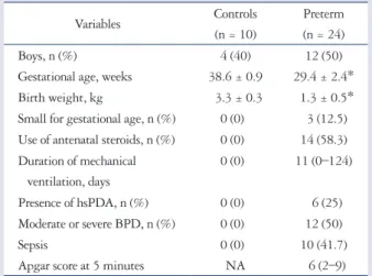
Table 1. Demographic and clinical data of preterm children and controls
Variables Controls
(n = 10)
Preterm (n = 24)
Boys, n (%) 4 (40) 12 (50)
Gestational age, weeks 38.6 ± 0.9 29.4 ± 2.4*
Birth weight, kg 3.3 ± 0.3 1.3 ± 0.5*
Small for gestational age, n (%) 0 (0) 3 (12.5) Use of antenatal steroids, n (%) 0 (0) 14 (58.3) Duration of mechanical
ventilation, days
0 (0) 11 (0–124)
Presence of hsPDA, n (%) 0 (0) 6 (25)
Moderate or severe BPD, n (%) 0 (0) 12 (50)
Sepsis 0 (0) 10 (41.7)
Apgar score at 5 minutes NA 6 (2–9)
Data are presented as mean ± standard deviation, or number (percentage).
*p < 0.05 vs. controls. hsPDA: hemodynamically significant patent ductus arteriosus, BPD: bronchopulmonary dysplasia, NA: not available
children (20.8%), RVSD was not detected during the first exam, but progression to RVSD was noted during the second exam.
Totally, in seven preterm children (29.2%), RVSD was detect- ed at 24-month corrected age. The demographic, clinical, and
echocardiographic data of preterm children with and without RVSD at 24-month CA are shown in Table 3.
Intraobserver and interobserver variability of RV deformation indices
The mean percentage error for intraobserver and interobserv- er variabilities in RV LPSS was 20% and 21%, respectively.
Correlation of RV LPSS with clinical variables In preterm children, small birth weight was negatively cor- related significantly with RV LPSS during the first exam (r = -0.446, p = 0.029). GA, sex, systolic and diastolic blood pres-
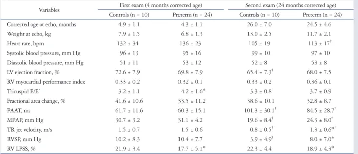
Table 2. Echocardiographic data of preterm children and controls
Variables First exam (4 months corrected age) Second exam (24 months corrected age) Controls (n = 10) Preterm (n = 24) Controls (n = 10) Preterm (n = 24)
Corrected age at echo, months 4.9 ± 1.1 4.3 ± 1.1 26.0 ± 7.0 24.5 ± 4.6
Weight at echo, kg 7.9 ± 1.5 6.8 ± 1.3 13.0 ± 2.5 11.7 ± 2.1
Heart rate, bpm 132 ± 34 136 ± 23 105 ± 19 113 ± 17†
Systolic blood pressure, mm Hg 96 ± 13 95 ± 16 99 ± 10 97 ± 10
Diastolic blood pressure, mm Hg 51 ± 11 53 ± 12 52 ± 8 53 ± 8
LV ejection fraction, % 72.6 ± 7.9 69.8 ± 7.9 65.4 ± 7.3† 68.0 ± 7.5
RV myocardial performance index 0.33 ± 0.2 0.32 ± 0.1 0.33 ± 0.2 0.36 ± 0.1
Tricuspid E/E’ 3.2 ± 1.1 4.2 ± 1.6* 3.3 ± 0.8 3.7 ± 0.9
Fractional area change, % 41.6 ± 10.6 33.5 ± 11.2 38.6 ± 10.1 32.8 ± 8.7
PAAT, ms 61.7 ± 11.6 60.3 ± 15.1 101.3 ± 30.1† 84.5 ± 28.7†
MPAP, mm Hg 30.7 ± 3.2 31.1 ± 4.2 19.6 ± 8.4† 24.3 ± 8.0†
TR jet velocity, m/s 1.5 ± 0.7 1.5 ± 0.6 0.8 ± 0.5† 1.3 ± 0.6*†
RVSP, mm Hg 10.2 ± 8.3 10.4 ± 7.7 3.9 ± 4.9† 8.0 ± 7.0*
RV LPSS, % 21.9 ± 3.4 17.7 ± 5.1* 22.3 ± 4.4 18.9 ± 4.3*
Data are presented as mean ± standard deviation, or number (percentage). Strain values are expressed as absolute values. *p < 0.05 vs. controls, †p < 0.05 vs.
first exam. bpm: beats per minute, LV: left ventricular, RV: right ventricular, E/E’: ratio between tricuspid inflow Doppler velocity during early diastole (E) and tricuspid annular tissue Doppler velocity during early diastole (E’), PAAT: pulmonary artery acceleration time, MPAP: mean pulmonary artery pressure, TR:
tricuspid regurgitation, RVSP: right ventricular systolic pressure, RV LPSS: right ventricular longitudinal peak systolic strain
Fig. 2. Change over time in right ventricular longitudinal peak systolic strain (RV LPSS) in preterm children and controls from a mean 4- to 24-month corrected age. LPSS1: RV LPSS at a mean 4 month corrected age, LPSS2: RV LPSS at a mean 24 month corrected age.
26
24
22
20
18
16
14
LPSS1 LPSS2
RV LPSS (%)
Controls Preterms
Fig. 3. Change over time in right ventricular longitudinal peak systolic strain (RV LPSS) in individual preterm children with right ventricular systolic dysfunction at a mean 24 month corrected age. LPSS1: RV LPSS at a mean 4 month corrected age, LPSS2: RV LPSS at a mean 24 month corrected age.
28 26 24 22 20 18 16 14 12 10
LPSS1 LPSS2
RV LPSS (%)
sures, history of severity of BPD, presence of hsPDA, use of an- tenatal steroids, and duration of mechanical ventilation all did not show a significant correlation with RV LPSS in preterm chil- dren at both time periods.
Discussion
In our study, the overall mean RV LPSS of preterm children showed no significant change from a mean 4–24-month cor- rected age over time but remained decreased at both time peri- ods compared with controls. The mean TR jet velocity de- creased over time in preterm children but remained increased compared with controls at mean 24-month corrected age. In seven of 24 preterm children (29.2%), RVSD, defined as RV LPSS < 16%, was detected at a mean of 24-month corrected age.
In five of seven children with RVSD at 24-month corrected age (20.8% of all preterm children), no RVSD was detected at mean 4-month corrected age, and RVSD progressed after a mean of 4-month corrected age. Low birth weight was negatively corre- lated with RV LPSS in preterm children at a mean of 4-month corrected age; however, the severity of BPD, duration of me-
chanical ventilation, presence of hsPDA, and use of antenatal steroids all did not correlate with RV LPSS. Age and weight during the exam did not correlate with RV LPSS in preterm children at both time periods.
Our study is the first to sequentially analyze RV LPSS in pre- term children from a mean 4–24-month corrected age. Our results of the mean RV LPSS remaining decreased compared with controls from 4- to 24-month corrected age may be due to elevated pulmonary vascular resistance in the preterm children compared with controls, as TR jet velocity was elevated in the preterm children compared with controls at 24-month correct- ed age.
However, TR jet velocity, in contrast to RV LPSS, was not significantly different between preterm children and controls at 4 months corrected age in our study. Since detection of TR jet velocity has not always been possible for children with chronic lung disease,6) RV LPSS might be a more sensitive parameter of RV dysfunction due to potentially elevated pulmonary vas- cular resistance in preterm children. In our study, during the second exam, RVSD was detected in 7 of 24 preterm children
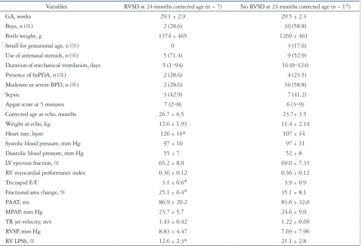
Table 3. Demographic, clinical, and echocardiographic data of preterm children with and without RVSD at a mean 24 months corrected age Variables RVSD at 24 months corrected age (n = 7) No RVSD at 24 months corrected age (n = 17)
GA, weeks 29.1 ± 2.9 29.5 ± 2.3
Boys, n (%) 2 (28.6) 10 (58.8)
Birth weight, g 1374 ± 465 1269 ± 461
Small for gestational age, n (%) 0 3 (17.6)
Use of antenatal steroids, n (%) 5 (71.4) 9 (52.9)
Duration of mechanical ventilation, days 5 (1–94) 16 (0–124)
Presence of hsPDA, n (%) 2 (28.6) 4 (23.5)
Moderate or severe BPD, n (%) 2 (28.6) 10 (58.8)
Sepsis 3 (42.9) 7 (41.2)
Apgar score at 5 minutes 7 (2–8) 6 (3–9)
Corrected age at echo, months 26.7 ± 6.5 23.7± 3.5
Weight at echo, kg 12.6 ± 1.93 11.4 ± 2.14
Heart rate, bpm 126 ± 16* 107 ± 14
Systolic blood pressure, mm Hg 97 ± 10 97 ± 11
Diastolic blood pressure, mm Hg 55 ± 7 52 ± 8
LV ejection fraction, % 65.2 ± 8.0 69.0 ± 7.33
RV myocardial performance index 0.36 ± 0.12 0.36 ± 0.12
Tricuspid E/E’ 3.1 ± 0.6* 3.9 ± 0.9
Fractional area change, % 25.1 ± 6.4* 35.1 ± 8.1
PAAT, ms 86.9 ± 20.2 83.6 ± 32.0
MPAP, mm Hg 23.7 ± 5.7 24.6 ± 9.0
TR jet velocity, m/s 1.43 ± 0.42 1.22 ± 0.69
RVSP, mm Hg 8.83 ± 4.47 7.69 ± 7.96
RV LPSS, % 12.6 ± 2.3* 21.1 ± 2.8
Data are presented as mean ± standard deviation, median (range), or number (percentage). Strain values are expressed as absolute values. *p < 0.05 vs. children without RVSD at 24-month corrected age. RVSD: right ventricular systolic dysfunction, GA: gestational age, hsPDA: hemodynamically significant patent ductus arteriosus, BPD: bronchopulmonary dysplasia, bpm: beats per minute, LV: left ventricular, RV: right ventricular, E/E’: ratio between tricuspid inflow Doppler velocity during early diastole (E) and tricuspid annular tissue Doppler velocity during early diastole (E’), PAAT: pulmonary artery acceleration time, MPAP: mean pulmonary artery pressure, TR: tricuspid regurgitation, RVSP: right ventricular systolic pressure, RV LPSS: right ventricular peak longitudinal systolic strain
(29.2%). In these children, RVSP was significantly higher com- pared with the controls during the second exam, although not reaching PH levels. As only two of these seven children had a history of moderate or severe BPD, we speculate that in at least five of these children primary lung vascular injury not associ- ated with BPD may have occurred over time,3)5) thus acting as an afterload to RV and causing RVSD over time. In five of these seven children who showed RVSD during the second exam (20.8% of preterm children), no RVSD was observed during the first exam. We speculate that this progression to RVSD in preterm children who initially did not show RVSD at a mean 4 month corrected age may have resulted from repeated epi- sodes of undetected hypoxemia, causing the progressive wors- ening of lung function, and vascular remodeling in preterm chil- dren, which could act as an afterload to the RV, causing RVSD.2)22) Our results are also in accord with other studies on the presence of late-onset PH detected by TR jet velocity and other con- ventional echocardiographic parameters.2)3)
Since RV LPSS detected using VVI is a relatively angle-in- dependent parameter compared with tricuspid E/E’, we specu- late that RV LPSS would enable a more sensitive detection of subtle RVSD in asymptomatic preterm children, in addition to conventional echocardiographic parameters such as tricus- pid E/E’.1) Tricuspid E/E’, a parameter that identifies RV dia- stolic dysfunction,16) has been reported to determine the sever- ity of BPD16) by evaluating RV function in preterm children.
However, in our study, we found significant elevations in the mean tricuspid E/E’ in preterm children compared with those in controls at the first exam but not at the second exam. Addi- tionally, we did not observe differences in mean FAC among preterm children compared with controls at both time periods, although FAC has been useful in differentiating RVSD among patients with BPD.16) In our study, five of the seven preterm children who showed RVSD during the second exam had a history of no or only mild BPD, and this might explain why the use of antenatal steroids, severity of BPD, and duration of me- chanical ventilation did not correlate with RV LPSS at both time periods. Similar results have been reported by Schubert et al.23) who noted no significant correlations between RV de- formation indices and the severity of BPD. RV LPSS correlated negatively with birth weight in preterm children during the first exam in our study, which is in accord with a previous study reporting growth restriction at birth and a greater risk of PH in preterm infants.2) Additionally, the presence of hsPDA did not correlate with RV LPSS at both time periods in our study.
Others were also unable to demonstrate the association of hsP- DA with cardiac function in preterm infants.23)
The limitations of this study were the heterogeneity in pa- tients, the retrospective design that could not provide causali- ty, the wide intraobserver and interobserver variability, and the small number of patients, which may have affected our statisti- cal results. Strain and strain rate are both frame rate-sensitive,24) and the higher heart rates of preterm children than those of con-
trols during the second exam may have affected VVI analysis because of out-of-plane motion.24) Considering the relatively low frame rate of 70 frames per second applied in our study, we focused on RV LPSS only because strain rate is known to be more frame rate-sensitive.24) RV LPSS was seen from the apical four-chamber view only; apical three-chamber and two-cham- ber views were not studied, probably accounting for the low reproducibility of our RV LPSS data.25)
Conclusion
RV performance, which is assessed using myocardial defor- mation imaging, remained significantly decreased over time in asymptomatic preterm infants compared with controls from 4- to 24-month corrected age. Subtle RVSD could be detected in a subgroup of preterm children at a mean 24-month corrected age, and furthermore, in 20.8% of asymptomatic preterm children, RVSD could develop at later than 4-month correct- ed age and progress up to 24-month corrected age, irrespective of BPD history. Long term screening with VVI in low-risk as- ymptomatic preterm children may help in detecting subtle RVSD and aid in determining prognosis. Further studies in- volving a larger group of patients will be necessary to validate these findings.
References
1. Breatnach CR, Levy PT, James AT, Franklin O, El-Khuffash A. Nov- el echocardiography methods in the functional assessment of the newborn heart. Neonatology 2016;110:248-60.
2. Bhat R, Salas AA, Foster C, Carlo WA, Ambalavanan N. Prospective analysis of pulmonary hypertension in extremely low birth weight infants. Pe- diatrics 2012;129:e682-9.
3. Mourani PM, Sontag MK, Younoszai A, Miller JI, Kinsella JP, Baker CD, Poindexter BB, Ingram DA, Abman SH. Early pulmonary vascu- lar disease in preterm infants at risk for bronchopulmonary dysplasia. Am J Respir Crit Care Med 2015;191:87-95.
4. Levy PT, El-Khuffash A, Patel MD, Breatnach CR, James AT, San- chez AA, Abuchabe C, Rogal SR, Holland MR, McNamara PJ, Jain A, Franklin O, Mertens L, Hamvas A, Singh GK. Maturational pat- terns of systolic ventricular deformation mechanics by two-dimensional speckle- tracking echocardiography in preterm infants over the first year of age. J Am Soc Echocardiogr 2017;30:685-98.e1.
5. Farrow KN, Steinhorn RH. Pulmonary hypertension in premature infants.
Sharpening the tools of detection. Am J Respir Crit Care Med 2015;191:
12-4.
6. Mourani PM, Sontag MK, Younoszai A, Ivy DD, Abman SH. Clini- cal utility of echocardiography for the diagnosis and management of pulmonary vascular disease in young children with chronic lung disease. Pediatrics 2008;
121:317-25.
7. Petitjean C, Rougon N, Cluzel P. Assessment of myocardial function: a review of quantification methods and results using tagged MRI. J Cardiovasc Magn Reson 2005;7:501-16.
8. Levy PT, Holland MR, Sekarski TJ, Hamvas A, Singh GK. Feasibili- ty and reproducibility of systolic right ventricular strain measurement by speck- le-tracking echocardiography in premature infants. J Am Soc Echocardiogr 2013;26:1201-13.
9. Haque U, Stiver C, Rivera BK, Richards B, Ma N, Cua CL, Smith CV, Backes CH. Right ventricular performance using myocardial deforma-
tion imaging in infants with bronchopulmonary dysplasia. J Perinatol 2017;
37:81-7.
10. Nestaas E, Støylen A, Brunvand L, Fugelseth D. Longitudinal strain and strain rate by tissue Doppler are more sensitive indices than fractional shortening for assessing the reduced myocardial function in asphyxiated neo- nates. Cardiol Young 2011;21:1-7.
11. Pirat B, Khoury DS, Hartley CJ, Tiller L, Rao L, Schulz DG, Nagueh SF, Zoghbi WA. A novel feature-tracking echocardiographic method for the quantitation of regional myocardial function: validation in an animal model of ischemia-reperfusion. J Am Coll Cardiol 2008;51:651-9.
12. Engle WA; American Academy of Pediatrics Committee on Fetus and Newborn. Age terminology during the perinatal period. Pediatrics 2004;114:1362-4.
13. Jobe AH, Bancalari E. Bronchopulmonary dysplasia. Am J Respir Crit Care Med 2001;163:1723-9.
14. Lai WW, Geva T, Shirali GS, Frommelt PC, Humes RA, Brook MM, Pignatelli RH, Rychik J; Task Force of the Pediatric Council of the American Society of Echocardiography; Pediatric Council of the American Society of Echocardiography. Guidelines and standards for performance of a pediatric echocardiogram: a report from the Task Force of the Pediatric Council of the American Society of Echocardiography. J Am Soc Echocardiogr 2006;19:1413-30.
15. Lang RM, Bierig M, Devereux RB, Flachskampf FA, Foster E, Pel- likka PA, Picard MH, Roman MJ, Seward J, Shanewise JS, Solomon SD, Spencer KT, Sutton MS, Stewart WJ; Chamber Quantification Writing Group; American Society of Echocardiography’s Guidelines and Standards Committee; European Association of Echocardiogra- phy. Recommendations for chamber quantification: a report from the Ameri- can Society of Echocardiography’s Guidelines and Standards Committee and the Chamber Quantification Writing Group, developed in conjunction with the European Association of Echocardiography, a branch of the European So- ciety of Cardiology. J Am Soc Echocardiogr 2005;18:1440-63.
16. Sehgal A, Malikiwi A, Paul E, Tan K, Menahem S. Right ventricular function in infants with bronchopulmonary dysplasia: association with respi- ratory sequelae. Neonatology 2016;109:289-96.
17. Lopez L, Colan SD, Frommelt PC, Ensing GJ, Kendall K, Younoszai AK, Lai WW, Geva T. Recommendations for quantification methods dur- ing the performance of a pediatric echocardiogram: a report from the Pediatric Measurements Writing Group of the American Society of Echocardiography
Pediatric and Congenital Heart Disease Council. J Am Soc Echocardiogr 2010;
23:465-95.
18. Lang RM, Badano LP, Mor-Avi V, Afilalo J, Armstrong A, Ernande L, Flachskampf FA, Foster E, Goldstein SA, Kuznetsova T, Lancellotti P, Muraru D, Picard MH, Rietzschel ER, Rudski L, Spencer KT, Tsang W, Voigt JU. Recommendations for cardiac chamber quantification by echo- cardiography in adults: an update from the American Society of Echocardiog- raphy and the European Association of Cardiovascular Imaging. J Am Soc Echocardiogr 2015;28:1-39.e14.
19. Levy PT, Patel MD, Groh G, Choudhry S, Murphy J, Holland MR, Hamvas A, Grady MR, Singh GK. Pulmonary artery acceleration time provides a reliable estimate of invasive pulmonary hemodynamics in children.
J Am Soc Echocardiogr 2016;29:1056-65.
20. Voigt JU, Pedrizzetti G, Lysyansky P, Marwick TH, Houle H, Bau- mann R, Pedri S, Ito Y, Abe Y, Metz S, Song JH, Hamilton J, Sen- gupta PP, Kolias TJ, d’Hooge J, Aurigemma GP, Thomas JD, Badano LP. Definitions for a common standard for 2D speckle tracking echocardiography: consensus document of the EACVI/ASE/Industry Task Force to standardize deformation imaging. J Am Soc Echocardiogr 2015;28:
183-93.
21. Kutty S, Padiyath A, Li L, Peng Q, Rangamani S, Schuster A, Dan- ford DA. Functional maturation of left and right atrial systolic and diastolic performance in infants, children, and adolescents. J Am Soc Echocardiogr 2013;26:398-409.e2.
22. Poets CF, Stebbens VA, Richard D, Southall DP. Prolonged episodes of hypoxemia in preterm infants undetectable by cardiorespiratory monitors. Pe- diatrics 1995;95:860-3.
23. Schubert U, Müller M, Abdul-Khaliq H, Norman M. Preterm birth is associated with altered myocardial function in infancy. J Am Soc Echocar- diogr 2016;29:670-8.
24. Sanchez AA, Levy PT, Sekarski TJ, Hamvas A, Holland MR, Singh GK. Effects of frame rate on two-dimensional speckle tracking-derived mea- surements of myocardial deformation in premature infants. Echocardiography 2015;32:839-47.
25. Levy PT, Sanchez Mejia AA, Machefsky A, Fowler S, Holland MR, Singh GK. Normal ranges of right ventricular systolic and diastolic strain measures in children: a systematic review and meta-analysis. J Am Soc Echo- cardiogr 2014;27:549-60.e3.