Cerebral ischemia is caused by arterial occlusion due to a thrombus or an embolus. Such oc- clusion induces multiple and concomitant pathophysiological processes that involve bioener- getic failure, acidosis, loss of cell homeostasis, excitotoxicity, and disruption of the blood-brain barrier. All of these mechanisms contribute to neuronal death, mainly via apoptosis or necro- sis. The immune system is involved in this process in the early phases after brain injury, which contributes to potential enlargement of the infarct size and involves the penumbra area. Where- as inflammation and the immune system both exert deleterious effects, they also contribute to brain protection by stimulating a preconditioning status and to the concomitant repair of the injured parenchyma. This review describes the main phases of the inflammatory process occurring after arterial cerebral occlusion, with an emphasis on the role of single mediators.
Key Wordszz ischemic stroke, inflammation, immune response.
Postischemic Inflammation in Acute Stroke
INTRODUCTION
Stroke is a leading cause of disability in adults that has a heavy social burden worldwide.
This disease is the third highest cause of mortality, resulting in approximately six million deaths annually.1 Acute cerebral ischemia accounts for more than 80% of all strokes and is due to brain arterial occlusion resulting from a thrombus or embolus. The pathophysiologi- cal processes following ischemic stroke are complex, involving bioenergetic failure, acidosis, loss of cell homeostasis, excitotoxicity, activation of neuronal and glial cells, and disruption of the blood-brain barrier (BBB) with infiltration of leukocytes.2 There is evidence that fac- tors of the immune system are involved in all stages of acute cerebral ischemia (Fig. 1).3 The ischemic brain promotes a potent suppressive effect on lymphoid organs via the autonomic nervous system, which increases the risk of the poststroke infections that are major deter- minants of morbidity and mortality.4 On the other hand, the innate immune system con- tributes to subsequent repair of the damaged cerebral tissue.5
In this review we describe the main phases of the inflammatory processes during the ear- ly postischemic period, with an emphasis on the role of single mediators.
INFLAMMATION, ENDOTHELIUM, AND CLOT FORMATION
A growing amount of attention is being paid to the mechanisms of clot formation, particu- larly in the field of endovascular treatment of acute ischemic stroke. Although most of the focus has been on intervention devices (with developments from first- to second-generation devices, thrombus aspiration, and balloon-occlusion guiding catheters), some research groups have studied the physiopathological mechanisms of clot formation. The relation be- tween inflammation and clot formation has been described previously, and it indicates that Simone Vidalea
Arturo Consolib Marco Arnaboldia Domenico Consolic
a Department of Neurology and Stroke Unit, Sant’Anna Hospital, Como, Italy
b Department of Interventional Neurovascular Unit, Careggi University Hospital, Florence, Italy
c Department of Neurology,
G. Jazzolino Hospital, Vibo Valentia, Italy
pISSN 1738-6586 / eISSN 2005-5013 / J Clin Neurol 2017;13(1):1-9 / https://doi.org/10.3988/jcn.2017.13.1.1
Received September 12, 2016 Revised October 30, 2016 Accepted October 31, 2016 Correspondence Simone Vidale, MD
Department of Neurology and Stroke Unit, Sant’Anna Hospital, Via Napoleona 60, 22100 Como, Italy Tel +39-0315859282
Fax +39-0315854989
E-mail simone.vidale@asst-lariana.it
cc This is an Open Access article distributed under the terms of the Creative Commons Attribution Non-Com- mercial License (http://creativecommons.org/licenses/by-nc/3.0) which permits unrestricted non-commercial use, distribution, and reproduction in any medium, provided the original work is properly cited.
JCN
Open Access REVIEWInflammation in Acute Ischemic Stroke
JCN
some cytokines (e.g., the RANTES)6 are responsible for the activation of a biochemical cascade or, indirectly, supports the concept that infections/inflammation promote athero- genesis and that some endothelial modifications that can lead to a prothrombotic status.7 Several research groups are currently focusing on the clot structure, with many findings supporting the concept of an inflammation-induced process.
In particular, fibrinogen is susceptible to oxidation, and chronic exposure to oxidative stress supported by inflamma- tion may lead to prothrombotic alterations in fibrin forma- tion and architecture. Previous studies concerning air pollu- tion have shown that particulate matter contributes to modulation of the fibrin structure.8-11 Furthermore, some in- teresting studies that have used electron microscopy to inves- tigate the clot surface (in myocardial infarction) highlight the less-investigated issue of the relation between the clot and the endothelium.12
EARLY POSTISCHEMIA TIME:
THE ISCHEMIC CASCADE
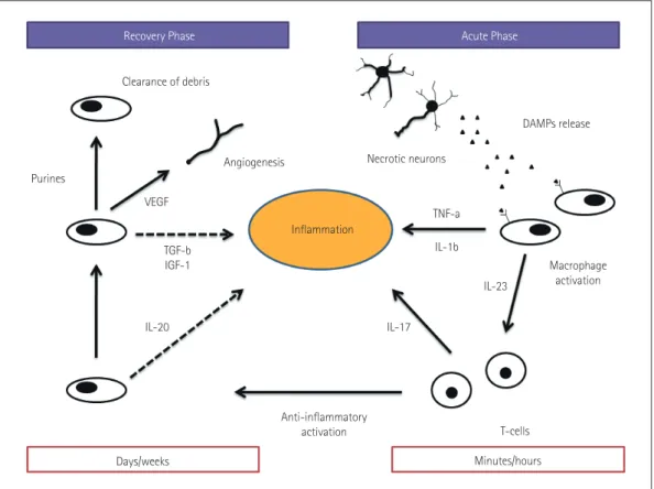
The ischemic cascade is represented by a complex series of interlinked molecular and cellular mechanisms that contrib- ute to ischemic cell death via necrosis or apoptosis (Fig. 2).
The primary insult after arterial occlusion is hypoperfusion, which dramatically reduces the availability of both glucose and oxygen in the brain, with particular vulnerability to isch- emic injury being evident in specific regions: the caudate body, putamen, insular ribbon, paracentral lobule, precen- tral, and middle and inferior frontal gyri.13 This situation contributes to bioenergetic failure by stopping or slowing ad- enosine triphosphate (ATP) production.14,15 A few minutes after an arterial occlusion, an ionic imbalance occurs with the abnormal influx of Na+ and efflux of K+, contributing to a widespread anoxic depolarization in the membranes of neu- rons and glial cells.16 The increased influx of Na+ into neu- rons causes the osmotic transport of water into cells that leads to cytotoxic edema, cell lysis, and necrosis. A recent
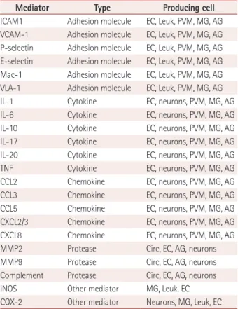
Fig. 1. Postischemic inflammation. Necrotic neurons release damage-associated molecular patterns (DAMPs), and these molecules activate mac- rophages via pattern-recognition receptors and inflammasomes. The activated macrophages contribute to enhance the inflammatory process via the release of proinflammatory cytokines and recruiting T-cells that contribute to maintain inflammation by interleukin (IL)-17. At several days af- ter the acute injury, the cellular elements of the innate immune system change to an anti-inflammatory phenotype, contributing to inhibit the in- flammation (dashed lines). In particular, anti-inflammatory cytokines (e.g., IL-10) are released. During this phase, the postischemic inflammation is resolved by the clearance of debris as well as angiogenesis supported by the release of growth factors. IGF: insulin-like growth factor, TGF, trans- forming growth factor, TNF: tumor necrosis factor, VEGF: vascular endothelial growth factor.
Clearance of debris
DAMPs release Necrotic neurons
TNF-a IL-1b
IL-17
IL-23
Macrophage activation
T-cells Minutes/hours Inflammation
Purines
VEGF
Angiogenesis
TGF-b IGF-1
IL-20
Days/weeks
Anti-inflammatory activation
Recovery Phase Acute Phase
Vidale S et al.
JCN
neuroimaging study using 23Na MRI and quantitative histo- chemical K+ staining revealed heterogeneity in the rate of Na concentration increase and in the K+ distribution within the ischemic core.17 The reduced ATP production following Na/
K imbalance (due to Na/K ATPase) also contributes to re- duce the reuptake of glutamate, which is the main excitatory neurotransmitter.18 This condition overstimulates the gluta- mate receptors so as to influence the Ca2+ influx, producing a series of nuclear and cytoplasmic events (excitotoxicity) that lead to mitochondrial failure and apoptosis.19,20 At the same time, the Ca2+ influx triggers the activation of catabolic en- zymes with the production of arachidonic acid, and increases the formation of reactive oxygen species (ROS), mainly in neurons rather than astrocytes.21
Previous studies found that mitochondrial failure could be predicted from the K+ concentration. Indeed, the mitochon- drial ATP-dependent K+ (mitoKATP) channel plays a critical role in the neuroprotective action, contributing to the so- called ischemic pre- and postconditioning states.22 The open- ing of mitoKATP channels attenuates the Ca2+ overload and
inhibits the formation of free radicals and ROS that contrib- ute to necrotic or apoptotic cell death.23,24 The depolarization of other neurons produces a further Ca2+ influx and addi- tional glutamate release, leading to local amplification of the ischemic damage.25 Contemporary with those processes, the persistence of arterial occlusion contributes to a critical re- duction of pO2 and a concomitant increase in pCO2. In the case of hypercapnia, the tissue pH could fall to around 6.6 or lower if severe ischemia and tissue hypoxia occur; in the last situation, anaerobic glycolysis leads to lactic acid accumula- tion with signs of irreversible injury identifiable in the cell morphology.26 The acidosis state increases necrosis and cell death via a mechanism called acidotoxicity and mediated by Ca2+-permeable acid-sensing ion channels.27-29 Other delete- rious effects of acidosis influence the synthesis and degrada- tion of cellular constituents, the mitochondrial function, the cell volume control, the postischemic flow, and the stimula- tion of ROS production, all conditions that occur also in the ischemic penumbra.30 Acidosis and ROS contribute to trig- ger a subsequent and concomitant phase represented by the
Fig. 2. Cerebral ischemic cascade. AMPA: α-amino-3-hydroxy-5-methyl-4-isoxazolepropionic acid, BAD: Bcl-2-associated death promoter, BBB:
blood-brain barrier, COX: cyclo-oxigenase, IL: interleukin, NMDA: N-methyl-D-aspartate, TNF: tumor necrosis factor.
Arterial occlusion
Hypoperfusion
Anaerobic glycolisis
Na/K-ATPase pump failure
Cell membrane depolarization
Excitotoxicity glutamate release
Activation of NMDA/AMPA
Activation kinase and proteinases
NO synthase activation
H2O accumulation Acidosis
Membrane degradation
Cell adhesion molecules expression
Leukocyte infiltration
Activation COX2
BBB disruption
BAD, BAX activation
Arachidonic acid production
Cytotoxic edema
Cell lysis
Production cytokines (IL-6, TNF-a)
Apoptosis
Necrosis NO
Mithocondrial failure
Intracellular
Na, Ca Free radicals
Oxidative stress
Inflammation
Inflammation in Acute Ischemic Stroke
JCN
activation of innate immunity and involving both resident cells (microglia) and circulating cells.
INFLAMMATORY AND INNATE IMMUNITY ACTIVATIONS
Inflammation in the ischemic brain:
cell infiltration and damage
Postischemic inflammation begins in the vascular compart- ment immediately after arterial occlusion. The production of ROS leads to an increase in the procoagulant state involving the activation of complement, platelet, and endothelial cells.31,32 The increased activity of cyclooxygenase-2 in in- flammatory cells and neurons may lead to tissue damage due to excessive ROS production and toxic prostanoids.33,34 ROS contribute also to reduce the availability of NO, leading to platelet aggregation and the adhesion of leukocytes, which aggravate the ischemic damage.35 There is evidence of iNOS (the inducible isoform of NO) being a critical effector and amplifier of tissue damage related to postischemic inflamma- tion (Table 1).36 The oxidative stress and inflammatory medi- ators affect the permeability of the BBB, impairing the so- called neurovascular unit that includes endothelial cells,
astrocytes, and neurons [involving matrix metalloproteinases (MMPs) or ROS], and allowing the extravasation of proteins and endothelial cells along with the activation of macro- phages and mast cells via the ischemia and reperfusion mech- anisms.37 This extravasation is also supported by a break- down of the BBB−secondary to the pericyte death−leading to a long-lasting decrease in capillary blood flow.38
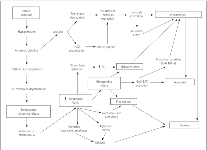
A few minutes after arterial occlusion and the associated in- tra- and extracellular modifications, the acute local damage is detected also by pattern-recognition receptors (PRR) (includ- ing Toll-like receptors) that respond to microbial structures (pathogen-associated molecular patterns) and host-derived danger signals (damage-associated molecular patterns).39-41 These molecules can be released by stressed cells, such as dur- ing the ischemic cascade. There is new evidence that the PRR of neurons and glial cells can play a fundamental role in acti- vating intracellular signaling pathways so as to enhance the proinflammatory expression of different genes (Fig. 3).42,43 This mechanism activates the immune system elements, resulting in mast cells releasing vasoactive mediators (e.g., histamine), proteases, and tumor necrosis factor (TNF), while macro- phages release proinflammatory cytokines.44
Adhesion-molecule P- and E-selectins (Table 2) and inter-
Fig. 3. DAMP receptors and signaling pathways. Cells detect DAMPs via DAMP receptors in two ways: (1) activation of a type of pattern-recogni- tion receptor [Toll-like receptor (TLR)] and (2) activation of inflammasomes. The first mechanism involves proinflammatory factors being released by the nuclear gene expression mediated by transcriptional mediators activated by TLR. The second mechanism involves the activation of cas- pase-1 leading to the clivation of the proinflammatory cytokines IL-1 and IL-18, converting them into their activated forms. DAMP: damage-asso- ciated molecular pattern, IL: interleukin, NLRP: nod-like receptor pyrin.
DAMPs
TLR
Transcriptional mediators
Pro-inflammatory gene expression Pro-caspase 1
Nucleus
Pro-IL-1 Pro-IL-18
IL-1 IL-18 NLRP and 3
Caspase 1
Inflammasome
Vidale S et al.
JCN
cellular adhesion molecule-1 are expressed on the membrane after their intracellular translocation and with the rapid gen- eration of proinflammatory signals.45,46 The adhesion recep- tors mediate interactions between adhesion molecules and integrins, contributing to an initial rolling mechanism of leu- kocytes and leading to adhesion to the endothelium and to a subsequent transmigration into the brain parenchyma (leu- kocyte infiltration). Following ischemia, these cells rapidly release proinflammatory mediators into the area, which pro- motes ischemic injury via different pathways: 1) the cerebral no-reflow phenomenon by impeding the flow of red blood cells, 2) increased production of ROS and proteases at the endothelium surface, 3) platelet aggregation by leukotriene, prostaglandin, or eicosanoid production due to activation of leukocyte phospholipases, and 4) deleterious activity of pro- inflammatory cytokines in the penumbra area. In particular, during the acute phase of brain ischemia, microglia and macrophages release interleukin (IL)-1b, IL-6, and IL-18 from the transcriptional intracellular pathways activated by
nucleotides from injured cells.47 These cytokines contribute to leukocyte infiltration in the damaged tissue, and they acti- vate the presentation of antigens between dendritic cells and T-cells.48 T-cells lead to tissue damage by innate immunity, through interferon-gamma and ROS. T-cells activated by IL- 23 released from microglia and macrophages produce IL-17, which worsens the acute ischemic cerebral injury.49 This un- balanced interplay between the immune and sympathetic nervous systems contributes to an early down-regulation of systemic cellular immune responses that leads to a functional deactivating of monocytes, T-helper cells, and invariant nat- ural-killer T-cells.50
Previous studies have demonstrated the predictive role of certain acute immune and stress biomarkers on clinical out- comes: copeptin and mid-regional proatrial natriuretic pep- tide.51,52 Even if the immune system is activated with recruit- ing elements in the focal ischemic area, specific injured cerebral sites contribute to different down-regulating respons- es being exhibited by the autonomous nervous system. In particular, involvement of the right frontoparietal cortex, in- sula, or brainstem could lead to increased cerebral inflamma- tion and concomitant systemic immunosuppression.53 This process is characterized by the increased apoptosis of lym- phocytes, suppression of peripheral cytokine release, and fi- Table 2. Mediators of initial postischemic inflammation
Mediator Type Producing cell
P-selectin Adhesion molecule EC, PLT
IL-1b Cytokine MG, PVM, MC
IL-1a Cytokine PLT
TNF-a Cytokine MC
CCL5 Chemokine
CXCL4 Chemokine
CXCL7 Chemokine PLT
CX3CL1 Chemokine Neurons
Elastase Protease
MMP8 Protease
MMP9 Protease
MT6-MMP Protease Leuk
Clotting factors Protease Plasma
Complement Protease Plasma, EC, neuron
Prostanoids Small molecule EC, PLT, MG, neurons Leukotrienes Small molecule EC, PLT, MG, neurons
ATP Small molecule Plasma, neurons
Free radicals Small molecule EC, PLT, Leuk, PVM, MG, neurons
Modified from Iadecola et al. Nat Med 2011;17:796-808, with permis- sion of Springer Nature.45
ATP: adenosine triphosphate, CCL: chemokine ligand, CX: d-chemokine ligand, EC: endothelial cell, IL: interleukin, Leuk: leukocytes, MC: mast cells, MG: microglia, MMP: matrix metalloproteinase, PLT: platelets, PVM: perivascular macrophages, TNF: tumor necrosis factor.
Table 1. Mediators of amplification of postischemic inflammation
Mediator Type Producing cell
ICAM1 Adhesion molecule EC, Leuk, PVM, MG, AG VCAM-1 Adhesion molecule EC, Leuk, PVM, MG, AG P-selectin Adhesion molecule EC, Leuk, PVM, MG, AG E-selectin Adhesion molecule EC, Leuk, PVM, MG, AG Mac-1 Adhesion molecule EC, Leuk, PVM, MG, AG VLA-1 Adhesion molecule EC, Leuk, PVM, MG, AG
IL-1 Cytokine EC, neurons, PVM, MG, AG
IL-6 Cytokine EC, neurons, PVM, MG, AG
IL-10 Cytokine EC, neurons, PVM, MG, AG
IL-17 Cytokine EC, neurons, PVM, MG, AG
IL-20 Cytokine EC, neurons, PVM, MG, AG
TNF Cytokine EC, neurons, PVM, MG, AG
CCL2 Chemokine EC, neurons, PVM, MG, AG
CCL3 Chemokine EC, neurons, PVM, MG, AG
CCL5 Chemokine EC, neurons, PVM, MG, AG
CXCL2/3 Chemokine EC, neurons, PVM, MG, AG
CXCL8 Chemokine EC, neurons, PVM, MG, AG
MMP2 Protease Circ, EC, AG, neurons
MMP9 Protease Circ, EC, AG, neurons
Complement Protease Circ, EC, AG, neurons
iNOS Other mediator MG, Leuk, EC
COX-2 Other mediator Neurons, MG, Leuk, EC Modified from Iadecola et al. Nat Med 2011;17:796-808, with permis- sion of Springer Nature.45
AG: astroglia, CCL: chemokine ligand, Circ: plasma, COX: cyclo-oxige- nase, CX: d- chemokine ligand, EC: endothelial cell, ICAM: intercellular adhesion molecule, IL: interleukin, iNOS: inducible isoform of NO, Mac:
macrophage antigen, MG: microglia, MMP: matrix metalloproteinase, PVM: perivascular macrophages, TNF: tumor necrosis factor, VCAM:
vascular cell adhesion molecule, VLA: very late antigen.
Inflammation in Acute Ischemic Stroke
JCN
nally the inhibition of T-helper-1 cells and alteration of the T-helper-1/T-helper-2 ratio.54 The stroke-induced immuno- depression contributes to increasing the risk of infection (in- fluenced also by comorbidities such as age, diabetes mellitus, and atrial fibrillation) and consequently a poor functional outcome.
The activity of microglia
The microglia represent the resident immune cells of the central nervous system and can be activated by local and sys- temic infections, neurodegenerative conditions, and injury.
Microglia are able to modify their morphology from a rest- ing (ramified) to an active (amoeboid) state.55 Microglia re- spond rapidly to ischemic stroke and other injuring condi- tions. By entering the ischemic core within 60 minutes after the induction of focal ischemia without reperfusion, microg- lia significantly increase the number of their processes, while 24 hours later they are reduced in both number and distance from the insult. Previous studies have detected various forms of activated microglia.56 M1 is the proinflammatory type and is able to release TNF-a, IL-1b, IL-18, and IL-6. On the other hand, the M2 type is the healing cell involved in neuropro- tection and repair, producing transforming growth factor (TGF)-b, nerve growth factor, and IL-4.57 The M1 type is ob- served for the first 24 hours in the ischemic core, and it in- creases in number over the first 2 weeks after the ischemic injury.58 The M2 type has been found at the end of the first 24 hours, entering the area during the first week before de- clining in number.55
Recent studies have provided evidence of microglia activa- tion in the penumbra area. Indeed, the pattern of activity dif- fers between the peri-infarct zone microglia and the ischemic core. During the first week, the M2 type was found to pre- dominate in the ischemic core while the M1 type predomi- nated in the peri-infarct zone.59 These observations suggest that the peri-infarct region is dominated by proinflammatory and activated microglia whose abundance increases during the first days after ischemic injury.60 The spatial distribution of the microglia phenotypes changes over time, which suggests the enlargement of injured and damaged cerebral tissue.
The role of inflammasomes
The above-mentioned inflammatory response leads to the production of proinflammatory cytokines and neuronal and glial cell death mediated by large intracellular multiprotein complexes called inflammasomes.45 Recent studies have demonstrated that the nod-like receptor pyrin (NLRP) and NLRP3 inflammasomes in neurons and glial cells allow de- tection of cellular damage and mediation of the inflammato- ry responses to aseptic tissue injury during cerebral ischemia.42
An increased activity of inflammasomes is also associated with neuron and glial cell death. Experimental studies and human data suggest that the NLRP1 and NLRP3 inflamma- somes in brain cells could activate pro-caspase-1 (cleaving to caspase-1) to produce IL-1b and IL-18, which are proinflam- matory cytokines, and lead to a particular type of cell death called pyroptosis (Fig. 3).61 In this way, the inflammasomes contribute to activating and supporting the innate immunity but also to worsening the tissue injury.
RESOLUTION OF INFLAMMATION AND REPAIR OF TISSUE
The inflammatory postischemia process is self-limiting, and its resolution is mediated by numerous factors that suppress the immune activity (Table 3). The termination of inflamma- tion triggers structural and functional reorganization of the injured brain. The first mechanism involved in this phase is the removal of dead cells, which is performed by microglia and infiltrating macrophages, mainly comprising phago- cytes.59,62 The principal factors driving these cells to the isch- emic site are purines released from injured cells and chemo- kines. Immunoglobulins directed against antigens of the central nervous system may also promote the release of IL- Table 3. Mediators of resolution of postischemic inflammation
Mediator Type Producing cell
BDNF Growth factor EC, Macr, AG, PVM, neurons EPO Growth factor EC, Macr, AG, PVM, neurons FGF Growth factor EC, Macr, AG, PVM, neurons G-CSF Growth factor EC, Macr, AG, PVM, neurons IGF-1 Growth factor EC, Macr, AG, PVM, neurons NGF Growth factor EC, Macr, AG, PVM, neurons VEGF Growth factor EC, Macr, AG, PVM, neurons
TGF-b Cytokine MG, Macr, AG
IL-10 Cytokine MG, Macr, AG
IL-17 Cytokine MG, Macr, AG
IL-23 Cytokine MG, Macr, AG
MMP9 Protease AG, neurons
Complement Protease Circ, EC, AG, neurons Prostaglandin Small molecule
Lipoxin Small molecule Docosanoid Small molecule
Modified from Iadecola et al. Nat Med 2011;17:796-808, with permis- sion of Springer Nature.45
AG: astroglia, BDNF: brain-derived neurotrophic factor, Circ: plasma, EC: endothelial cell, EPO: erythropoietin, FGF: fibroblast growth factor, G-CSF: granulocyte colony-stimulating factor, IGF: insulin-like growth factor, IL: interleukin, Macr: macrophages, MG: microglia, MMP: matrix metalloproteinase, NGF: nerve growth factor, PVM: perivascular mac- rophages, TGF: transforming growth factor, VEGF: vascular endothelial growth factor.
Vidale S et al.
JCN
10 and TGF-b, which contribute to suppressing the immune process and to inhibiting the expression of adhesion mole- cules and the production of proinflammatory cytokines (Fig.
1).63 These pleiotropic immunoregulatory cytokines can fa- cilitate tissue repair after promoting the resolution of inflam- mation, and they exert cytoprotective effects on the surviving cells in the ischemic area.35 Concomitant growth factors re- leased by inflammatory cells, neurons, and astrocytes64 sup- port cell sprouting, neurogenesis, and angiogenesis as well as matrix reorganization after ischemic injury. Insulin-like growth factor-1 is a critical factor in the sprouting of neurons after cerebral ischemia, while the reactivity of astrocytes is mandatory for the functional recovery of damaged tissue.65 Concomitant actions of vascular endothelial growth factor and neutrophil MMPs are required in angiogenesis, support- ing the need for the combined activity of inflammatory cells and astrocytes.66
NEURONAL PRECONDITIONING AND INFLAMMATORY MEDIATORS
The brain is the most metabolically active organ in the body, consuming about 25% of total glucose and oxygen, and so a high oxidative stress is generated by its metabolism. Neurons would therefore be expected to respond actively to stress, have low capacities for replacement, and strong potential strategies for protection and repair. While most of the under- lying neuroprotective and repairing pathways are still under investigation, this adaptive response to an insult by the acti- vating intracellular signals is well known, and called precon- ditioning.67 Preconditioning occurs in two distinct phases.
The first happens early and is associated with posttransla- tional modifications of proteins. This period lasts from min- utes to 1–2 hours, and involves certain protective proteins (mainly protein chaperones) being rapidly released from stressed cells. The second phase requires the synthesis of new proteins. During this phase, which lasts several days, the pro- inflammatory cytokines IL-1b and TNF activate intracellular signaling pathways that lead to a tolerant state by mediating the stress responses. In particular, ROS and cytokines are able to activate Ras, Raf, and kinases of the cellular mem- brane, with the subsequent intracellular transcription and production of survival proteins. These last mechanisms con- tribute to preserving the endothelium via vascular protection and angiogenesis [mitogen-activated protein kinase p38 and extracellular signal-regulated kinase 1/2 (ERK 1/2)], the neu- rons via neuroprotection and neurogenesis [ERK 1/2 and nuclear factor kB (NF-kB)], and the glia via an anti-inflam- matory action (serine/threonine kinase).68 Other proteins, called heat-shock proteins (Hsp), contribute to cellular main-
tenance.69 In particular, Hsp70 leads to neuronal survival, mitochondrial stabilization, and cell-death blocking, mainly by facilitating the activation of transcription factor and NF- kB.70,71 NF-kB is affected also by the phosphorylation of Akt secondary to the activation of phosphoinositide-3-kinase72 This enzyme is also able to attenuate cellular apoptosis in the neurovascular unit, but it is inhibited after ischemia or reper- fusion so as to induce cell death.73 Several studies have indi- cated that any stimulus that modifies brain function appears to increase the resistance of the brain to further injuries, in- cluding to different types of injury.74 The results of some studies involving patients with previous transient ischemic attacks suggest that brain preconditioning and tolerance oc- cur because these patients have a more favorable clinical out- come and smaller infarcts.75,76
CONCLUSIONS
Inflammatory responses during the acute stage of ischemic stroke have effects ranging from deleterious to beneficial. A first reaction is negative due to cytotoxicity, which leads to tissue damage by cellular death. At the same time, inflamma- tion exerts protective effects by stimulating a preconditioning status that preserves brain tissue by adapting the brain itself after an insult. Finally, inflammatory mediators contribute to autolimit the pathological process and to repair the injured parenchyma by remodeling the tissue.
Future observations will contribute to a better understanding of other inflammatory mechanisms involved in these early stages of ischemic stroke, and this information will potential- ly lead to the development of effective neuroprotective agents.
SEARCH STRATEGY AND SELECTION CRITERIA
References for use in this review were identified by searching the PubMed and EMBASE databases for articles published between 1976 and June 2016, as well as references in relevant articles. The search terms used were “inflammation,” “im- mune system,” “immunity,” “brain ischemia,” “cerebral isch- emia,” “stroke,” “cerebral occlusion,” and “cerebrovascular disease.” There were no language restrictions. The final refer- ence list was generated based on the relevance to the topics covered in this review.
Conflicts of Interest
The authors have no financial conflicts of interest.
Inflammation in Acute Ischemic Stroke
JCN
REFERENCES
1. Writing Group Members, Mozaffarian D, Benjamin EJ, Go AS, Ar- nett DK, Blaha MJ, et al. Executive summary: heart disease and stroke statistics--2016 update: a report from the American Heart As- sociation. Circulation 2016;133:447-454.
2. Woodruff TM, Thundyil J, Tang SC, Sobey CG, Taylor SM, Arumu- gam TV. Pathophysiology, treatment, and animal and cellular models of human ischemic stroke. Mol Neurodegener 2011;6:11.
3. Meisel C, Schwab JM, Prass K, Meisel A, Dirnagl U. Central nervous system injury-induced immune deficiency syndrome. Nat Rev Neu- rosci 2005;6:775-786.
4. Famakin BM. The immune response to acute focal cerebral ischemia and associated post-stroke immunodepression: a focused review. Ag- ing Dis 2014;5:307-326.
5. Wattananit S, Tornero D, Graubardt N, Memanishvili T, Monni E, Tatarishvili J, et al. Monocyte-derived macrophages contribute to spontaneous long-term functional recovery after stroke in mice. J Neurosci 2016;36:4182-4195.
6. Libby P, Simon DI. Inflammation and thrombosis: the clot thickens.
Circulation 2001;103:1718-1720.
7. Libby P, Aikawa M, Jain MK. Vascular endothelium and atheroscle- rosis. Handb Exp Pharmacol 2006;(176 Pt 2):285-306.
8. Undas A, Ariëns RA. Fibrin clot structure and function: a role in the pathophysiology of arterial and venous thromboembolic diseases.
Arterioscler Thromb Vasc Biol 2011;31:e88-e99.
9. Mehta BP, Nogueira RG. Should clot composition affect choice of endovascular therapy? Neurology 2012;79(13 Suppl 1):S63-S67.
10. Yuki I, Kan I, Vinters HV, Kim RH, Golshan A, Vinuela FA, et al.
The impact of thromboemboli histology on the performance of a mechanical thrombectomy device. AJNR Am J Neuroradiol 2012;33:
643-648.
11. Singh P, Kaur R, Kaur A. Clot composition and treatment approach to acute ischemic stroke: the road so far. Ann Indian Acad Neurol 2013;16:494-497.
12. Becatti M, Marcucci R, Bruschi G, Taddei N, Bani D, Gori AM, et al.
Oxidative modification of fibrinogen is associated with altered func- tion and structure in the subacute phase of myocardial infarction.
Arterioscler Thromb Vasc Biol 2014;34:1355-1361.
13. Payabvash S, Souza LC, Wang Y, Schaefer PW, Furie KL, Halpern EF, et al. Regional ischemic vulnerability of the brain to hypoperfusion:
the need for location specific computed tomography perfusion thresh- olds in acute stroke patients. Stroke 2011;42:1255-1260.
14. Hertz L. Bioenergetics of cerebral ischemia: a cellular perspective.
Neuropharmacology 2008;55:289-309.
15. Rossi DJ, Brady JD, Mohr C. Astrocyte metabolism and signaling during brain ischemia. Nat Neurosci 2007;10:1377-1386.
16. Kaplan JH. Biochemistry of Na,K-ATPase. Annu Rev Biochem 2002;71:
511-535.
17. Yushmanov VE, Kharlamov A, Yanovski B, LaVerde G, Boada FE, Jones SC. Correlated sodium and potassium imbalances within the ischemic core in experimental stroke: a 23Na MRI and histochemi- cal imaging study. Brain Res 2013;1527:199-208.
18. Swanson RA, Farrell K, Simon RP. Acidosis causes failure of astrocyte glutamate uptake during hypoxia. J Cereb Blood Flow Metab 1995;15:
417-424.
19. Li XM, Yang JM, Hu DH, Hou FQ, Zhao M, Zhu XH, et al. Contri- bution of downregulation of L-type calcium currents to delayed neu- ronal death in rat hippocampus after global cerebral ischemia and reperfusion. J Neurosci 2007;27:5249-5259.
20. Zhang QG, Xu YL, Li HC, Han D, Zhang GY. NMDA receptor/L- VGCC-dependent expression and AMPA/KA receptor-dependent activation of c-Jun induced by cerebral ischemia in rat hippocampus.
Neurosci Lett 2006;398:268-273.
21. Kahlert S, Zündorf G, Reiser G. Glutamate-mediated influx of extra-
cellular Ca2+ is coupled with reactive oxygen species generation in cultured hippocampal neurons but not in astrocytes. J Neurosci Res 2005;79:262-271.
22. Robin E, Simerabet M, Hassoun SM, Adamczyk S, Tavernier B, Val- let B, et al. Postconditioning in focal cerebral ischemia: role of the mi- tochondrial ATP-dependent potassium channel. Brain Res 2011;1375:
137-146.
23. Teshima Y, Akao M, Li RA, Chong TH, Baumgartner WA, Johnston MV, et al. Mitochondrial ATP-sensitive potassium channel activation protects cerebellar granule neurons from apoptosis induced by oxi- dative stress. Stroke 2003;34:1796-1802.
24. McCabe RD, Bakarich MA, Srivastava K, Young DB. Potassium in- hibits free radical formation. Hypertension 1994;24:77-82.
25. Sattler R, Tymianski M. Molecular mechanisms of calcium-depen- dent excitotoxicity. J Mol Med (Berl) 2000;78:3-13.
26. Rehncrona S. Brain acidosis. Ann Emerg Med 1985;14:770-776.
27. Sherwood TW, Lee KG, Gormley MG, Askwith CC. Heteromeric ac- id-sensing ion channels (ASICs) composed of ASIC2b and ASIC1a display novel channel properties and contribute to acidosis-induced neuronal death. J Neurosci 2011;31:9723-9734.
28. Xiang Z, Yuan M, Hassen GW, Gampel M, Bergold PJ. Lactate in- duced excitotoxicity in hippocampal slice cultures. Exp Neurol 2004;
186:70-77.
29. Xiong ZG, Zhu XM, Chu XP, Minami M, Hey J, Wei WL, et al. Neu- roprotection in ischemia: blocking calcium-permeable acid-sensing ion channels. Cell 2004;118:687-698.
30. Anderson RE, Tan WK, Meyer FB. Brain acidosis, cerebral blood flow, capillary bed density, and mitochondrial function in the isch- emic penumbra. J Stroke Cerebrovasc Dis 1999;8:368-379.
31. Carden DL, Granger DN. Pathophysiology of ischaemia-reperfusion injury. J Pathol 2000;190:255-266.
32. Peerschke EI, Yin W, Ghebrehiwet B. Complement activation on platelets: implications for vascular inflammation and thrombosis.
Mol Immunol 2010;47:2170-2175.
33. Hurley SD, Olschowka JA, O’Banion MK. Cyclooxygenase inhibition as a strategy to ameliorate brain injury. J Neurotrauma 2002;19:1-15.
34. Minghetti L. Role of COX-2 in inflammatory and degenerative brain diseases. Subcell Biochem 2007;42:127-141.
35. Iadecola C, Abe T, Kunz A, Hallembeck J. Cerebral ischemia and in- flammation. In: Mohr JP, Wolf PA, Grotta JC, Moskowitz MA, May- berg M, von Kummer R, editors. Stroke: Pathophysiology, Diagnosis and Management. 5th ed. Philadelphia: Elsevier Health Sciences, 2011;138-153.
36. Garcia-Bonilla L, Moore JM, Racchumi G, Zhou P, Butler JM, Iadec- ola C, et al. Inducible nitric oxide synthase in neutrophils and endo- thelium contributes to ischemic brain injury in mice. J Immunol 2014;
193:2531-2537.
37. Kim JY, Kawabori M, Yenari MA. Innate inflammatory responses in stroke: mechanisms and potential therapeutic targets. Curr Med Chem 2014;21:2076-2097.
38. Hall CN, Reynell C, Gesslein B, Hamilton NB, Mishra A, Sutherland BA, et al. Capillary pericytes regulate cerebral blood flow in health and disease. Nature 2014;508:55-60.
39. Zhang Q, Raoof M, Chen Y, Sumi Y, Sursal T, Junger W, et al. Circu- lating mitochondrial DAMPs cause inflammatory responses to inju- ry. Nature 2010;464:104-107.
40. Matzinger P. The evolution of the danger theory. Interview by Lauren Constable, commissioning editor. Expert Rev Clin Immunol 2012;8:
311-317.
41. Caso JR, Pradillo JM, Hurtado O, Lorenzo P, Moro MA, Lizasoain I.
Toll-like receptor 4 is involved in brain damage and inflammation after experimental stroke. Circulation 2007;115:1599-1608.
42. Savage CD, Lopez-Castejon G, Denes A, Brough D. NLRP3-Inflam- masome activating DAMPs stimulate an inflammatory response in glia in the absence of priming which contributes to brain inflamma-
Vidale S et al.
JCN
tion after injury. Front Immunol 2012;3:288.
43. Fann DY, Lee SY, Manzanero S, Chunduri P, Sobey CG, Arumugam TV. Pathogenesis of acute stroke and the role of inflammasomes.
Ageing Res Rev 2013;12:941-966.
44. Strbian D, Karjalainen-Lindsberg ML, Tatlisumak T, Lindsberg PJ.
Cerebral mast cells regulate early ischemic brain swelling and neu- trophil accumulation. J Cereb Blood Flow Metab 2006;26:605-612.
45. Iadecola C, Anrather J. The immunology of stroke: from mecha- nisms to translation. Nat Med 2011;17:796-808.
46. Huang J, Choudhri TF, Winfree CJ, McTaggart RA, Kiss S, Mocco J, et al. Postischemic cerebrovascular E-selectin expression mediates tissue injury in murine stroke. Stroke 2000;31:3047-3053.
47. Allan SM, Rothwell NJ. Cytokines and acute neurodegeneration. Nat Rev Neurosci 2001;2:734-744.
48. Burnstock G. Purinergic signalling and disorders of the central ner- vous system. Nat Rev Drug Discov 2008;7:575-590.
49. Shichita T, Sugiyama Y, Ooboshi H, Sugimori H, Nakagawa R, Taka- da I, et al. Pivotal role of cerebral interleukin-17-producing gam- madeltaT cells in the delayed phase of ischemic brain injury. Nat Med 2009;15:946-950.
50. Meisel A, Meisel C, Harms H, Hartmann O, Ulm L. Predicting post- stroke infections and outcome with blood-based immune and stress markers. Cerebrovasc Dis 2012;33:580-588.
51. Katan M, Fluri F, Morgenthaler NG, Schuetz P, Zweifel C, Bingisser R, et al. Copeptin: a novel, independent prognostic marker in pa- tients with ischemic stroke. Ann Neurol 2009;66:799-808.
52. Katan M, Fluri F, Schuetz P, Morgenthaler NG, Zweifel C, Bingisser R, et al. Midregional pro-atrial natriuretic peptide and outcome in patients with acute ischemic stroke. J Am Coll Cardiol 2010;56:1045- 1053.
53. Günther A, Witte OW, Hoyer D. Autonomic dysfunction and risk stratification assessed from heart rate pattern. Open Neurol J 2010;4:
39-49.
54. Hannawi Y, Hannawi B, Rao CP, Suarez JI, Bershad EM. Stroke-asso- ciated pneumonia: major advances and obstacles. Cerebrovasc Dis 2013;35:430-443.
55. Taylor RA, Sansing LH. Microglial responses after ischemic stroke and intracerebral hemorrhage. Clin Dev Immunol 2013;2013:746068.
56. Starossom SC, Mascanfroni ID, Imitola J, Cao L, Raddassi K, Her- nandez SF, et al. Galectin-1 deactivates classically activated microglia and protects from inflammation-induced neurodegeneration. Immu- nity 2012;37:249-263.
57. Kawanokuchi J, Shimizu K, Nitta A, Yamada K, Mizuno T, Takeuchi H, et al. Production and functions of IL-17 in microglia. J Neuroim- munol 2008;194:54-61.
58. Morrison HW, Filosa JA. A quantitative spatiotemporal analysis of microglia morphology during ischemic stroke and reperfusion. J Neuroinflammation 2013;10:4.
59. Denes A, Vidyasagar R, Feng J, Narvainen J, McColl BW, Kauppinen RA, et al. Proliferating resident microglia after focal cerebral isch- aemia in mice. J Cereb Blood Flow Metab 2007;27:1941-1953.
60. Villarreal A, Rosciszewski G, Murta V, Cadena V, Usach V, Dodes- Traian MM, et al. Isolation and characterization of ischemia-derived astrocytes (IDAs) with ability to transactivate quiescent astrocytes.
Front Cell Neurosci 2016;10:139.
61. Fink SL, Cookson BT. Caspase-1-dependent pore formation during pyroptosis leads to osmotic lysis of infected host macrophages. Cell Microbiol 2006;8:1812-1825.
62. Schilling M, Besselmann M, Müller M, Strecker JK, Ringelstein EB, Kiefer R. Predominant phagocytic activity of resident microglia over hematogenous macrophages following transient focal cerebral isch- emia: an investigation using green fluorescent protein transgenic bone marrow chimeric mice. Exp Neurol 2005;196:290-297.
63. Nathan C, Ding A. Nonresolving inflammation. Cell 2010;140:871- 64. Li S, Overman JJ, Katsman D, Kozlov SV, Donnelly CJ, Twiss JL, et al. 882.
An age-related sprouting transcriptome provides molecular control of axonal sprouting after stroke. Nat Neurosci 2010;13:1496-1504.
65. Hayakawa K, Nakano T, Irie K, Higuchi S, Fujioka M, Orito K, et al.
Inhibition of reactive astrocytes with fluorocitrate retards neurovas- cular remodeling and recovery after focal cerebral ischemia in mice.
J Cereb Blood Flow Metab 2010;30:871-882.
66. Hao Q, Chen Y, Zhu Y, Fan Y, Palmer D, Su H, et al. Neutrophil de- pletion decreases VEGF-induced focal angiogenesis in the mature mouse brain. J Cereb Blood Flow Metab 2007;27:1853-1860.
67. Obrenovitch TP. Molecular physiology of preconditioning-induced brain tolerance to ischemia. Physiol Rev 2008;88:211-247.
68. Dawson DA, Furuya K, Gotoh J, Nakao Y, Hallenbeck JM. Cerebro- vascular hemodynamics and ischemic tolerance: lipopolysaccharide- induced resistance to focal cerebral ischemia is not due to changes in severity of the initial ischemic insult, but is associated with preserva- tion of microvascular perfusion. J Cereb Blood Flow Metab 1999;19:
616-623.
69. Daugaard M, Rohde M, Jäättelä M. The heat shock protein 70 family:
highly homologous proteins with overlapping and distinct functions.
FEBS Lett 2007;581:3702-3710.
70. Giffard RG, Han RQ, Emery JF, Duan M, Pittet JF. Regulation of apoptotic and inflammatory cell signaling in cerebral ischemia: the complex roles of heat shock protein 70. Anesthesiology 2008;109:339- 348.
71. Yenari MA, Liu J, Zheng Z, Vexler ZS, Lee JE, Giffard RG. Antiapop- totic and anti-inflammatory mechanisms of heat-shock protein pro- tection. Ann N Y Acad Sci 2005;1053:74-83.
72. Koh SH, Lo EH. The role of the PI3K pathway in the regeneration of the damaged brain by neural stem cells after cerebral infarction. J Clin Neurol 2015;11:297-304.
73. Lan R, Xiang J, Zhang Y, Wang GH, Bao J, Li WW, et al. PI3K/Akt pathway contributes to neurovascular unit protection of Xiao-Xu- Ming decoction against focal cerebral ischemia and reperfusion in- jury in rats. Evid Based Complement Alternat Med 2013;2013:459467.
74. Schaller B. Ischemic preconditioning as induction of ischemic toler- ance after transient ischemic attacks in human brain: its clinical rele- vance. Neurosci Lett 2005;377:206-211.
75. Fu Y, Sun JL, Ma JF, Geng X, Sun J, Liu JR, et al. The neuroprotection of prodromal transient ischaemic attack on cerebral infarction. Eur J Neurol 2008;15:797-801.
76. Moncayo J, de Freitas GR, Bogousslavsky J, Altieri M, van Melle G.
Do transient ischemic attacks have a neuroprotective effect? Neurol- ogy 2000;54:2089-2094.
![Fig. 3. DAMP receptors and signaling pathways. Cells detect DAMPs via DAMP receptors in two ways: (1) activation of a type of pattern-recogni- pattern-recogni-tion receptor [Toll-like receptor (TLR)] and (2) activapattern-recogni-tion of inflammasomes](https://thumb-ap.123doks.com/thumbv2/123dokinfo/5264070.140099/4.892.149.745.578.998/receptors-signaling-pathways-receptors-activation-receptor-activapattern-inflammasomes.webp)