http://dx.doi.org/10.3988/jcn.2013.9.1.1 J Clin Neurol 2013;9:1-8
Autonomic Function Tests: Some Clinical Applications
Phillip A. Low,a Victoria A. Tomalia,a Ki-Jong Parkb
aDepartment of Neurology, Mayo Clinic, Rochester, MN, USA
bDepartment of Neurology, Gyeongsang Institute of Health Science, School of Medicine, Gyeongsang National University, Jinju, Korea
Received September 11, 2012 Revised September 28, 2012 Accepted September 28, 2012 Correspondence Phillip A. Low, MD Department of Neurology, Mayo Clinic, 200 First Street SW Rochester, MN 55905, USA Tel +1-507-284-3375 Fax +1-507-284-3133 E-mail [email protected]
Modern autonomic function tests can non-invasively evaluate the severity and distribution of au- tonomic failure. They have sufficient sensitivity to detect even subclinical dysautonomia. Stan- dard laboratory testing evaluates cardiovagal, sudomotor and adrenergic autonomic functions.
Cardiovagal function is typically evaluated by testing heart rate response to deep breathing at a defined rate and to the Valsalva maneuver. Sudomotor function can be evaluated with the quanti- tative sudomotor axon reflex test and the thermoregulatory sweat test. Adrenergic function is evaluated by the blood pressure and heart rate responses to the Valsalva maneuver and to head- up tilt. Tests are useful in defining the presence of autonomic failure, their natural history, and response to treatment. They can also define patterns of dysautonomia that are useful in helping the clinician diagnose certain autonomic conditions. For example, the tests are useful in the di- agnosis of the autonomic neuropathies and distal small fiber neuropathy. The autonomic neu- ropathies (such as those due to diabetes or amyloidosis) are characterized by severe generalized autonomic failure. Distal small fiber neuropathy is characterized by an absence of autonomic failure except for distal sudomotor failure. Selective autonomic failure (which only one system is affected) can be diagnosed by autonomic testing. An example is chronic idiopathic anhidrosis, where only sudomotor function is affected. Among the synucleinopathies, autonomic function tests can distinguish Parkinson’s disease (PD) from multiple system atrophy (MSA). There is a gradation of autonomic failure. PD is characterized by mild autonomic failure and a length-de- pendent pattern of sudomotor involvement. MSA and pure autonomic failure have severe gen- eralized autonomic failure while DLB is intermediate. J Clin Neurol 2013;9:1-8 Key Wordszz cardiovagal, sudomotor, adrenergic, dysautonomia.
Open Access
cc This is an Open Access article distributed under the terms of the Cre- ative Commons Attribution Non-Commercial License (http://creative- commons.org/licenses/by-nc/3.0) which permits unrestricted non-com- mercial use, distribution, and reproduction in any medium, provided the ori- ginal work is properly cited.
Introduction
The autonomic nervous system regulates such important func- tions as blood pressure (BP), heart rate, thermoregulation, res- piration, gastrointestinal, bladder, and sexual function. Auto- nomic dysfunction can occur as a result of many diseases that affect autonomic pathways. The clinician’s role is to seek out symptoms of dysautonomia, but it is then necessary to deter- mine if these symptoms are really due to involvement of auto- nomic systems. In the past, methods to evaluate autonomic function has been unavailable or too invasive. Recent advanc- es in technology and the development/selection of autonomic
function tests have resulted in the availability of quantitative, non-invasive, and reproducible tests and have made autonom- ic function testing accessible to the clinician. The focus of this review is to briefly describe a number of tests that are available to the clinician and how they might help in clinical situations.
Autonomic Function Tests
The goals of autonomic function tests are summarized in Table 1. In clinical terms, they help the clinician diagnose the pres- ence of dysautonomia, its distribution and severity, and since they are quantitative, whether it is getting better or worse. Goal
#1 is to evaluate the severity and distribution of sudomotor, cardiovagal, and adrenergic function using non-invasive quan- titative tests (described below). Goal #2 is to diagnose limited or restricted autonomic failure. When autonomic testing first
began, the goal was to diagnose generalized autonomic failure alone. With increasing sophistication, we can now diagnose dysautonomia confined to a single system or area. One exam- ple is distal small fiber neuropathy (DSFN), where unmyelinat- ed fibers to the toes and feet are affected, causing loss of sweat- ing and pain. Another example is chronic idiopathic anhidrosis,1 where widespread sudomotor failure occurs and adrenergic and cardiovagal functions remain intact. Goal #3 is to diagnose and evaluate orthostatic intolerance. Head-up tilt (HUT) will allow the laboratory to diagnose orthostatic hypotension (OH). It now is recognized that more subtle alterations, such as the postural tachycardia syndrome (POTS), can be diagnosed on HUT by an excessive heart rate response. Goal #4 is to monitor the co- urse of dysautonomia. The laboratory permits the clinician to quantitatively determine if the condition is getting better or worse and the rate of change. For instance, the rate of change is quite slow in Parkinson’s disease and much more rapid in mul- tiple system atrophy (MSA). Goal #5 is to monitor response to treatment either clinically or in research (Goal #6). Autonomic testing is very useful when incorporated into clinical trials. It is possible to provide a quantity of autonomic failure and deter- mine whether treatment is making this number get better or worse. We have used this approach in evaluating if IVIG will
work in treatment of autoimmune autonomic ganglionopathy.
Tests of sudomotor function
The tests are summarized in Table 2, the system evaluated, neural pathways involved, and clinical interpretation. We pro- vide here a brief description of each test.
Quantitative sudomotor axon reflex test (QSART) QSART evaluates the functional integrity of the postganglion- ic sympathetic sudomotor axon. Acetylcholine is iontopho- resed into the skin, activating axon terminal. Impulses travel along the postganglionic sudomotor axon, initially antidromi- cally, reaching a branch point, then orthodromically (hence an axon reflex) to release acetylcholine at nerve terminal, releas- ing acetylcholine, which activates muscarinic receptors on ec- crine sweat gland.2 The resulting sweat response is recorded routinely from four sites (forearm, proximal leg, distal leg, and foot). The results are then interpreted by comparison with nor- mative data derived from studies on 223 healthy subjects aged 10-83 years.3
Thermoregulatory sweat test (TST)
The TST is a test where sweating is induced by thermoregula- tory warming resulting in a rise of core temperature. Sweating occurs when core temperature exceeds thermoregulatory set point at the hypothalamus. This tests the intactness of thermo- regulatory sympathetic pathways from the hypothalamus to eventually the eccrine sweat gland. Therefore, this is a test of lesions anywhere from the hypothalamus to the sweat gland.
An unclothed subject lies supine and his or her exposed body surface is covered with an indicator powder mixture.4,5 The body is warmed to a core temperature of 38°C; sweat is recog- Table 1. Clinical goals in the evaluation of autonomic function
1. To evaluate the severity and distribution of autonomic function
2. To diagnose limited autonomic neuropathy 3. To diagnose and evaluate orthostatic intolerance 4. To monitor the course of dysautonomia
5. To monitor response to treatment 6. As an instrument in research studies
Table 2. Tests of autonomic function
Test System evaluated Pathways Interpretation
QSART Postganglionic sudomotor Axon Reflex Defines distribution of sweat loss
TST Sudomotor Central, preganglionic, postganglionic
pathwasys and eccrine sweat gland
Provides accurate patterns of anhidrosis; pattern can suggest site of lesion
HRV Cardiovagal function Vagal afferent and efferent pathways Normal or impaired cardiovagal function
Valsalva ratio Cardiovagal function Vagal pathway mediating baroreflex function
Normal or impaired cardiovagal function
BP responses to Valsalva Maneuver
Adrenergic function and baroreflex sensitivity
Baroreflex afferent and efferents Baroreflex function
HUT Baroreflex function Baroreflex afferents and efferents Detection of OH Plasma NE supine/
standing
Adrenergic terminals and baroreflexes
Baroreflexes and adrenergic terminals NE response to standing Cardiac MIBG Adrenergic function Postganglionic innervation of the heart Postganglionic adrenergic
denervation
HRV: heart rate variability, HUT: head-up tilt, MIBG: meta-iodobenzylguanidine, NE: norepinephrine, QSART: quantitative sudomotor axon reflex test, TST: thermoregulatory sweat test.
nized by a change in color in the indicator. The sweat distribu- tion is documented by digital photography. The sweat pattern provides a pattern of intact sweating and of anhidrosis. Some patterns are highly characteristic, for instance of neuropathy, ganglionopathy, or of generalized autonomic failure. The dig- ital images are processed by a pixel counter to derive an accu- mulative value for the area and percentage of anhidrosis. The percent anhidrosis has provided a useful number to follow quantitatively the course of an autonomic disorder.
Tests of cardiovagal function
Cardiovagal function can be evaluated by a number of meth- ods. In the time domain, the commonly used and most reliable approach is to quantify heart rate response to deep breathing and to the Valsalva maneuver. The subject breathes at 6 breaths per minute and the magnitude of heart rate variation (maximal heart rate minus minimum heart rate) is averaged and provides an index of cardiovagal function. There are a number of alter- native approaches, including mean circular resultant.6
The Valsalva ratio is derived from the maximum heart rate generated by the Valsalva maneuver divided by the lowest heart rate occurring within 30 seconds of the peak heart rate.2,4,7 Our studies suggest that 40 mm Hg for a duration of 15 sec- onds should be used as the standard since it has yielded the most reproducible results.8
Cardiovagal function can also be quantified in the frequen- cy domain. Spectral analysis of resting heart rate produces several peaks. The highest frequency peak (>0.15 Hz) reflects oscillations of heart rate due to respiratory sinus arrhythmia and is considered to be a measure of cardiovagal function.6 Tests of adrenergic function
1. BP and heart rate responses to the Valsalva maneuver 2. Head-up tilt study
3. Plasma norepinephrine 4. MIBG, cardiac uptake
The evaluation of adrenergic function, specifically the vagal
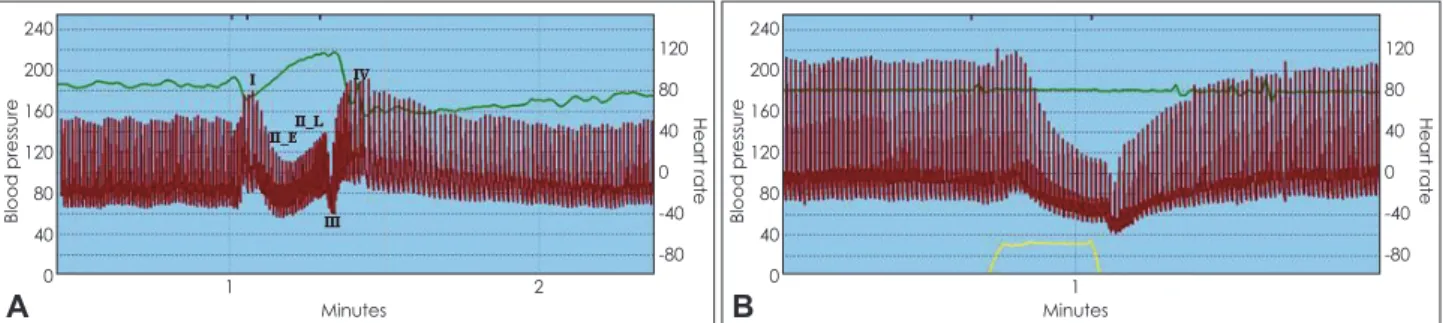
and adrenergic components of the baroreflex, can be made by studying dynamic alterations during the Valsalva maneuver.9 There are four main phases in the Valsalva maneuver (Fig. 1).
Phase I is a transient rise in BP due to the act of blowing. Ear- ly phase II is due to reduced preload (venous return). The baro- reflex then results in efferent sympathetic discharge to muscle resulting in a rise in BP. This sustained rise in BP is interrupt- ed by a transient fall in BP, phase III, which like phase I, is mechanical, lasting 1-2 seconds, during which BP falls (be- cause the subject stops the maneuver). This rise in BP is de- scribed as phase II_L, BP recovery following phase III and phase IV, reflects the increase in total peripheral resistance (Fig. 1A). Evaluation of the baroreflex can be done non-inva- sively from the Valsalva maneuver. The vagal component of the baroreflex is evaluated by relating the beat-to-beat BP dur- ing phase II_E to the heart period (reciprocal of heart rate) providing baroreflex sensitivity in msec/mm Hg.10 The adren- ergic component of the baroreflex can be evaluated by BP re- covery time (PRT), which is the time (in seconds) that the BP takes to recover from phase III to baseline.11 Fig. 1B is a Val- salva maneuver from a patient with diabetic autonomic neu- ropathy. Note that, in contrast with a normal response (IA), heart rate response to the fall in BP (phase II_E) is blunted (in- dicating impaired vagal baroreflex function). Adrenergic baro- reflex function is markedly impaired. This is manifested by the absence of either II_L and IV (absence of reflex vasoconstric- tion) and prolonged PRT.
HUT complements the beat-to-beat BP responses to the Val- salva maneuver. HUT provides an evaluation of the BP and heart rate response to tilt. A normal response consists of a mod- est rise in heart rate (by 5-20 bpm) and BP remains relatively constant (modest fall in systolic BP by <10 mm Hg and mod- est diastolic rise). HUT is done to detect if OH is present. Fig.
2 are examples of HUT showing a normal response (Fig. 2A), neurogenic OH (Fig. 2B), POTS in Fig. 2C, and syncope (Fig.
2D). In Fig. 2B, there is a marked fall in BP with a blunted heart rate response, typical of neurogenic OH. This was re-
Fig. 1. Valsalva maneuver from a normal subject (A) and a patient with diabetic autonomic neuropathy (B). The heart rate and beat-to-beat blood pressure responses to the Valsalva maneuver are shown. Expiratory pressure is shown at the bottom. The phases of the Valsalva maneuver (I, II_E, II_L, III, IV) are indicated in the Fig. 1A. Autonomic neuropathy with adrenergic failure is manifested as a loss of phases II_
L and IV and delayed blood pressure recovery. Impairment of the vagal component of the baroreflex is manifested as blunting of heart rate response to changes in blood pressure.
240 200 160 120 80 40
0 1
Minutes 2
120 80 40 0 -40 -80
Blood pressure Heart rate
A
240 200 160 120 80 40
0 1
Minutes
120 80 40 0 -40 -80
Blood pressure Heart rate
B
corded in a patient with MSA. Fig. 2C is a recording from a patient with POTS, showing orthostatic tachycardia without OH. Heart rate increment is >30 bpm and eventually exceeds 120 bpm. Fig. 2D shows a patient with vasodepressor synco- pe. To HUT (first mark) there was a transient increase in BP (sympathetic surge) followed by abrupt fall in BP.
Plasma norepinephrine measured with the subject supine and after a period of standing provides another method of studying adrenergic function.12 A normal response consists of doubling of NE on standing. The patient with generalized postganglion- ic adrenergic failure, as in pure autonomic failure (PAF), will have low supine NE. The patient with preganglionic lesion, as in MSA, will typically have normal supine values (since the postganglionic fibers are intact) but a failure to increment on standing.13
The quantitative uptake of the radiopharmaceutical [123I]
MIBG (iodine-123 meta-iodobenzylguanidine), a norepineph- rine analog, can be measured by single photon emission com- puted tomography. A newer approach, using 6-[18F]fluorodo- pamine positron-emission tomography and neurochemical an- alyses provide better resolution. These approaches provide an index of the functional integrity of presynaptic postganglionic adrenergic sympathetic terminals in the heart. Cardiac adrener- gic imaging is reduced in a postganglionic lesion as in PAF or PD and is usually normal in MSA, although not invariably.14
Applications of Autonomic Testing
The Mayo Autonomic Laboratory was established in 1983. It
now performs autonomic studies on over 4000 patients per year. A number of applications have been well-documented and have stood the test of time. We shall summarize examples of such applications.
Distal small fiber neuropathy
One of the most common and distressing neurologic com- plaints is DSFN. The patient complains of a burning sensation in their feet, especially toes and plantar aspect of their feet.
There is typically troublesome allodynia, where a non painful stimulus elicits pain. Sensory examination may demonstrate a loss of sharp/dull or temperature perception at the toes. Motor function is usually normal. Common causes are diabetic and inherited neuropathy, but the most common cause is idiopath- ic. Nerve conduction studies are often normal since this is a small fiber and not large fiber neuropathy. This condition causes a length-dependent involvement of unmyelinated fibers, both somatic and autonomic. Sympathetic sudomotor fibers are affected, so that QSART will show abnormalities at the feet and normal sweating more proximally (Table 3) in about 3 out of 4 patients tested. Similarly, the thermoregulatory sweat test shows anhidrosis that is confined to the distal feet. Skin biop- sy and evaluation of intraepidermal fiber density on the same subjects and the same sites have been studied prospectively in patients with autonomic neuropathy.15 Both skin biopsy and QSART will accurately diagnose DSFN but their agreement is imperfect, since they measure different populations of unmy- elinated fibers.15 QSART is also helpful in following the nat- ural history of DSFN and to determine if the neuropathy re- Fig. 2. Head-up tilt (HUT) responses in a normal subject (A), neurogenic OH (B), POTS (C), and syncope (D). Neurogenic OH is manifest- ed as a pronounced fall in blood pressure (BP) with a blunted heart rate (HR) response. POTS (C) is manifested as an exaggerated HR re- sponse without OH. D shows vasodepressor syncope manifested as an abrupt fall in BP. OH: orthostatic hypotension, POTS: postural tachycardia syndrome.
240 200 160 120 80 40
0 5 6 7 8 9
Minutes 10 11
120 80 40 0 -40 -80
Blood pressure Heart rate
A
240 200 160 120 80 40
0 3 4 5 6 7 8 9 10 11 12 13 14 15 16 17 Minutes
120 80 40 0 -40 -80
Blood pressure Heart rate
C
240 200 160 120 80 40
0 4 5 6 7 8 9
Minutes 10 11 12
120 80 40 0 -40 -80
Blood pressure Heart rate
B
240 200 160 120 80 40
0 3 4 5 6 7 8 9 10 11 12 13 14 15 16 17 18 19 20 21 Minutes
120 80 40 0 -40 -80
Blood pressure Heart rate
D
mains confined or becomes generalized. This capability is of importance since neuropathies due to diabetes or amyloidosis can start with DSFN and progress, while others do not. It is of- ten difficult to make this determination with clinical examina- tion alone. Table 3 provides a summary of experience with au- tonomic function tests in DSFN. Either TST of QSART will detect distal denervation in >70% of cases and together show an abnormality in 9/10 cases. As expected, adrenergic and car- diovagal abnormalities are less common. EMG is normal in
>70% of cases.
Generalized autonomic failure
Physicians have learnt to recognize symptoms of autonomic failure. These include orthostatic lightheadedness, syncope, erectile dysfunction, and symptoms suggestive of neurogenic bladder and bowel. However, it is not possible to quantitate the severity and distribution of such abnormalities to help the phy- sician determine if the patient is getting better or getting worse or if they are responding to therapy. With the availability of au- tonomic testing, it is possible to determine if generalized auto- nomic failure is present. Generalized autonomic failure refers to autonomic failure that is diffuse (multiple regions) and in- volves more than one autonomic system. Examples of gener- alized autonomic failure are the autonomic neuropathies16 and multiple system atrophy. Examples of autonomic neuropathies are diabetic autonomic neuropathy, amyloid neuropathy,17 and autoimmune autonomic ganglionopathy.18 In severe diabetic autonomic neuropathy,19 there is widespread loss of sweating, cardiovagal failure is present, and OH with impaired barore- flexes is seen. In the synucleinopathies (Parkinson’s disease, multiple system atrophy, Lewy body dementia), generalized autonomic failure tends to be present in MSA, mild in PD, and intermediate in DLB.20
Typically, for a comprehensive evaluation of autonomic fun- ction, we undertake an autonomic reflex screen and thermoreg-
ulatory sweat test as a routine. We often measure norepineph- rine supine and standing to determine if supine values are re- duced (indicating widespread postganglionic adrenergic de- nervation) and if an orthostatic increment (doubling) occurs. We will typically do additional studies to seek a cause. These in- clude fat aspirate or nerve biopsy for amyloidosis17 or an anti- body panel seeking especially for presence of ganglionic anti- body.21 Detailed descriptions are available for a comprehensive coverage.12
Selective autonomic failure
The astute clinician can suspect that autonomic failure may be restricted rather than generalized. This suspicion can be con- firmed and quantified with autonomic testing. Autonomic tests can confirm that a specific autonomic function is affected and that other systems are intact. We provide 3 examples. A patient, aged 30 years, feels hot, weak, dizzy and uncomfortable when ambient temperature rises. BP supine and standing is normal.
He looks hot and flushed but has dry skin. Orthostatic BP re- cording is normal without evidence of OH. The clinician may suspect this is restricted autonomic failure but testing is neces- sary. Autonomic testing demonstrates normal cardiovagal and adrenergic function. However, QSART and TST show global anhidrosis. This is a condition called chronic idiopathic anhi- drosis.1 Another example is a patient with gastroparesis. This patient has postprandial bloating and weight loss. He vomits up food that is 3 days old. Autonomic testing shows normal sudo- motor and adrenergic function. Gastric transit studies show marked delay in gastric transit. A third patient has Adies pupils.
Tests can be done to confirm the entity, but autonomic testing is needed to demonstrate that the deficits are confined and not generalized.
The synucleinopathies
The term synucleinopathies is used to describe several neuro- Table 3. Comparison of current study with earlier studies33
Stewart et al.30 Tobin et al.31 Novak et al.32
Current study
p value*
Total Abnormal EMG
Normal EMG
Number of patients 40 15 92 125 47 78
QSART abnormalities 32/40 (80%) 12/15 (80%) 67/92 (73%) 96/125 (77%) 35/47 (74%) 61/78 (78%) 0.79 TST distal patterns of anhidrosis 18/25 (72%) n/a n/a 93/125 (74%) 35/47 (74%) 58/78 (74%) 0.84
TST any abnormality 24/25 (96%) n/a n/a 114/125 (91%) 44/47 (94%) 70/78 (90%) 0.53
Either TST or QSART abnormality 36/40 (90%) n/a n/a 123/125 (98%) 46/47 (98%) 77/78 (99%) >0.99 Cardiovagal abnormality 11/40 (28%) 9/12 (75%) 59/92 (64%) 43/125 (35%) 15/46 (33%) 28/76 (37%) 0.78 Adrenergic abnormality
(*some studies just included OH)
0/40* (0%) 2/12* (17%) 0/92* (0%) 53/125 (43%) 21/46 (46%) 32/77 (42%) 0.80 We used Pearson chi-squared test with Yates continuity correction except for “TST any abnormality” and “Either TST or QSART abnor- mality” which are based on Fisher exact test due to small numbers in some cells of the 2×2 table.
*Comparing current study with previous studies.
EMG: electromyogram, OH: orthostatic hypotension, QSART: quantitative sudomotor axon reflex test, TST: thermoregulatory sweat test.
degenerative disorders characterized by fibrillary aggregates of alpha-synuclein protein in oligodendroglia (in MSA) and in se- lective population of neurons. These disorders include Parkin- son’s disease (PD), dementia with Lewy bodies (DLB), PAF, and MSA. Although it is recognized that autonomic failure, manifest as erectile dysfunction, neurogenic bladder, constipa- tion, and OH occur in the synucleinopathies, there has been lit- tle emphasis on the use of autonomic function tests in the dif- ferential diagnosis of these conditions. Based on our research
on the involvement of autonomic structures in brain and spinal cord, we hypothesized that the severity and distribution of au- tonomic failure was different among synucleinopathies.
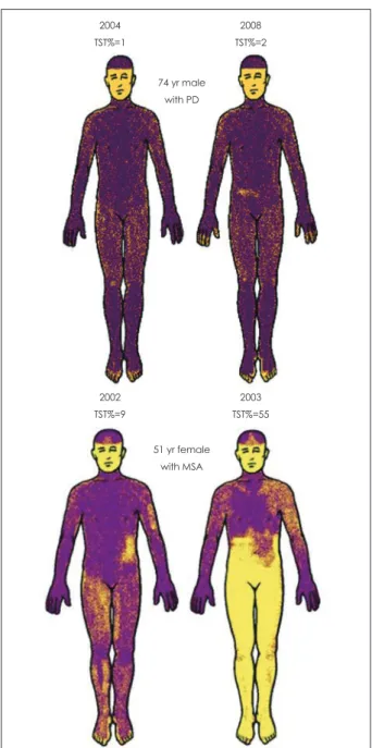
PD and MSA share clinical features of hypomimia, hyperto- nia, dysarthria, micrographia, and gait impairment. PD is more likely associated with rest tremor and is typically levodopa-re- sponsive. However, MSA can also have rest tremor and le- vodopa responsiveness, albeit less robust.22 Recent studies in- dicate that MSA is distinguishable from PD using autonomic tests. PD is characterized by a length-dependent involvement of postganglionic sudomotor fibers, whereas MSA is character- ized by widespread, early and preganglionic autonomic fail- ure.23 MIBG or fluorodopa scan of the heart, which images postganglionic adrenergic innervation, is typically defective in PD and normal in MSA.13,14 However, denervation can occur in 25-30% of MSA patients.14 Another useful test is the TST. TST is either normal or shows very distal anhidrosis, typically af- fecting the toes (Fig. 3). In Fig. 3, the PD case showed very distal anhidrosis, affecting only parts of the toes, and did not progress over time. In contrast, MSA causes widespread anhi- drosis.23 If both QSART and TST are performed, normal QSART volume in an anhidrotic region indicates that the le- sion is preganglionic in site (Fig. 3). When the autonomic re- flex screen is performed and the aggregate score, the compos- ite autonomic severity score (CASS) calculated, CASS sepa- rates MSA from PD with sensitivity and specificity. The auto- nomic and clinical characteristics of the synucleinopathies are distinctive (Table 4). PAF is characterized by severe OH, with- out clinical central involvement. Supine plasma norepinephrine is often markedly reduced and CASS is >6 and TST% >40%.
PD is associated with normal NE, CASS <6, and TST% <40%.
MSA is characterized by CASS >6 and TST% >40%. DLB has intermediate autonomic failure.
Orthostatic intolerance
Orthostatic intolerance refers to the development of symptoms after assuming the standing posture that clears on sitting or ly- Fig. 3. Thermoregulatory sweat test (TST) showing characteristic
patterns in the synucleinopathies. In Parkinson’s disease (top panel), anhidrosis is distal and percent anhidrosis remained <5%.
In contrast, in multiple system atrophy (MSA), anhidrosis is re- gional and percent anhidrosis is greater and progressed more rapidly.
2004 TST%=1
2002 TST%=9
2008 TST%=2
2003 TST%=55 74 yr male
with PD
51 yr female with MSA
Table 4. Autonomic scores in the synucleinopathies
Variable PD DLB MSA PAF
Age 71.5±8.5*
(53-84)
74.4±7.2 (52-86)
73.0±5.5 (65-82)
59.9±16
N 20 20 20 20
CASS 2.2±1.2 (0-5)
5.2±2.0 (2-10)
8.1±1.3 (5-10)
9.4±1.2 TST% 19.3±35.5
(0-97)
26.4±30.0 (0-85)
69.1±26.9 (12-99)
73.1±36.3
*All values expressed as SD and range in brackets.
CASS: composite autonomic severity score, DLB: dementia with Lewy bodies, MSA: multiple system atrophy, PAF: pure autonom- ic failure, PD: Parkinson’s disease, TST: thermoregulatory sweat test.
ing back down. Typical symptoms are lightheadedness, blurred vision, cognitive difficulties, or tiredness. Some patients also have symptoms of sympathetic activation, such as tremulous- ness and palpitations. In the category of orthostatic intolerance, we consider OH,9 postural tachycardia syndrome,24 and neuro- cardiogenic syncope (vasovagal and vasodepressor).25
Orthostatic hypotension is defined as an orthostatic fall in SBP by >20 mm Hg,26 although in the laboratory, we have rou- tinely required a fall of >30 mm Hg9,27 and this latter criterion is more robust in the diagnosis of MSA.28 Orthostatic hypoten- sion can occur in the absence of autonomic failure and the role of the laboratory is to determine if it is neurogenic; i.e., if au- tonomic reflexes, especially the baroreflexes are impaired.
Typically in neurogenic OH, the fall in BP is associated with attenuated heart rate increment (Fig. 2B).
POTS is defined as an increase on tilt of heart rate >30 bpm within 5 minutes in adults and associated with symptoms of or- thostatic intolerance.24,29 Autonomic laboratory testing is essen- tial to make the diagnosis of POTS to demonstrate this heart rate increment and to rule out OH (Fig. 2C).
Neurocardiogenic syncope typically occurs in subjects who have normal autonomic reflexes. The most common type is va- sovagal syncope. There is a sudden fall in BP and heart rate.
There is often an antecedent sympathetic surge as might occur in a subject who gets an injection or sees something unpleas- ant. Hence patients with POTS, who typically have an incre- ased sympathetic tone, are excessively prone to vasovagal syn- cope. Vasodepressor syncope refers to an event where there is this fall in BP without an associated fall in heart rate (Fig. 2D).
The autonomic laboratory will often capture syncope, although most autonomic laboratories do not attempt to provoke syn- cope.
Concluding Thoughts
Clinical management of the dysautonomias depends on good clinical acumen. The role of autonomic testing is to increase sensitivity and specificity in the detection of autonomic failure.
The two work hand in glove and each enhances the other.
There are limitations of clinical autonomic testing. The non-in- vasive approach is appropriate but imperfect. For instance, the beat-to-beat BP devices sometimes give erroneous signals, so that judgment is important in the interpretation of autonomic tests. Autonomic testing is a growing and evolving field so that guidelines will and should change over time.
Conflicts of Interest
The authors have no financial conflicts of interest.
Acknowledgements
This work was supported in part by National Institutes of Health (NS
44233 Pathogenesis and Diagnosis of Multiple System Atrophy, U54 NS065736 Autonomic Rare Disease Clinical Consortium), Mayo CTSA (UL1 TR000135), and Mayo Funds.
The Autonomic Diseases Consortium is a part of the NIH Rare Diseases Clinical Research Network (RDCRN). Funding and/or programmatic support for this project has been provided by U54 NS065736 from the National Institute of Neurological Diseases and Stroke (NINDS) and the NIH Office of Rare Diseases Research (ORDR).
The content is solely the responsibility of the authors and does not nec- essarily represent the official views of the National Institute of Neuro- logical Disorders and Stroke or the National Institutes of Health.
REFERENCES
1. Low PA, Fealey RD, Sheps SG, Su WP, Trautmann JC, Kuntz NL.
Chronic idiopathic anhidrosis. Ann Neurol 1985;18:344-348.
2. Low PA. Autonomic nervous system function. J Clin Neurophysiol 1993;10:14-27.
3. Low PA, Denq JC, Opfer-Gehrking TL, Dyck PJ, O’Brien PC, Slezak JM. Effect of age and gender on sudomotor and cardiovagal function and blood pressure response to tilt in normal subjects. Muscle Nerve 1997;20:1561-1568.
4. Low PA, Walsh JC, Huang CY, McLeod JG. The sympathetic nervous system in diabetic neuropathy. A clinical and pathological study. Brain 1975;98:341-356.
5. Fealey RD, Low PA, Thomas JE. Thermoregulatory sweating abnor- malities in diabetes mellitus. Mayo Clin Proc 1989;64:617-628.
6. Freeman RL. Noninvasive evaluation of heart rate: time and frequency domains. In: Low PA, Benarroch EE. Clinical Autonomic Disorders.
3rd ed. Philadelphia: Lippincott Williams & Wilkins, 2008;185-197.
7. Low PA, Zimmerman BR, Dyck PJ. Comparison of distal sympathet- ic with vagal function in diabetic neuropathy. Muscle Nerve 1986;9:
592-596.
8. Benarroch EE, Opfer-Gehrking TL, Low PA. Use of the photoplethys- mographic technique to analyze the Valsalva maneuver in normal man.
Muscle Nerve 1991;14:1165-1172.
9. Low PA, Singer W. Management of neurogenic orthostatic hypoten- sion: an update. Lancet Neurol 2008;7:451-458.
10. Schrezenmaier C, Singer W, Swift NM, Sletten D, Tanabe J, Low PA.
Adrenergic and vagal baroreflex sensitivity in autonomic failure. Arch Neurol 2007;64:381-386.
11. Vogel ER, Sandroni P, Low PA. Blood pressure recovery from Valsal- va maneuver in patients with autonomic failure. Neurology 2005;65:
1533-1537.
12. Low PA, Sletten DM. Laboratory evaluation of autonomic failure. In:
Low PA, Benarroch EE. Clinical Autonomic Disorders. 3rd ed. Phila- delphia: Lippincott Williams & Wilkins, 2008;130-163.
13. Goldstein DS, Polinsky RJ, Garty M, Robertson D, Brown RT, Biag- gioni I, et al. Patterns of plasma levels of catechols in neurogenic or- thostatic hypotension. Ann Neurol 1989;26:558-563.
14. Kimpinski K, Iodice V, Burton DD, Camilleri M, Mullan BP, Lipp A, et al. The role of autonomic testing in the differentiation of Parkinson’s disease from multiple system atrophy. J Neurol Sci 2012;317:92-96.
15. Singer W, Spies JM, McArthur J, Low J, Griffin JW, Nickander KK, et al. Prospective evaluation of somatic and autonomic small fibers in se- lected autonomic neuropathies. Neurology 2004;62:612-618.
16. Low PA. Autonomic neuropathies. Curr Opin Neurol 2002;15:605-609.
17. Wang AK, Fealey RD, Gehrking TL, Low PA. Patterns of neuropathy and autonomic failure in patients with amyloidosis. Mayo Clin Proc 2008;83:1226-1230.
18. Klein CM, Vernino S, Lennon VA, Sandroni P, Fealey RD, Benrud- Larson L, et al. The spectrum of autoimmune autonomic neuropathies.
Ann Neurol 2003;53:752-758.
19. Low PA. Diabetic autonomic neuropathy. Semin Neurol 1996;16:143- 151.
20. Thaisetthawatkul P, Boeve BF, Benarroch EE, Sandroni P, Ferman TJ, Petersen R, et al. Autonomic dysfunction in dementia with Lewy bod- ies. Neurology 2004;62:1804-1809.
21. Vernino S, Low PA, Fealey RD, Stewart JD, Farrugia G, Lennon VA.
Autoantibodies to ganglionic acetylcholine receptors in autoimmune autonomic neuropathies. N Engl J Med 2000;343:847-855.
22. Iodice V, Lipp A, Ahlskog JE, Sandroni P, Fealey RD, Parisi JE, et al.
Autopsy confirmed multiple system atrophy cases: Mayo experience and role of autonomic function tests. J Neurol Neurosurg Psychiatry 2012;83:453-459.
23. Lipp A, Sandroni P, Ahlskog JE, Fealey RD, Kimpinski K, Iodice V, et al. Prospective differentiation of multiple system atrophy from Parkin- son disease, with and without autonomic failure. Arch Neurol 2009;66:
742-750.
24. Thieben MJ, Sandroni P, Sletten DM, Benrud-Larson LM, Fealey RD, Vernino S, et al. Postural orthostatic tachycardia syndrome: the Mayo clinic experience. Mayo Clin Proc 2007;82:308-313.
25. Shen WK, Low PA, Rea RF, Lohse CM, Hodge DO, Hammill SC.
Distinct hemodynamic profiles in patients with vasovagal syncope: a heterogeneous population. J Am Coll Cardiol 2000;35:1470-1477.
26. Consensus statement on the definition of orthostatic hypotension, pure autonomic failure, and multiple system atrophy. The Consensus Com- mittee of the American Autonomic Society and the American Academy
of Neurology. Neurology 1996;46:1470.
27. Schrezenmaier C, Gehrking JA, Hines SM, Low PA, Benrud-Larson LM, Sandroni P. Evaluation of orthostatic hypotension: relationship of a new self-report instrument to laboratory-based measures. Mayo Clin Proc 2005;80:330-334.
28. Gilman S, Low PA, Quinn N, Albanese A, Ben-Shlomo Y, Fowler CJ, et al. Consensus statement on the diagnosis of multiple system atrophy.
J Neurol Sci 1999;163:94-98.
29. Schondorf R, Low PA. Idiopathic postural orthostatic tachycardia syn- drome: an attenuated form of acute pandysautonomia? Neurology 1993;
43:132-137.
30. Stewart JD, Low PA, Fealey RD. Distal small fiber neuropathy: results of tests of sweating and autonomic cardiovascular reflexes. Muscle Nerve 1992;15:661-665.
31. Tobin K, Giuliani MJ, Lacomis D. Comparison of different modalities for detection of small fiber neuropathy. Clin Neurophysiol 1999;110:
1909-1912.
32. Novak V, Freimer ML, Kissel JT, Sahenk Z, Periquet IM, Nash SM, et al. Autonomic impairment in painful neuropathy. Neurology 2001;56:
861-868.
33. Low VA, Sandroni P, Fealey RD, Low PA. Detection of small-fiber neuropathy by sudomotor testing. Muscle Nerve 2006;34:57-61.