INTRODUCTION
Carotid artery stenosis accounts for 20 percent of all ischemic strokes in Western countries.11) Carotid ar- tery stsenting (CAS) is emerging as an alternative to
classic carotid endarterectomy (CEA) to treat carotid artery stenosis.1)11) Additionally, improvements in en- dovascular technique and the development of pro- tective devices have dramatically reduced CAS complications. Postoperative complications such as in-
Neurologic complications in patients with carotid artery stenting
Na Young Kim, Jong Wook Choi, Kum Whang, Sung Min Cho, Youn Moo Koo, Jong Yeon Kim
Department of Neurosurgery, Yonsei University Wonju College of Medicine, Wonju Severance Christian Hospital, Wonju, Korea
Objective : Carotid artery stenting is helpful in patients with carotid artery stenosis and is a common method of treatment. However, data on the neurological consequences that might arise from, especially Asian patients after CAS is not enough. The purpose of this study was to investigate the outcome and prognostic factors affecting CAS patients.
Methods : From January 2013 to June 2018 it was enrolled 97 patients who underwent CAS with severe carotid artery stenosis in a single institution. We retrospectively reviewed neurologic complications such as restenosis, ipsilateral or contralateral stroke, and hyperperfusion during the 6-month follow-up period.
Results : There were no complication occured during the procedure in all 97 patients. Neurologic complications occurred in 30 patients (30.9%) af- ter the procedure, and ipsilateral stroke 6 (6.2%), contralateral stroke 9 (9.4%), restenosis 2 (2.1%) and hyperperfusion 13 respectively. One of them had died (1.0%), of which the rest were discharged after symptoms improve. On univariate analysis, DM and pre-op NIHSS score was asso- ciated with the risk of CAS complication, exclusively. On the binary logis- tic regression for risk factors, DM (OR 0.144, 95% CI [0.029-0.718]), his- tory of radiotheraphy (OR 36.103, 95% CI [1.009-1291.789]) and pre- operative NIHSS (OR 1.266, 95% CI [1.059-1.513]) showed independent risk factors associated with post procedural neurological complications, statistically.
Conclusion : Carotid artery stenting is a relatively safe and reliable long-term outcome for patients with carotid artery stenosis. However, careful observation should be taken after procedure immediately for any possible complications.
J Cerebrovasc Endovasc Neurosurg.
2019 June;21(2):86-93 Received : 6 June 2019 Revised : 16 June 2019 Accepted : 19 June 2019 Correspondence to Jong Yeon Kim Department of Neurosurgery, Yonsei University Wonju College of Medicine, Wonju Severance Christian Hospital, 20 Ilsan-ro, Wonju, Gangwon-do, 26426, Korea
Tel : +82-10-3425-8934 Fax : +82-33-746-2287 E-mail : [email protected]
ORCID : http://orcid.org/0000-0002-9407-0127
This is an Open Access article distributed under the terms of the Creative Commons Attribution Non- Commercial License (http://creativecommons.org/li- censes/by-nc/3.0) which permits unrestricted non- commercial use, distribution, and reproduction in any medium, provided the original work is properly cited.
Keywords Carotid Stenosis, Stents, Endovascular Procedures, Postoperative Complications
tracranial hemorrhage (ICH), cerebral hyperperfusion syndrome (CHS), stroke, and restenosis after CEA have been well documented, but few reports of neu- rological outcomes have been made in patients with CAS especially in Asian population. Therefore, the purpose of this study is to study the neurological re- sults of patients receiving CAS from a single institution.1)23)
MATERIALS AND METHODS
Patient selection
From January 2013 to June 2018, 97 patients who re- ceived CAS for carotid stenosis registered for the study from a single institution. Neurologists and vas- cular surgeons selected patients suitable for CAS. We performed digital subtraction angiography (DSA) to estimate the degree of stenosis. According to ECST criteria, the indications for CAS were more than 70%
(n = 65 (63.16%) with symptom (transient ischemic at- tack (TIA) or stroke at the site supplied by the sten- otic artery) or in asymptomatic cases, more than 70%
(n = 32 (36.84%)) on DSA. Our institution's routine douplex ultrasound tests conducted follow-up man- agement regularly six months after stent insertion.
After CAS, neurological complications such as ipsi- lateral or contralateral stroke, resenosis, and hyper- perfusion were recorded, analyzed. And the charac- teristics and conditions before and after procedure was also recorded and reviewed as well.
In Nov. 1999, our CAS program was started at the single neurovascular center. For internal quality con- trol, we have created a computer-based database that includes all patients with carotid stenosis with CAS or carotid vascularization. Patient data was utilized until June 2018 for current analysis. All patients provided consent based on information on procedures and anonymous data collection and processing. All pa- tients with or without symptoms were treated from the time of registration. The anatomical qualification criteria have been tested in all patients by CT angiog- raphy or MRA in our institution. If not already given
with any anticoagulants, all patients were treated with 300 mg of clopidogrel and 500 mg of acetylsali- cylic acid at least 24 hours prior to the procedure.
After the procedure, 100 mg of acetylsalicylic acid and 75 mg of clopidogrel were given continuously for more than four weeks. Patients were then advised to take 100 milligrams of acetyl salicylate for life.
Procedure
In our institution, only local anesthesia was used when performing CAS. Bolus of heparin (5000 IU) was administered in a standard manner. Whenever anatomically possible, we used an embolic protection device for periprocedural neuroprotection. All proce- dures were performed by a single neurointerventionist with at least 5 years of experience. Also, seven days before the procedure, patients received aspirin and Plavix for optimal treatment in addition to CAS. And within 24 hours prior to and after procedure, all pa- tients were received neurological assessment by neurologist. An independent neurologist evaluated with the National Institutes of Health Stroke Scale and the modified Rankin Scale. First, entryways for angiography were secured through the right femoral artery and 5-F sheath was used. If you secure the fore-aft-side of the CCA through the catheter se- lection, and if stenosis is verified, we replaced it with guided-wire long enough that can reach the statistical CCA and ECA. Special care must be taken to avoid inappropriate contact between the stenosis and wire when the tip of the guided wired reaches near the CCA. If the Micro-wire and Microcatheter are success- fully located in the stenosis, the delivery device must be over the microwire and past the stenosis. The length of the stent should be sufficient to extend from the CCA to the ICA in most cases to fully cover the stenosis. The diameter of the stent must match the CCA in order to properly match the wall adaptation of all carotid segments. The stent should be posi- tioned slightly further than the desired stenosis posi- tion and should be pulled out before the position to be placed is reached, in order to reduce the possibility
of a forward swivel movement. Atropine should be prepared for bradycardia at all times. Angiography should be performed on both cervical ICAs and intra- cranial circulation to evaluate stenosis, vasospasm, dissection, and cerebral blood flow. In this case, it is important to compare with the angiogram performed before the operation because the distal emboli are dif- ficult to detect. After performing CAS through this procedure, it is necessary to pay attention to the com- plications that may arise from enlarging the diameter of the lesion by dilating the stent.
Statistically analysis
Data are expressed according to the properties of the variable. Continuous variables are presented as mean and standard deviation. Categorical variables are presented as frequency and percentage. In order to compare two groups, we performed the two-simple t-test or chi-square test (Fisher`s exact test) as appropriate. Logistic regression analysis was used to identify the factors to predict neurologic complica- tions after CAS and the result were expressed as odds ratio (OR) with 95% confidence interval (CI). A p-val- ue less than 0.05 was considered statistically sig- nificant and all statistical analyses were conducted us- ing SPSS (version 24; IBM, Armonk, NY, USA).
RESULTS
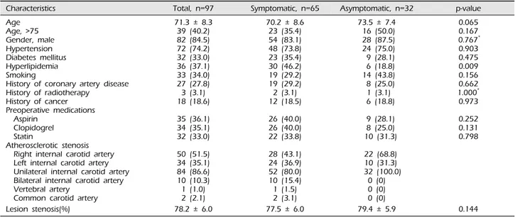
We retrospectively analyzed 97 patients who under- went CAS with carotid artery stenosis from 2013 to 2018. The total number of patients enrolled in the study was 97 (15 women (15.5%) and 82 men (84.5%)) and the average age was 71.3 year old. Procedure was successfully performed in all 97 patients and pro- tection devices were used in all cases. 65 patients were diagnosed with TIA or cerebral infarction before CAS, and 32 patients who received CAS were diag- nosed with stenosis from regular health check-ups without any symptoms. The occurrence of selected patient characteristics is shown in Table 1, which lists the general risk factors of the study population. We
discovered that the most frequently found medical history is hypertension in 72 (74.2%) patients, history of stroke in 55 (56.7 %) patients, and hyperlipidemia in 36 (37.1%) patients. Furthermore, the medical his- tory showed that 27 (27.8%) patients had history of coronary artery disease, 13 (13.4%) had history of transient ischemic attack, 32 (33.0%) patients had dia- betes mellitus (DM), 33 (34.0%) patients were smok- ers, 18 (18.6%) had a history of cancer and 3 (3.1%) patients and a history of radiotherapy. In this study, only hyperlipidemia was a risk factor associated with carotid artery stenosis, statistically (p=0.009). Other characteristics were not related to the occurrence of the symptoms (Table 1).
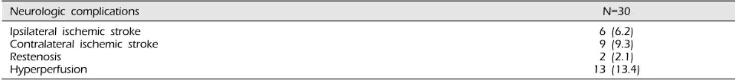
Neurologic complications were observed in 30 pa- tients (30.9%) after the procedure, and ipsilateral stroke 6 (6.2%), contralateral stroke 9 (9.4%), reste- nosis 2 (2.1%) and hyperperfusion 13 (13.4%) occurred (Table 2). One of them died from ICH caused by se- vere hyperperfusion (Figure 1), while the rest were discharged after symptoms improved. Two patients (2.1%) with restenosis were identified by Duplex Ultrasonography during follow up period.
On univariate analysis, gender, age, hypertension, hyperlipidemia, and presence of coronary artery dis- ease, history of radiotherapy, or smoking did not show a significant association with procedural neuro- logical events (Table 3). DM and pre-op NIHSS score was associated with the risk of CAS complication, exclusively. On the binary logistic regression for risk factors, DM (OR 0.144, 95% CI [0.029-0.718]), history of radiotheraphy (OR 36.103, 95% CI [1.009-1291.789]) and preoperative NIHSS (OR 1.266, 95% CI [1.059-1.513]) showed independent risk factors associated with post procedural neurological complications, statistically.
Interestingly, DM showed association with the risk of CAS complications in negative OR (Table 3).
DISCUSSION
According to American Guidelines for the Prevention
of Stroke, carotid recanalization, CEA, or carotid ar- tery stenting, is recommended for patients who expe- rience ischemic stroke or transient cerebral ischemic stroke within 6 months (symptomatic patient) under- lying ipsilateral carotid stenosis as demonstrated by catheter-based angiography or noninvasive imaging.20) CAS has recently been widely used as a reliable treat-
ment that can replace CEA, and there are significantly more CAS-based treatments in our institution com- pared to CEA in patients with carotid artery stenosis.
In particular, for asymptomatic carotid artery stenosis in stroke patients, recanalization is recommended to reduce the surgical risk to 3% or less. In spite of these great advantages, it sometimes provides a disastrous
A B C
Fig. 1. A 79-year-old male was admitted for left side weakness due to right border zone infarction (A). After performing digital sub- traction angiography, he was diagnosed with a 78.4% stenosis of the right internal carotid artery (B). He underwent right CAS pro- cedure without periprocedural complication (C). Three hours following CAS, a computerized tomography of the brain reveals a large right-hemisphere hematoma, with midline shift and intraventricular hemorrhage (D).
Characteristics Total, n=97 Symptomatic, n=65 Asymptomatic, n=32 p-value
AgeAge, >75 Gender, male Hypertension Diabetes mellitus Hyperlipidemia Smoking
History of coronary artery disease History of radiotherapy
History of cancer Preoperative medications Aspirin
Clopidogrel Statin
Atherosclerotic stenosis Right internal carotid artery Left internal carotid artery Unilateral internal carotid artery Bilateral internal carotid artery Vertebral artery
Common carotid artery Lesion stenosis(%)
71.3 ± 8.3 39 (40.2) 82 (84.5) 72 (74.2) 32 (33.0) 36 (37.1) 33 (34.0) 27 (27.8) 3 (3.1) 18 (18.6) 35 (36.1) 34 (35.1) 32 (33.0) 50 (51.5) 34 (35.1) 84 (86.6) 10 (10.3) 1 (1.0) 2 (2.1) 78.2 ± 6.0
70.2 ± 8.6 23 (35.4) 54 (83.1) 48 (73.8) 23 (35.4) 30 (46.2) 19 (29.2) 19 (29.2) 2 (3.1) 12 (18.5) 26 (40.0) 26 (40.0) 22 (33.8) 28 (43.1) 24 (36.9) 52 (80.0) 10 (15.4) 1 (1.5) 2 (3.1) 77.5 ± 6.0
73.5 ± 7.4 16 (50.0) 28 (87.5) 24 (75.0) 9 (28.1) 6 (18.8) 14 (43.8) 8 (25.0) 1 (3.1) 6 (18.8) 9 (28.1) 8 (25.0) 10 (31.3) 22 (68.8) 10 (31.3) 32 (100.0)
0 (0) 0 (0) 0 (0) 79.4 ± 5.9
0.065 0.167 0.767* 0.903 0.475 0.009 0.156 0.662 1.000* 0.973 0.252 0.131 0.798
0.144
* Fisher’s exact test
Table 1. Patients characteristics with and without symptoms
cerebral deficit such as contrast-induced encephalop- athy, ischemic stroke, and CHS.20) CAS was used to expand the diameter of a stenotic lesion using a stent, and pre- and post-operative data on neurological events including stroke, seizure, ICH, CHS, or changes in neurological status were collected.
Cerebral hyperperfusion syndrome and ICH
Many authors have described CHS as a primary complication of carotid recanalization procedure.17) CHS is defined in several ways such as: 1) Severe headache, seizures, mental deterioration, and / or de- velopment of local neurological symptoms; 2) no ad- ditional ischemic lesion in imaging diagnosis of the brain; 3) Increased cerebral blood flow in the ipsi- lateral hemisphere, increased blood flow in the con- tralateral hemisphere, or increased cerebral artery flow rate value of > 100% compared with pre- operative velocity, all postoperatively.7)14) According to Moulakakis et al, the incidence of CHS and ICH af- ter CAS was 1.16% (range, 0.44% to 11.7%) and 0.74%
(range, 0.36% to 4.5%), respectively and CHS may be fatal once an intracranial hemorrhage occurs.17) In this
study, 13 cases (13.4%) were shown in all cases con- forming to the above definition of HPS, but HPS with fetal ICH reported was 1 case (1.1%) in our institution.
This increase in morbidity is associated with in- creased blood flow through the cerebral artery after the procedure and inadequate arterial blood pressure control.2) Although we found this phenomenon to be present after CEA for the first time, many authors have reported that post carotid angioplasty and stent- ing have similar results.7)21)
The most common time period for occurrence of CHS is several hours to several days after the procedure.20) Ogasawara suggested that the onset time of CHS among patients who received CAS reached its peak within 12 hours after the procedure. Mental de- terioration, confusion, and headache are the most common symptoms of CHS, among which headache usually occurs on the ipsilateral side of the arterial ar- tery, often degree of moderate to severe, and charac- terized by pulsatile and migraine symptoms.4) Cerebral edema is a neurological deficit of secondary CHS, which is usually transient. These are mostly de- rived from the cerebral cortex, including hemiplegia,
Neurologic complications N=30
Ipsilateral ischemic stroke Contralateral ischemic stroke Restenosis
Hyperperfusion
6 (6.2) 9 (9.3) 2 (2.1) 13 (13.4) Table 2. Neurologic complications after carotid artery stenting
Parameters Univariate* Multivariate OR (95%CI)
Age Age group Gender, male Hypertension Diabetes mellitus Hyperlipidemia Smoking
History of coronary artery disease History of transient ischemic attack History of stroke
Symptomatic
History of radiotherapy History of cancer Preoperative NIHSS
0.774 0.544 0.339† 0.558 0.020 0.081 0.930 0.830 1.000† 0.345 1.000† 0.139† 1.000† 0.028
0.351 (0.098-1.264) 1.680 (0.374-7.537) 0.965 (0.256-3.638) 0.144 (0.029-0.718) 0.288 (0.073-1.135) 0.560 (0.141-2.229) 1.686 (0.450-6.311)
1.354 (0.392-4.676) 36.103 (1.009-1291.789)
0.500 (0.084-2.982) 1.266 (1.059-1.513)
*statistical significance based on two-sample t-test or chi-square test
† Fisher’ exact test
Table 3. Univariate and multivariate analysis of risk factors and neurologic complications
hemiparesis, hemianopsia, aphasia, and obtundation, and also epileptic disorders such as generalized or fo- cal seizures. Other symptoms such as movement dis- orders, visual impairment, cognitive impairment, and psychotic disorders are rare, but have reported in CHS patients as well.19) And some of the untreated patients may die.17)
The pathogenesis and risk factors of CHS are not fully known, but uncontrolled hypertension seems to be the most important cause of CHS.5) It stimulates the occipital-parietal region more noticeably because of the sympathetic nerves that innervate very little on the vertebrobasilar circulation.22) This suggests that impaired cerebral autoregulation, baroreceptor-reflex breakdown, and an axon-like trigeminovascular reflex contribute to the pathogenesis of CHS.16)17) Another serious concern is the intake of antiplatelet and sta- tins, which is recommended for patients with carotid stenosis before and after procedure.5) These drugs can increase the risk of cerebral hemorrhage and CHS, es- pecially after carotid stenting.20)
ICH is the most disasteric event secondary to hyper- perfusion, which is also associated with CAS.17) ICH can cause vomiting or change of mentality because of increased pressure in the cranium.1)
It is important to note that ICH after CAS is difficult to prevent because it appears to occur within few hours without symptoms and mostly unavoidable and even fatal.17) There are several risk factors, including preoperative hypertension, bilateral carotid disease or contralateral carotid occlusion as well as impaired cerebrovascular reserve, which were associated with the formation of ICH after the procedure.7) It is known that ICH, which appears immediately after CAS, is caused by the rupture of the perforating ar- tery in the basal ganglia hence the sudden exposure of normal perfusion pressure pathophysiologically.17) Rate of morbidity and mortality are high in both pa- tients who treated with open surgery or endovascular repair, therefore it is crucial to prevent such a cata- strophic event after the procedure.3)7)
Stroke complications
Stroke occurs more commonly after CAS than after CEA. The periprocedural strokes in CREST were most commonly minor, ipsilateral to the treated artery, and ischemic in type and occurred twice as frequently in the CAS arm.9) Major stroke occurred in 0.6%
(13/2272), indicative of the very low overall complica- tion rate observed in the trial.8)9) According to Michael et al, complications that occurred during the carotid stent placement or within a 30-day period following placement were recorded and the definitions were de- fined as follows: (1) TIA, any neurological deficit (either ocular or cerebral) that resolved completely within 24 hours; (2) minor stroke, any new neuro- logical deficit (either ocular or cerebral) that persisted for 24 hours and that either resolved completely with- in 7 days or increased the National Institutes of Health stroke scale 3 points; (3) major stroke, any new neurological deficit that persisted after 30 days and increased the National Institutes of Health stroke scale by 3 points.12)
There is evidence of a higher risk of stroke and mortality after CAS in patients with symptomatic pa- tients than in asymptomatic patients, but it has not yet been studied how certain symptoms affect pre- viously symptomatic patients after CAS procedure.12) Risk factors of CAS
In this study, we analyzed all patients who under- went CAS to identify potential clinical risk factors for neurologic complications after CAS procedure. The significance of this study is that no previous studies analyzing the effects of CAS on post-procedural re- sults have been reported, particularly in Asia, and based on evidence that the risk of stroke and death after CEA depends not only on symptoms but also on the type of presenting event.11)12) In some studies, age was identified as an independent risk factor for neu- rologic complications after CAS procedure, but DM and preoperative NIHSS were found to be related to our study. Hyperlipidemia has also been described as a risk factor in patients with complication after CAS
in a number of studies. Comparable with recent stud- ies in Qureshi et al., with that in univariate analysis, we found no association between the hyperlipidemia and postprocedural neurological deficit. Diabetes (DM) is an independent risk factor for cardiovascular disease and stroke and has a high incidence of diabetes among people undergoing carotid revascularization.12)13)15) However, it is well known that the risk of morbidity and mortality or the risk of late stroke is not greater after CAS in diabetic patients.12) Renato et al. reported that the presence of diabetes was associated with an increased periprocedural risk, but did not show any additional risk as a result of long-term follow-up and had a higher rate of restenosis in diabetic patients.18) Therefore, these studies are not comparable to our results. Studies suggesting that post-surgical risk for DM patients may be higher after CEA.6) And this may be helpful in selecting the appropriate technique for carotid revascularization in patients best suited to the procedure type. In some studies, clinical symptoms have also been known to be potential risk factors for periprocedural complications following CAS, but not in accord with our study. However, this association is reported to be consistently insignificant in other stud- ies as well, therefore we could come to a conclusion that it is not significant.10) The cause of this conflicting result may be a small number of patients.
Study limitation
Several limitations should be pointed out. There may be some neurological complications that the even patient has not noticed would have been missed. The size of the data was small. Without a randomized control group, no clear comparison of CEA or other medical treatment can be made. Since the current data is collected as a single center experience, the results cannot be generalized in a simple way.
CONCLUSION
CAS is a relatively safe and reliable procedure that provides long-term outcome for patients with carotid
artery stenosis. However, it is necessary to recognize the incidence of complications after CAS procedure in carotid artery stenosis patients, and careful ob- servation of possible complications after procedure is necessary for patients with a history of pretreatment radiotherapy or high NIHSS score.
Disclosure
The authors report no conflict of interest concerning the materials or methods used in this study or the findings specified in this paper.
REFERENCES
1. Abou-Chebl A, Yadav JS, Reginelli JP, Bajzer C, Bhatt D, Krieger DW. Intracranial hemorrhage and hyper- perfusion syndrome following carotid artery stenting:
risk factors, prevention, and treatment. J Am Coll Cardiol.
2004 May 5;43(9):1596-601.
2. Bouri S, Thapar A, Shalhoub J, Jayasooriya G, Fernando A, Franklin IJ, et al. Hypertension and the post-carotid endarterectomy cerebral hyperperfusion syndrome. Eur J Vasc Endovasc Surg. 2011 Feb;41(2):229-37.
3. Cheung RT, Eliasziw M, Meldrum HE, Fox AJ, Barnett HJ, North American Symptomatic Carotid Endarterectomy Trial G. Risk, types, and severity of intracranial hemor- rhage in patients with symptomatic carotid artery stenosis.
Stroke. 2003 Aug;34(8):1847-51.
4. Coutts SB, Hill MD, Hu WY. Hyperperfusion syndrome:
toward a stricter definition. Neurosurgery. 2003 Nov;53(5):
1053-58; discussion 8-60.
5. De Rango P. Cerebral hyperperfusion syndrome: the dark side of carotid endarterectomy. Eur J Vasc Endovasc Surg. 2012 Apr;43(4):377.
6. G P, P DR, Cieri E VF, G G, G S, G I, et al. Diabetes is not a predictor of outcome for carotid revasculariza- tion with stenting as it may be for carotid endarterectomy. J Vasc Surg. 2012 January 2012;55(1):79-89.
7. Galyfos G, Sianou A, Filis K. Cerebral hyperperfusion syndrome and intracranial hemorrhage after carotid en- darterectomy or carotid stenting: A meta-analysis. J Neurol Sci. 2017 Oct 15;381:74-82.
8. Hill M, Brooks W, Mackey A, Clark WM, Meschia JF, Morrish WF, et al. Stroke After Carotid Stenting and Endarterectomy in the Carotid Revascularization Endarterectomy Versus Stenting Trial (CREST). Circulation.
2012 JULY 01, 2018;17(7):587-96.
9. Hill MD, Brooks W, Mackey A, Clark WM, Meschia JF, Morrish WF, et al. Stroke after carotid stenting and en- darterectomy in the Carotid Revascularization Endarterectomy versus Stenting Trial (CREST). Circulation. 2012 Dec 18;126(25):3054-61.
10. Hofmann R, Niessner A, Kypta A, Steinwender C, Kammler J, Kerschner K, et al. Risk score for peri-inter-
ventional complications of carotid artery stenting. Stroke.
2006 Oct;37(10):2557-61.
11. Hung CS, Lin MS, Chen YH, Huang CC, Li HY, Kao HL. Prognostic Factors for Neurologic Outcome in Patients with Carotid Artery Stenting. Acta Cardiol Sin.
2016 Mar;32(2):205-14.
12. Kastrup A, Groschel K, Schulz JB, Nagele T, Ernemann U. Clinical predictors of transient ischemic attack, stroke, or death within 30 days of carotid angioplasty and stenting. Stroke. 2005 Apr;36(4):787-91.
13. Knur R. Cerebral Hyperperfusion Syndrome following Protected Carotid Artery Stenting. Case Rep Vasc Med.
2013;2013:207602.
14. Lieb M, Shah U, Hines GL. Cerebral hyperperfusion syndrome after carotid intervention: a review. Cardiol Rev. 2012 Mar-Apr;20(2):84-9.
15. Mathur A, Roubin GS, Iyer SS, Piamsonboon C, Liu MW, Gomez CR, et al. Predictors of stroke complicating carotid artery stenting. Circulation. 1998 Apr 7;97(13):1239-45.
16. McCabe DJ, Brown MM, Clifton A. Fatal cerebral re- perfusion hemorrhage after carotid stenting. Stroke. 1999 Nov;30(11):2483-6.
17. Moulakakis KG, Mylonas SN, Sfyroeras GS, Andrikopoulos V. Hyperperfusion syndrome after car- otid revascularization. J Vasc Surg. 2009 Apr;49(4):1060-8.
18. O A, Martin K, Otuada D, T A. Diabetes Mellitus with
Chronic Complications in Relation to Carotid Endarterectomy and Carotid Artery Stenting Outcomes.
Journal of Stroke and Cerebrovascular Diseases. 2017 January 2017;26(1):217-24.
19. Ogasawara K, Yamadate K, Kobayashi M, Endo H, Fukuda T, Yoshida K, et al. Postoperative cerebral hy- perperfusion associated with impaired cognitive function in patients undergoing carotid endarterectomy. J Neurosurg.
2005 Jan;102(1):38-44.
20. Siroos B, Harirchian MH, Kazemi Khaledi A, Ghaffarpour M, Golshani S. Cerebral Hyperperfusion Syndrome, an Unusual but Disastrous Complication of Carotid Recanalization: A Case Report. J Stroke Cerebrovasc Dis.
2018 Feb;27(2):e17-e9.
21. Tan GS, Phatouros CC. Cerebral hyperperfusion syn- drome post-carotid artery stenting. J Med Imaging Radiat Oncol. 2009 Feb;53(1):81-6.
22. van Mook WN, Rennenberg RJ, Schurink GW, van Oostenbrugge RJ, Mess WH, Hofman PA, et al. Cerebral hyperperfusion syndrome. Lancet Neurol. 2005 Dec;4(12):
877-88.
23. Wholey MH, Wholey M, Mathias K, Roubin GS, Diethrich EB, Henry M, et al. Global experience in cer- vical carotid artery stent placement. Catheter Cardiovasc Interv. 2000 Jun;50(2):160-7.