INTRODUCTION
The success of adhesive bonding of a ceramic restoration depends on a number of factors such as the ceramic system, luting agent, curing light characteristics, and surface treatments.
Among those, adequate polymerization of the resin-based luting cement is one of the critical factors for the stability and clinical performance of the ceramic restoration. Adequate curing of a resin cement under a ceramic restoration is also a very important factor in obtaining adequate physical1 and biologic properties.2
Dual-cured resin cements have been developed in an attempt to combine the most desirable properties of the chemical-cur- ing and light-curing materials, thereby providing adequate poly- merization in deeper areas, reduced inhibition zone, and shorter setting time.3Although the additional use of a chem- ically curing catalyst may compensate for these problems, it
is also reported that the incorporation of the necessary catalyst has the potential for discoloration of the luting resin and dis- advantage which causes lower bond strength in interference with some self-etching adhesives.4,5Therefore, Calamia and Calamia6recommended the use of a sorely light-curing luting composite resin due to certain clinical advantages.
To obtain the most out of the mechanical and chemical properties of photo-activated light or dual polymerization cements, the light should reach the porcelain subjacent cement in an intensity that is capable of polymerizing its light activation component.7Strang et al.8reported that dental ceramic absorbs 40% to 50% of the curing light intensity and that increased ceram- ic thickness requires increased exposure times beyond 30 to 40 seconds exposure for adequate resin curing. Blackman et al.9found that the thickness of the ceramic affected the curing of luting resins because of light absorption of the ceramic.
Mechanical properties of resinous materials are dependent
Curing efficiency of various resin-based materials polymerized through different ceramic thicknesses
and curing time
Jung-Won Lee, DDS, MSD, Hyun-Suk Cha, DDS, MSD, PhD, Joo-Hee Lee*, DDS, MSD, PhD Department of Dentistry, College of Medicine, University of Ulsan, Ulsan, Korea
PURPOSE. The aim of this in vitro study was to examine the curing efficiency of various resin-based materials polymerized through ceram- ic restorations with 3 different thicknesses. Curing efficiency was evaluated by determining the surface microhardness (VHN) of the resin spec- imens. MATERIALS AND METHODS. Four kinds of resin materials were used. Z350 (3M ESPE FiltekTMZ350: A2 Shade), Z250 (3M ESPE FiltekTMZ250: A2 Shade) and Variolink�II (VL: Ivoclar vivadent, base: transparent) either with or without a self-curing catalyst (VLC: Ivoclar vivadent, catalyst: low viscosity/transparent) were filled into the silicone mold (10 mm diameter, 1 mm thick). They were cured through ceramic discs (IPS e.max Press MO-0 ingot ivoclar vivadent, 10 mm diameter, 0.5, 1 and 2 mm thicknesses) by LED light-curing units for 20 and 40 seconds.
Vicker’s microhardness numbers (VHNs) were measured on the bottom surfaces by a microhardness tester. Data were analyzed using a 3- way analysis of variance (ANOVA) at a significance level of 0.05. RESULTS. The thickness of ceramic disc increased, the VHNs of all four resin types were decreased (P<.05). The mean VHN values of the resins light cured for 40 seconds were significantly higher than that of LED for 20 seconds in all four resin materials (P<.05). VLC showed significantly higher VHN values than VL regardless of other conditions (P<.05). Z350 and Z250 showed higher values than VL or VLC (P<.01). CONCLUSION. Thinner ceramic disc with increased curing time resulted higher VHN values of all resin materials. The use of a catalyst produced a greater hardness with all polymerization methods. Restorative resin materials (Z350, Z250) showed higher VHN values than resin cement materials (VL, VLC). [J Adv Prosthodont 2011;3:126-31]
KEY WORDS: Dental ceramic; VHN; Resin-based material; Light curing
Corresponding author: Joo-Hee Lee
Department of Dentistry, College of Medicine, University of Ulsan 388-1 Pungnap-2dong, Songpa-gu, Seoul, 138-736, Korea Tel. 82 2 3010 3831:e-mail, [email protected]
Received April 25, 2011 / Last Revison May 24, 2011 / Accepted June 7, 2011
ⓒ 2011 The Korean Academy of Prosthodontics
This is an Open Access article distributed under the terms of the Creative Commons Attribution Non-Commercial License (http://creativecommons.org/licenses/by- nc/3.0) which permits unrestricted non-commercial use, distribution, and reproduction in any medium, provided the original work is properly cited.
on the degree of conversion of the resin matrix.1The degree of conversion can be assessed by direct and indirect methods.
Direct methods such as FT-IR1,10or laser Raman spectroscopy11 are the most sensitive techniques, but they are very time consuming and expensive.12Economic alternatives are the indi- rect methods, such as depth of cure13and microhardness test- ing.13,14These indirect methods were reported to be easy to per- form and demonstrated differences between different exposure situation.15Hardness testing at various situations is the most common technique used for measuring the degree of conversion for resin cements. Rueggeberg et al.16,17pointed out that sur- face hardness measurements showed results similar to those obtained with FT-IR spectroscopy.
There are many recent researches which show the rela- tionships between porcelain thickness and light curing time and units. However the efficiency of light transmission in poly- merization of different kinds of resins and resin-based cements under different thickness of ceramic restorations has not been fully investigated.14
This study was undertaken to ascertain the way in which the thickness of porcelain samples affects the completeness of cure of an underlying light cured resin based materials of uni- form thickness by using the VHN to evaluate resin poly- merization.
MATERIALS AND METHODS
A pressable lithium disilicate-based glass ceramic (IPS e.max MO-0 ingot, Ivoclar vivadent, Liechtenstein) was selected as for ceramic disc fabrication in this study. Four resin materials were examined in this study (Table 1). Two restora- tive resins (Z350: 3M ESPE FiltekTMflowable, A2 Shade, St.
Paul, MN, USA; Z250: 3M ESPE FiltekTM universial, A2 Shade, St. Paul, MN, USA) and a dual cured resin cement (VL:
Variolink�II ivoclar vivadent base: transparent, Liechtenstein) either with or without a self-curing catalyst (VLC: Variolink� II ivoclar vivadent catalyst: low viscosity /transparent, Liechtenstein) were used as specimens.
Light curing of the resin materials was carried out with a LED light (WBL-100 Santafe DLX LED Lamp 936 mW/cm2 S-Denti, Seoul, Korea) for 20 and 40 seconds. Power density of light- curing units was monitored using a radiometer (Cure-RiteTM Dentsply International, NY, USA).
Ceramic disc preparation
Three glass-ceramic disc specimens of 0.5 mm, 1 mm and 2 mm thickness with 10 mm diameter were fabricated accord- ing to the manufacture’s instruction. In each specimen, 0.5 mm and 1 mm discs were made of framework material. In 2 mm thick disc, it was made of 1 mm framework material with lay- ering material (IPS e.max Ceram layering glass-ceramic BL- 2 shade ivoclar vivadent, Liechtenstein). The discs were trimmed with diamond burs to produce flat superior and inferior surfaces which were then polished using abrasive paper (DEER� silicon carbide water proof abrasive paper CC- 1200Cw). The abrasive paper was secured to a flat bench sur- face and the disc moved over the abrasive paper in a circular motion under light digital pressure until a smooth flat surface was obtained, and the thicknesses of the discs were mea- sured by digital caliper (Dial caliper D, Girrbach Dental GmbH, Durrenweg, Germany).
Resin specimen preparation
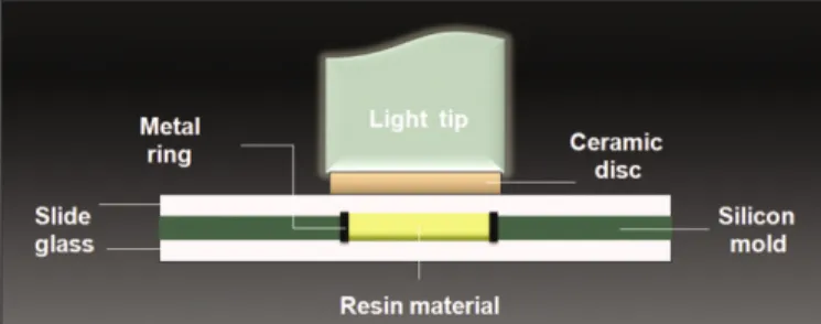
A silicone mold of 7 mm in diameter and 1 mm in height was placed on the glass slab. To ensure that each specimen of resin based material was of the same thickness (1 mm height), a met- al ring (0.2 mm thick, 1 mm height) was placed in the center of the mold.
For specimen preparation, a clear glass slab on top of a black background was used as a supporting surface and to decrease the reflectivity of the underlying surface toward the specimen.
Four resin materials were filled into the silicone mold. Then, the resin materials in control group were covered with a glass microscopic slide (1 mm) to obtain a flat polymerized surface. For the resin specimens curing beneath ceramic, the ceramic disc pre- viously described was placed on top of the thin slide glass (1 mm) to prevent adhesion of the resin materials to the disc (Fig. 1).
Table 1. Description of the resin materials used in this study
Resin material Shade Filler content (% by Weight) Manufacturer
Z350 A2 65% 3M ESPE FiltekTM
Z250 A2 60% 3M ESPE FiltekTM
Variolink II base (VL) Transparent 73.40% Ivoclar vivadent
Variolink II base + catalyst (VLC) Transparent 71.20% Ivoclar vivadent
Fig. 1. Schematic drawings of the test specimen set-up.
Curing was performed using a LED light for 20 or 40 sec- onds. The light outputs of the curing units were measured before each testing procedure using a radiometer. The light tips were in close contact with either the microscopic slide or ceram- ic disc. Using two light curing times, 4 types of resin based mate- rials, and 3 kinds of ceramic discs provided 32 subgroups (Table 2). Each experimental and control subgroup contained 5 specimens with 4 points measured.
All specimens were stored in light-proof containers in dis- tilled water at room temperature for 24 hours. The microhardness was measured using a microhardness tester (Model DMH-2 No.D9137 Matsuzawa Seiki, Japan) with a marker for Vicker’
s unit. Microhardness indentations were made on the bottom surface of each specimen. Four readings with 25 g load for 10 seconds were taken on each surface, and the average was con- verted into a Vicker’s hardness number (VHN).
Statistical analysis
Descriptive statistics of VHNs according to three different thicknesses of upper porcelain layer including one control group and four different types of lower resin layer at two curing time points were displayed as mean and standard deviation.
Considering four repeated measurements were obtained from
the same specimen, a mixed-level repeated measures three-way analysis of variance (ANOVA) was used to compare mean VHNs by different thicknesses of porcelain and different types of resin, applying the mixed procedure with compound symmetry covariance structure, in the SAS statistical package version 9.13 (SAS Inc., Cary, NC, USA). Type one error level 0.05 was used in determining statistical significance.
RESULTS
The mean VHNs for each specimen are shown in Table 3 and 4.
Four VHN values from each resin specimen were measured.
Although variations in the VHN values for these replicates were apparent, it was not found to be statistically significant in ANO- VA (P>.05). Tukey HDS indicated significant differences in mean VHN according to porcelain thickness, resin material, and time (P<.001).
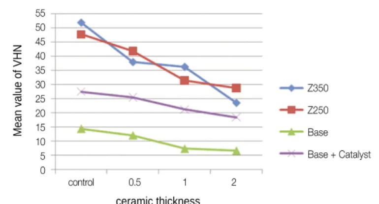
The results indicate that, for curing times of both 20 s and 40 s, the mean VHN decreased as the thickness of the overlaying porcelain increased in all resin types (Figs. 2 and 3). Z350 at control group revealed the highest values of VHN both in 20 and 40 seconds curing condition which was not significant- ly different with Z250 (P<.01). VHNs of resin specimens under 2 mm discs had shown the least values regardless of resin types Table 2. Description of the experimental groups used in this study
Resin material Curing time (sec) Ceramic thickness (mm)
Z350 20 Control 0.5 1.0 2.0
40 Control 0.5 1.0 2.0
Z250 20 Control 0.5 1.0 2.0
40 Control 0.5 1.0 2.0
Variolink II base (VL) 20 Control 0.5 1.0 2.0
40 Control 0.5 1.0 2.0
Variolink II base + catalyst (VLC) 20 Control 0.5 1.0 2.0
40 Control 0.5 1.0 2.0
Table 4. Mean Vickers hardness numbers for each specimen (40 sec)
Time = 20 sec Z350 Mean (P<.05) Z250 Variolink II base Variolink II base + catalyst
Control 51.9 (7.5) 47.7 (4.8) 14.4 (0.8) 27.5 (2.3)
0.5 mm 38.0 (3.6) 41.8 (3.1) 12.1 (0.8) 25.5 (1.4)
1 mm 36.3 (4.4) 31.5 (5.8) 7.4 (0.9) 21.2 (1.9)
2 mm 23.6 (3.5) 28.8 (2.6) 6.7 (0.8) 18.5 (2.3)
Table 3. Mean Vickers hardness numbers for each specimen (20 sec)
Time = 20 sec Z350 Mean (P<.05) Z250 Variolink II base Variolink II base + catalyst
Control 42.7 (7.2) 39.8 (4.4) 9.4 (0.8) 22.4 (2.9)
0.5 mm 29.0 (5.7) 31.2 (3.5) 6.8 (0.8) 18.8 (1.6)
1 mm 21.5 (3.2) 25.9 (2.7) 5.6 (1.2) 16.2 (2.5)
2 mm 17.0 (2.8) 20.5 (3.5) 3.3 (0.6) 12.4 (2.1)
or light curing time.
The increase in VHN of the underlying resin materials with increasing curing time of the light was again statistical- ly significant (P<.05).
As with the light curing, the hardness of all four resin materials increased with increasing light curing time from 20 to 40 seconds. In control group, both 20 and 40 seconds of cur- ing, the hardness of Z350 was significantly higher than VL and VLC, but not significant with Z250 (P<.01). The prolonged cur- ing time was more influential on VL under all ceramic thick- nesses which recorded the lowest values among all resin materials (P<.01).
The use of a catalyst produced significantly higher VHNs than VL groups regardless of the porcelain thickness or light cur- ing time. A significant difference was detected between the dual- cured and light-cured cements when photopolymerized under the same ceramic disc in both 20 and 40 seconds curing (P<.05).
Both universal and flowable restorative resins (Z350 and Z250) showed significantly higher VHN values than cement mate- rials (VL and VLC) (P<.05). However, between the two restorative resin groups (Z350 and Z250), no significant dif- ferences were detected (P<.01).
DISCUSSION
In the present study the influence of the factors (ceramic thick- ness, the influence of additional chemical curing, light curing time) upon the polymerization rate of resin materials was examined by determination of Vicker’s microhardness.
For highly esthetic ceramic prosthesis, Magne and Belser also recommended the minimum tooth reduction depth 0.3 - 0.5 mm, and for fully coverage more than 1.5 mm preparation is needed in incisal area.18 In consideration of the anterior esthetic ceramic prosthesis, IPS e.max Press was chosen and 0.5 mm, 1.0 mm, 2.0 mm ceramic discs were fabricated
which could simulate proper reduction depth.
To produce natural-looking restorations, proper thick veneer layering is inevitable. In 0.5 and 1 mm discs, measuring and building up the proper veneer ceramic was impossible. But in 2 mm disc, to simulate transparent crown third part, the 2 mm thickness consisted of a 1 mm thick framework material and 1 mm thick layering material.
After that to regulate the distance between the light tip and the surface of ceramic restoration, we used 1 mm thickness slide glass in control and experimental group so that we could control the influence of light source distance and avoid direct contact with the restorations.
In case of restorative resins (Z350 and Z250), 20 to 30 seconds of light curing was recommended and for Variolink II, 10 to 40 seconds curing time was recommended by the man- ufacturer. To compare the time factor, specimens were cured for 20 and 40 seconds with LED light in this study.
By far in a number of studies, the curing efficiency of halogen and LED curing units has been compared.15,19Generally, it was found that the decisive factor which determined the degree of monomer conversion was not the source of light but the inten- sity of light resulting in the expected physical and mechani- cal properties of resin composites.13,19
From our results, statistically significant differences in VHNs light cured through ceramic discs were found between the LED exposure time 20 and 40 seconds. Therefore, it can be concluded that the curing efficiency of the LED light with a longer exposure time of 40 seconds is significantly high- er than that with shorter exposure time of 20 seconds in all four resin types including dual curing resin cement. This is incon- sistent with other investigations that showed the ability of high- power LED or QTH lights to reduce exposure time in curing resin-based materials.20 Nevertheless, many studies have shown that sufficient curing time would result in higher degree of conversion, polymerization depth, and hardness values.21,22
Fig. 2. Graph of the mean square microhardness according to porcelain thickness (Light curing time for 20 seconds).
45 40 35 30 25 20 15 10 5 0
Mean value of VHN
control 0.5 1 2 ceramic thickness
Z350 Z250 Base Base + Catalyst
Fig. 3. Graph of the mean square microhardness according to ceramic thickness (Light curing time for 40 seconds).
55 50 45 40 35 30 25 20 15 10 5 0
Mean value of VHN
control 0.5 1 2 ceramic thickness
Z350 Z250 Base Base + Catalyst
In this study, regardless of light curing time and types of resin specimens, the mean VHN decreased significantly as the thickness of the overlaying porcelain increased, which is in agree- ment with Koch et al. who found that the transmission of light through dental ceramic is dependent on its thickness.13The thick- ness of porcelain veneer was considered as the primary factor determining the light transmittance available for polymer- ization.23,24Jung et al.19reported that dual polymerization of a resin luting agent produced greater hardness and polymerization for depths under 2 mm thick for Empress 2 ceramic restora- tions when compared to light polymerization alone. Hofmann et al.15obtained similar results by using 2.5 mm-thick Empress ceramic specimens. On the other hand, Akgungor et al.25 suggested that for Empress 2 ceramic, a thickness of less than 2 mm did not affect complete polymerization of the light-polymerized resin luting agent. This controversial result could be related to the longer light exposure time of 60 seconds used in that study. Similarly, some authors20,26suggested that a minimum 60 seconds of light exposure was necessary for ade- quate polymerization of the resin luting agents through ceram- ic or composite restorations with a thickness of less than 2 mm.
In this study recommended by manufacturer light curing time 20 and 40 seconds were compared. From the results of this study, resin materials under ceramic thickness of 2 mm require additional light curing time. The research with prolonged curing time more than 40 seconds will be needed.
The use of a catalyst resulted in a higher degree of poly- merization. Twenty seconds light curing with catalyst showed higher VHNs compared to 40 seconds light curing without cat- alyst, which emphasized the importance of the self-curing cat- alyst. This result is supported by El-Mowafy and Rubo3who showed that dual-curing resin cements reached an adequate degree of conversion even in the area that was less accessible to the curing lights. VHN values of this study indicate that the catalyst is recommendable in most cases, because statistical dif- ferences were found even in the control group.
It was also demonstrated that restorative resins (Z350 and Z250) showed higher VHN values than resin cements (VL and VLC) regardless of ceramic discs and light curing times.
Although many kinds of resin cements were studied through- out last few decades, no study had compared the restorative resins and the luting cements. Our results pointed out that restorative composites showed higher physical properties than luting resin cements throughout the all variable conditions.
Many kinds of commercially used restorative resin materi- als show high film thickness in room temperature, but a strong correlation was found between the consistency and the film thickness of the luting agents, which is supported by the temperature dependency on the film thickness.27If the flowa- bility of restorative resins obtaines a film thickness in room tem- perature, they could be considered as a luting resin cements.
In this study, the result implies that if the flow of restorative
resin permits, they can be considered as luting materials which show better mechanical properties than normally used resin cements. More researches will be needed in this part.
CONCLUSION
Within the limitations of this study, the following conclusions could be drawn.
1. The decrease in VHN of the underlying resin based materials with increasing thickness of the porcelain disc was statistically significant.
2. The increase in VHN with an increase in curing time was highly significant.
3. The use of a self-curing catalyst increased the VHN which showed stronger influence than doubled light cur- ing time.
4. Restorative composites showed significantly higher VHN values compare to resin cements.
REFERENCES
1. Asmussen E. Restorative resins: hardness and strength vs.
quantity of remaining double bonds. Scand J Dent Res 1982;
90:484-9.
2. Caughman WF, Caughman GB, Shiflett RA, Rueggeberg F, Schuster GS. Correlation of cytotoxicity, filler loading and curing time of dental composites. Biomaterials 1991;12:737-40.
3. El-Mowafy OM, Rubo MH. Influence of composite inlay/onlay thickness on hardening of dual-cured resin cements. J Can Dent Assoc 2000;66:147.
4. Cheong C, King NM, Pashley DH, Ferrari M, Toledano M, Tay FR. Incompatibility of self-etch adhesives with chemical/dual- cured composites: two-step vs one-step systems. Oper Dent 2003;28:747-55.
5. Suh BI, Feng L, Pashley DH, Tay FR. Factors contributing to the incompatibility between simplified-step adhesives and chemi- cally-cured or dual-cured composites. Part III. Effect of acidic resin monomers. J Adhes Dent 2003;5:267-82.
6. Calamia JR, Calamia CS. Porcelain laminate veneers: reasons for 25 years of success. Dent Clin North Am 2007;51:399-417.
7. Garber DA, Goldstein RE. Porcelain and composite inlays and onlays: esthetic posterior restorations. Chicago; Quintessence Publishing Co.; 1994.
8. Strang R, Macdonald I, O'Hagan S, Murray J, Stephen KW.
Variations in performance of curing light units by determination of composite resin setting time. Br Dent J 1987;162:63-5.
9. Blackman R, Barghi N, Duke E. Influence of ceramic thickness on the polymerization of light-cured resin cement. J Prosthet Dent 1990;63:295-300.
10. Peutzfeldt A, Asmussen E. The effect of postcuring on quantity of remaining double bonds, mechanical properties, and in vit- ro wear of two resin composites. J Dent 2000;28:447-52.
11. Pianelli C, Devaux J, Bebelman S, Leloup G. The micro- Raman spectroscopy, a useful tool to determine the degree of con- version of light-activated composite resins. J Biomed Mater Res 1999;48:675-81.
12. Shortall AC, Harrington E. Effect of light intensity on poly- merisation of three composite resins. Eur J Prosthodont Restor Dent 1996;4:71-6.
13. Koch A, Kroeger M, Hartung M, Manetsberger I, Hiller KA, Schmalz G, Friedl KH. Influence of ceramic translucency on cur- ing efficacy of different light-curing units. J Adhes Dent 2007;
9:449-62.
14. Hooshmand T, Mahmoodi N, Keshvad A. Microhardness of a resin cement polymerized by light-emitting diode and halogen lights through ceramic. J Prosthodont 2009;18:411-6.
15. Hofmann N, Papsthart G, Hugo B, Klaiber B. Comparison of pho- to-activation versus chemical or dual-curing of resin-based luting cements regarding flexural strength, modulus and surface hardness. J Oral Rehabil 2001;28:1022-8.
16. Rueggeberg FA, Craig RG. Correlation of parameters used to es- timate monomer conversion in a light-cured composite. J Dent Res 1988;67:932-7.
17. Rueggeberg FA, Ergle JW, Mettenburg DJ. Polymerization depths of contemporary light-curing units using microhard- ness. J Esthet Dent 2000;12:340-9.
18. Magne P, Belser U. Bonded porcelain restorations in the ante- rior dentition-a biomimetic approach. Chicago: Quintessence Publishing Co., 2002.
19. Jung H, Friedl KH, Hiller KA, Haller A, Schmalz G. Curing ef- ficiency of different polymerization methods through ceramic restorations. Clin Oral Investig 2001;5:156-61.
20. Rasetto FH, Driscoll CF, von Fraunhofer JA. Effect of light source and time on the polymerization of resin cement through ceramic veneers. J Prosthodont 2001;10:133-9.
21. Santos GC Jr, El-Mowafy O, Rubo JH, Santos MJ. Hardening of dual-cure resin cements and a resin composite restorative cured with QTH and LED curing units. J Can Dent Assoc 2004;70:
323-8.
22. Moon HJ, Lee YK, Lim BS, Kim CW. Effects of various light curing methods on the leachability of uncured substances and hard- ness of a composite resin. J Oral Rehabil 2004;31:258-64.
23. O'Keefe KL, Pease PL, Herrin HK. Variables affecting the spectral transmittance of light through porcelain veneer samples.
J Prosthet Dent 1991;66:434-8.
24. Brodbelt RH, O'Brien WJ, Fan PL. Translucency of dental porcelains. J Dent Res 1980;59:70-5.
25. Akgungor G, Akkayan B, Gaucher H. Influence of ceramic thick- ness and polymerization mode of a resin luting agent on early bond strength and durability with a lithium disilicate-based ceramic system. J Prosthet Dent 2005;94:234-41.
26. Rueggeberg FA, Caughman WF. The influence of light expo- sure on polymerization of dual-cure resin cements. Oper Dent 1993;18:48-55.
27. van Meerbeek B, Inokoshi S, Davidson CL, De Gee AJ, Lambrechts P, Braem M, Vanherle G. Dual cure luting composites- Part II: Clinically related properties. J Oral Rehabil 1994;21:57- 66.