PREVENTION RESEARCH □ ORIGINAL ARTICLE □
83 책임저자:최영현, 614-052, 부산시 진구 양정동 산 45번지
동의대학교 한의과대학 생화학교실 Tel: 051-850-7413, Fax: 051-853-4036 E-mail: [email protected]
접수일:2010년 2월 5일, 1차수정일: 2010년 2월 8일, 2차수정일: 2010년 2월 11일, 게재승인일:2010년 2월 16일
Correspondence to:Yung Hyun Choi
Department of Biochemistry, Dongeui University College of Oriental Medicine, San 45, Yangjung-dong, Busanjin-gu, Busan 614-052, Korea Tel: +82-51-850-7413, Fax: +82-51-853-4036
E-mail: [email protected]
상황버섯 추출물에 의한 인체혈구암세포의 세포사멸 및 Prostaglandin E2의 생성 저하
동의대학교 1블루바이오 소재개발센터, 2생활과학대학 식품영양학과, 3대학원 바이오물질제어학과,
4한의과대학 생화학교실, 5한국승강기대학 승강기보수과
박 철1ㆍ현숙경1,2ㆍ황혜진1,2ㆍ최성현5ㆍ최영현1,3,4
Induction of Apoptosis and Inhibition of Prostaglandin E
2Production by Proteoglycan of Phellinus linteus in U937 Human Leukemia Cells
Cheol Park1, Sook Kyung Hyun1,2, Hye Jin Hwang1,2, Sung-Hyun Choi5 and Yung Hyun Choi1,3,4
1Blue-Bio Industry RIC, 2Department of Food and Nutrition, College of Human Ecology, 3Department of Biomaterial Control (BK21 Program), Graduate School, 4Department of Biochemistry, College of Oriental Medicine,
Dong-Eui University, Busan 614-052, 5Korea Lift College, Geochang 670-802, Korea
Various mushroom polysaccharides and polysaccharides-protein complexes are known to possess anti-tumor and immunomodulating effects. Here, we demonstrated that proteoglycan extracted from Phellinus linteus has a potent anti-tumor and/or anti-inflammatory activity. Proteoglycan treatment resulted in a concentration-dependent growth inhibition by including apoptosis, which could be proved by DAPI staining, agarose gel electrophoresis and flow cytometry analysis. The increase in apoptosis induced by proteoglycan was correlated with up-regulation of pro-apoptotic Bax and Bad expression, and down- regulation of anti-apoptotic Bcl-2 and Bcl-xL expression. Proteoglycan inhibited the levels of IAP family members such as XIAP and cIAP-1 and induced the proteolytic activation of caspase-3, and a concomitant degradation of β-catenin. In addition, it was found that proteoglycan treatment markedly decreased the levels of cyclooxygenases (COX)-2 mRNA and protein expression without significant changes in the expression of COX-1, which was correlated with a decrease in prostaglandin E2 synthesis. Taken together, these findings provide important new insights into the possible molecular mechanisms of the anti-cancer activity of proteoglycan. (Cancer Prev Res 15, 83-91, 2010)
Key Words: Phellinus linteus, Proteoglycan, Apoptosis, Prostaglandin E2
서 론
Apoptosis는 개체의 발생단계나 DNA 손상, 바이러스 감염 등에 의한 유전적 조절 하에서 일어나는 정교한 인 체방어기전이란 점에서 necrosis와 구별된다.1,2) 또한 ap-
optosis는 개체보존수준에서 손상된 세포들의 제거를 위 한 중요한 수단이며, 정상적인 세포주기의 이탈이나 특 정 세포주기 조절인자 활성의 변화가 apoptosis의 주 원인 이 될 수 있다.3) Apoptosis의 유발에 Bcl-2 family 인자들과 연관된 caspase protease가 관여한다는 사실이 알려지면서 apoptosis와 연관된 분자적 기전이 최근 많이 밝혀지고 있
다.4) 따라서 항암 효능을 가지는 특정 후보물질에 의한 암세포의 apoptosis 유발 여부의 조사와 연관된 기전 연구 는 항암물질 개발을 위한 가장 기본적인 과정으로 인식 되어지고 있다.
한편 염증반응은 생체조직의 방어 반응 중 하나로써 암을 포함한 다양한 질환의 유발 원인으로 인식되어지 고 있다. 이 과정에서 중요한 역할을 하는 prostaglandin (PG)은 세포의 분열이나 증식을 조절함으로서 각종 인체 질병의 유발과 진행을 촉진하는 것으로 최근 밝혀지고 있다.5∼7) 다양한 종류의 PG의 합성은 cytosolic phospho- lipase A2 (PLA2)에 의해 membrane phospholipid로부터 ara- chidonic acid가 방출되면 cyclooxygenases (COXs)에 의해 방 출된 arachidonic acid가 PGG2와 PGH2로 전환이 되고 PG synthase 등에 의해 PGH2가 각각의 PGs로 전환된다. PG 합성에 중요한 역할을 하는 두 가지 중요한 COX 중, COX-1은 대부분의 조직에서 일정한 수준으로 발현되어 있으며 주로 인체의 항상성 유지에 관여하는 반면, COX-2는 다양한 cytokine에 의하여 유발되어 암을 포함 한 세포의 성장 및 분화와 연관된 각종 퇴행성 질환의 발병과 진행에 중요한 역할을 한다.8,9)
최근 연구 결과들에 의하면 버섯은 단순한 식품이상 의 약리적인 효능을 가지고 있는 것으로 나타나고 있으 며, 면역증강 및 항암활성에 큰 효능이 있음을 보여주고 있다.10) 이러한 효능의 대부분은 polysaccharide 또는 po- lysaccharides-protein complex로서, 특히 상황버섯(목질진흙 버섯, Phellinus linteus)에서 추출된 이들 물질은 강력한 im- munomodulating 효과 및 암세포 사멸 유도 효과가 있는 것으로 알려져 있다.10∼15) 본 연구실에서도 그동안 다양 한 상황버섯 추출물의 항암 및 면역증강 효과를 보고한 바 있으나, 여전히 구체적인 분자세포생물학적 기전은 완전히 밝혀지진 않았다.16∼18) 본 연구에서는 암세포의 강력한 면역증강 효과가 있었던 상황버섯 유래 proteo- glycan17)이 인체혈구암세포의 증식억제 과정에 연관된 ap- optosis 유도 및 PGE2 생성 저해 효과를 보고하고자 한다.
재료 및 방법
1. 암세포 배양, proteoglycan의 처리 및 생존율 조 사
실험에 사용한 U937 혈구암세포는 생명공학연구소 (KRIBB, Taejeon, Korea)에서 분양 받았으며 90%의 RPMI- 1640 배지, 10% fetal bovine serum (FBS)에 1%의 penicillin 및 streptomycin이 포함된 배지(Gibco BRL, Grand Island, NY, USA)를 사용하여 5% CO2, 37oC의 조건에서 배양하
였다. 상황버섯에서 추출된 proteoglycan17)은 dimethyl sul- foxide (DMSO)에 녹여 stock 용액으로 제조한 뒤 −20oC에 보관하였고 적정 농도로 배지에 희석하여 처리하였다.
Proteoglycan 처리에 따른 U937 세포의 생존율을 조사하 기 위하여 proteoglycan을 적정농도로 처리하여 48시간 배양하였다. 그 후 상층액을 제거하고 세포 부유액과 0.5% trypan blue (Gibco BRL)를 동량으로 넣어 2분간 처리 하고 도립 현미경(Carl Zeiss, Germany)을 이용하여 살아있 는 세포를 계수하였다.
2. DAPI staining에 의한 세포핵의 형태 관찰
핵의 형태적 변화를 관찰하기 위하여 37% formalde- hyde 용액과 PBS를 1:9의 비율로 섞은 fixing solution을 모아진 세포에 첨가하여 상온에서 10분 동안 고정한 후 cytospin을 이용하여 slide glass 위에 세포를 부착시켰다.
부착된 세포에 4',6-diamidino-2-phenylindole (DAPI, Sigma Chemical Co., St. Louis, MO, USA) 용액을 처리하여 염색시 키고 mounting solution을 처리한 후, 형광 현미경(Carl Zeiss)을 이용하여 400배의 배율로 핵의 형태 변화를 관 찰하였다.
3. Agarose gel 전기영동에 의한 DNA fragmen- tation의 분석
정상 및 proteoglycan이 처리된 배지에서 48시간 동안 배양된 세포를 모아 lysis buffer [5 mM Tris-HCl (pH 7.5), 5 mM EDTA, 0.5% Triton X-100]를 4oC에서 30분간 처 리한 다음, 그 상층액에 proteinase K (Sigma)를 0.5 mg/ml 의 농도로 처리하여 50oC에서 3시간 동안 반응시켰다.
그 후 phenol:chloroform:isoamyl alcohol 혼합 용액(25:
24:1, Sigma)을 첨가하고 30분간 반응시킨 후 원심 분리 하여 얻어진 상층액에 적정량의 isopropanol (Sigma)과 5 M NaCl를 첨가한 다음 24시간 정도 4oC에서 반응시켰다.
분리된 DNA pellet에 RNase A가 적당량 들어있는 TE buffer를 첨가하여 녹이고 1.5% agarose gel을 이용하여 전 기영동한 후 ethidium bromide (EtBr, Sigma)로 염색하고 ultra vilolet (UV) 하에서 관찰하였다.
4. Flow cytometry 분석
정상 및 proteoglycan이 함유된 배지에서 자란 암세포 들을 PBS로 두세 번 씻어내고, 고정액(70% ethyl alcohol, 0.5% Tween 20)을 첨가하여 4oC에서 고정시킨 후, 핵산에 특이적으로 결합하는 형광물질인 DNA intercalating dye propidium iodide (PI, concentration, 50μg/ml; Sigma)와 10 kunit의 RNase (Sigma)를 처리하고 4oC에서 1시간동안 염
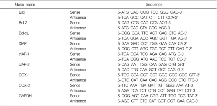
Table 1. Sequence of primers used for RT-PCR
Gene name Sequence
Bax Sense
Antisense
5'-ATG GAC GGG TCC GGG GAG-3' 5'-TCA GCC CAT CTT CTT CCA-3'
Bcl-2 Sense
Antisense
5'-CAG CTG CAC CTG ACG-3' 5'-ATG CAC CTA CCC AGC-3'
Bcl-xL Sense
Antisense
5'-CGG GCA TTC AGT GAC CTG AC-3' 5'-TCA GGA ACC AGC GGT TGA AG-3'
XIAP Sense
Antisense
5'-GAA GAC CCT TGG GAA CAA CA-3' 5'-CGC CTT AGC TGC TCT CTT CAG T-3'
cIAP-1 Sense
Antisense
5'-TGA GCA TGC AGA CAC ATG C-3' 5'-TGA CGG ATG AAC TCC TGT CC-3'
cIAP-2 Sense
Antisense
5'-CAG AAT TGG CAA GAG CTG G-3' 5'-CAC TTG CAA GCT GCT CAG G-3'
COX-1 Sence
Antisence
5'-TGC CCA GCT CCT GGC CCG CCG CTT-3' 5'-GTG CAT CAA CAC AGG CGC CTC TTC-3'
COX-2 Sence
Antisence
5'-TTC AAA TGA GAT TGT GGG AAA AT-3' 5'-AGA TCA TCT CTG CCT GAG TAT CTT-3
GAPDH Sence
Antisence
5'-CGG AGT CAA CGG ATT TGG TCG TAT-3' 5'-AGC CTT CTC CAT GGT GGT GAA GAC-3'
색하였다. 이를 다시 PBS로 두 번 씻어낸 후 DNA flow cytometry (Becton Dickinson, San Jose, CA, USA)에 적용시 켜 형광반응에 따른 histogram을 ModiFit LT (Becton Dic- kinson) 프로그램으로 분석하였다.
5. Reverse transcription (RT)-PCR 분석
동일한 조건에서 준비된 세포들을 대상으로 TRIzol B (Invitrogen, Carlsbad, CA, USA)를 이용하여 total RNA를 분 리하였다. 분리된 RNA를 정량한 후, ONE-STEP RT-PCR PreMix (iNtRON BIOTECHNOLOGY, Korea)를 이용하여 2 μg의 RNA에서 ss cDNA를 합성하였다. 이들 cDNA를 template로 사용하여 관찰 대상 유전자(Bioneer, Taejeon, Korea, Table 1)를 PCR로 증폭하였다. 이때 housekeeping 유전자인 glyceraldehyde-3-phosphate dehydrogenase (GAPDH) 유전자를 internal control로 사용하였다. 각 PCR 산물들을 1% agarose gel을 이용하여 전기영동하고 ethidium bro- mide (EtBr)로 염색한 후 UV 하에서 확인하였다.
6. 단백질의 분리, 전기영동 및 Western blotting
준비된 세포들을 lysis buffer로 용해한 후, 고속원심분 리로 세포 내 잔사물을 분리시킨 후 동량의 단백질을 SDS-polyacrylamide gel 전기영동으로 분리하였다. 분리된 단백질을 nitrocellulose membrane (Schleicher and Schuell, Keene, NH, USA)으로 전이시킨 후, 특정 단백질에 대한 항체와 그에 대한 이차 항체 반응을 실시한 후 enhanced chemiluminoesence (ECL) 용액(Amersham Life Science)을 적
용시킨 다음 X-ray film에 감광시켜 특정 단백질의 발현 양을 분석하였다. 이를 위해 사용된 항체는 CalBiochem (San Diego, CA, USA), 및 Santa Cruz Biotechnology Inc.
(Santa Cruz, CA, USA)에서 구입하였으며, immunoblotting 을 위해 2차 항체로 사용된 peroxidase-labeled donkey anti- rabbit 및 peroxidase-labeled sheep anti-mouse immunoglobulin 은 Amersham Life Science Corp. (Arlington Heights, IL, USA) 에서 구입하였다.
7. In vitro caspase-3의 활성 측정
Proteoglycan 처리에 의한 caspase-3의 활성 변화 여부를 조사하기 위하여 준비된 세포에서 단백질을 추출하고 정량하여 각각 150μg의 단백질을 fluorogenic peptide 기 질 100μM이 함유된 extraction buffer [40 mM HEPES (pH 7.4), 20% glycerol (v/v), 1 mM EDTA, 0.2% NP-40 and 10 mM DL-DTT] 50μl에 혼합하였으며, microtiter plate에 다 시 extraction buffer에 희석하여 각 sample 당 총 volume이 100μl가 되게 하였다. 준비된 plate를 3oC에서 3시간동안 반응 시킨 후 ELISA reader를 이용하여 405 nm의 흡광도 를 이용하여 반응의 정도를 측정하였다. 실험에 사용된 caspase-3의 기질은 Asp-Glu-Val-Asp (DEVD)-p-nitroaniline (pNA)이었다.
8. Prostaglandin E2의 측정
PGE2의 생성양의 측정은 Amersham Corp.의 enzyme immunoassay (EIA) kit를 사용하였으며, 준비된 세포를
Fig. 1. Growth inhibition and apoptosis induction of human leukemia U937 cells after treatment with proteoglycan isolated from P. linteus. (A) Cells were seeded as described in materials and methods, and the viable cells were counted after proteoglycan (PG) treatment for 48 h. Results are expressed as averages +/− SD form separate experiments. (B) Cells were treated with proteoglycan for 48 h and then stained with DAPI solution. After 10 min incubation at room temperature, the cells were washed with PBS and nuclear morphology was photographed with a fluorescent microscope using blue filter. Magnification, ×400. (C) For the analysis of DNA fragmentation, the cells were treated with various concentrations of proteoglycan for 48 h. The genomic DNA was extracted, separated on 1.0% agarose gel electrophoresis and visualized under UV light after staining with EtBr. M indicates a size marker of the DNA ladder. (D) The cells grown under same condition as (B) were stained with PI for flow cytometry analysis. The percentages of cells with hypodiploid DNA (sub-G1 phase) contents represent the fractions undergoing apoptotic DNA degradation. Data are expressed as mean of two independent experiments.
96-well plate에 well 당 160μl의 배지에 104 cell/well 정도 로 분주하여 24시간 동안 배양한 후, proteoglycan을 농도 별로 배지에 희석하여 처리하였다. 이때 마지막 배지의 양을 모두 180μl 통일시켰다. 48시간 후 배지에 2.5%의 dodecyltrimethylammonium bromide가 함유된 buffer를 20μl 첨가하여 총 배지의 양이 200μl 되게 한 후 lysis가 잘 일어나도록 pipetting을 수회 실시하였다. 상온에 약 10분 간 incubation 한 후 trypan blue를 이용하여 암세포의 수를 계수하였다. 약 50μl의 lysate를 취하여 kit에 준한 pro- tocol에 따라 EIA를 실시한 후 450 nm의 파장에서 얻어진
값을 기준으로 PGE2의 양을 추정하였다.
결과 및 고찰
1. Proteoglycan에 의한 U937 세포의 apoptosis 유 발
Proteoglycan 처리에 의한 U937 세포의 apoptosis 유발 및 COX-2-PGE2 연관 실험을 수행하기 위한 조건의 설정 을 위하여 48시간 동안 다양한 농도의 proteoglycan을 처 리한 후 U937 세포의 생존율을 조사하였다. Fig. 1A에 나
Fig. 2. Effects of proteoglycan treatment on the levels of Bcl-2 and IAP family members in human leukemia U937 cells. (A) After 48 h incubation with proteoglycan, total RNAs were isolated and reverse-transcribed. The resulting cDNAs were subjected to PCR with the indicated primers, and the reaction products were subjected to electrophoresis in a 1% agarose gel and visualized by EtBr staining. GAPDH was used as an internal control. (B) The cells were lysed and then cellular proteins were separated by SDS-polyacrylamide gels and transferred onto nitrocellulose membranes. The membranes were probed with the indicated antibodies. Proteins were visualized using an ECL detection system. Actin was used as an internal control.
타낸 결과에서 알 수 있듯이 조사된 U937 세포에서 proteoglycan 처리 농도의 증가에 따라 U937 세포의 생존 율이 매우 감소되어, 1.5 mg/ml 처리군에서 대조군에 비 하여 50% 이하로 낮아졌으며, 2.5 mg/ml 처리군에서 15% 이하로 감소되었다. Proteoglycan의 처리에 의한 생 존율 억제 효과가 apoptosis 유발과 상관성이 있는지를 조 사하기 위하여 DAPI 염색에 의한 핵의 형태적 변화, agarose gel 전기영동에 의한 DNA 단편화 및 flow cyto- metry 분석에 의한 sub-G1기 세포의 빈도 변화 등을 조사 하였다. 먼저 Fig. 1B에 나타낸 것과 같이 전형적인 ap- optosis가 유발된 세포에서 관찰되는 chromatin condensa- tion에 의한 apoptotic body의 출현이 proteoglycan 처리 농 도 의존적으로 증가됨을 확인할 수 있었고, Fig. 1C의 agarose gel 전기영동에 의하여 proteoglycan의 처리 농도 증가에 따라 DNA 단편화가 증가되었음을 알 수 있었다.
또한 Fig. 1C의 flow cytometry 결과에서 나타난 것과 같 이, 정상 배지에서 자란 U937 세포에 비하여 proteoglycan 이 처리된 배지에서 배양된 세포에서 처리 농도 의존적 으로 apoptosis가 일어난 세포에 해당되는 sub-G1기에 속 하는 세포의 빈도가 상당량 증가하는 것을 확인하였다.
이와 같은 결과는 proteoglycan의 처리에 의한 U937 세포 의 증식 억제 효과가 apoptosis의 유발과 직접적인 연관성 이 있음을 의미하여 주는 것이다.
2. Bcl-2 및 IAP family 인자들의 발현에 미치는 proteoglycan의 영향
Apoptosis 유발 여부의 조절인자 중 Bcl-2 family에 속하 는 인자들은 apoptosis 유발 조절에서 가장 대표적인 유전 자군로 알려져 있는데, 그 중 Bcl-2 및 Bcl-xL 등은 anti- apoptotic 분자로서 apoptosis의 유발을 억제하는 기능을 가지며, Bax는 pro-apoptotic 분자로 apoptosis의 유발과 관 계가 있다. 이들 두 유전자는 세포 내 소기관 중 mito- chondria로부터의 cytochrome c를 유리시켜 cysteine-related protease인 caspase 또는 DNA의 단편화와 연관된 endo- nuclease 등을 활성화시키며, 이들은 서로 dimer의 형태로 존재하며 그들의 발현 수준에 변화가 초래되면 apoptosis 가 유발되는 것으로 알려져 있다.19,20) 따라서 U937 세포 에서 proteoglycan에 의한 apoptosis 유발에 이들 유전자가 관련되어 있는지의 여부를 RT-PCR 및 Western blot 분석 으로 조사하였다. Fig. 2A에 나타낸 바와 같이 anti-
Fig. 3. Activation of caspase-3 and degradation of β-catenin proteins by proteoglycan treatment in human leukemia U937 cells. (A) After 48 h incubation with proteoglycan, the cells were lysed, and cellular proteins were separated by SDS-polyacryla- mide gel and transferred onto nitrocellulose membranes. The membranes were probed with anti-caspase-3 and β-catenin antibodies. Proteins were visualized using an ECL detection system. Actin was used as an internal control. (B) U937 cells were treated with indicated concentrations of proteoglycan for 48 h, collected and then lysed. Aliquots (150μg proteins) were incubated with DEVD-pNA for caspase-3 activity at 37oC for 3 h. The released fluorescent products were measured. The data shown are means of two independent experiments.
apoptotic 인자인 Bcl-2 및 Bcl-xL의 발현은 proteoglycan 처 리 농도의 증가에 따라 발현의 정도가 감소되었으나, pro-apoptotic 인자인 Bax 또는 Bad의 경우 proteoglycan 처 리 농도 증가에 따라 발현이 모두 증가되었다. 이는 proteoglycan의 처리에 의한 U937 세포의 생존율 저하에 따른 apoptosis 유발에 Bcl-2 family의 발현 변화가 중요한 역할을 하고 있음을 보여주는 결과이다.
Apoptosis 조절에 관여하는 또 다른 인자 중 IAP family 에 속하는 여러 인자들은 caspase와의 직접적인 결합을 통하여 그들의 apoptotic 활성을 억제할 수 있을 것으로 밝혀져 있다.21,22) 따라서 본 연구에서는 proteoglycan 처리 에 의한 U937 세포의 apoptosis 유발에 이들 IAP family가 관여하는지의 여부를 조사하였으며, 그 결과는 Fig. 2B에 나타난 바와 같다. 결과에서 알 수 있듯이 proteoglycan 처리에 따라서 조사된 IAP family의 3가지 인자(XIAP, cIAP-1 및 cIAP-2) 대부분이 proteoglycan의 처리 농도 증 가에 따라 전사 및 번역 수준에서 발현의 정도가 현저히 감소되었다. 이러한 결과는 proteoglycan의 처리에 의한 U937 세포의 apoptosis 유발에 IAP family 인자들의 발현 감소에 따른 caspase 효소들의 활성이 증가할 수 있음을 보여 주는 것이다.
3. Caspase-3의 활성에 미치는 proteoglycan의 영향
Caspase protease는 apoptosis 유발에 가장 중요한 조절인 자로서 작용하는데, 이들 family에 속하는 단백질들은 세 포에서 핵과 mitochondria의 외막에 불활성 상태로 존재 하며, Bcl-2/Bax family 발현의 변화에 따라 이들의 활성도 가 조절될 수 있다.23,24) 이들은 proenzyme 형태로 존재하 다가 apoptosis 유도를 활성화 시키는 신호에 의해 활성화 된 protease로 전환되어 직접 또는 간접적으로 세포 내에 존재하는 많은 표적 단백질의 분해에 관여하는 것으로 알려져 있다. 본 연구에서는 proteoglycan 처리에 의하여 caspase 중, apoptosis 유도의 종결에 가장 중요하게 작용하 는 caspase-3의 활성이 증가하는지의 여부를 조사하였다.
먼저 Western blotting에 의한 caspase-3 단백질 발현의 정 도를 비교한 결과는 Fig. 3A에 나타낸 바와 같이, pro- teoglycan 처리에 따라 활성형 caspase-3의 발현이 매우 증 가되었다. 이러한 단백질 수준에서의 결과를 재확인하 기 위하여 in vitro caspase 활성을 정량적으로 조사한 결 과, Fig. 3B에서 알 수 있듯이 proteoglycan의 처리 농도 증가에 따라 caspase-3의 활성이 점차 증가되었다.
한편 활성화된 caspase는 세포의 정상적인 생존에 필수 적인 세포 내 주요 단백질들을 분해할 수 있으며, 단편화
가 일어난 이들 단백질들은 apoptosis가 유발되었다는 표 지자로서 활용이 된다. 본 연구에서는 대표적인 caspase-3 의 기질 단백질에 해당되는 β-catenin의 발현 변화를 조 사하였는데, β-catenin 단백질은 cell-cell adhesion 및 Wnt signaling에 관여하는 E-cadherin–associated 단백질로서 세 포 내 골격 유지, 다양한 부착성 세포의 전사 조절 및 세포 유착에 관여하는 caspase의 표적 단백질이다.25) Fig.
3A에 나타낸 것처럼 β-catenin 단백질의 발현이 proteo- glycan 처리 농도의 증가에 따라 단편화의 정도가 증가하 였음을 알 수 있었는데, 이러한 결과는 proteoglycan 처리
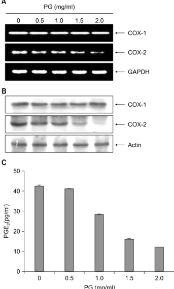
Fig. 4. Inhibition of COX-2 expression and PGE2 production in human leukemia U937 cells after exposure to proteoglycan.
(A) After 48 h incubation with proteoglycan, total RNAs were isolated and reverse-transcribed. The resulting cDNAs were subjected to PCR with COX-1 and COX-2 primers, and the reaction products were subjected to electrophoresis in a 1%
agarose gel and visualized by EtBr staining. GAPDH was used as an internal control. (B) The cells grown under same con- dition as (A) were lysed and then cellular proteins were separated by SDS-polyacrylamide gels and transferred onto nitrocellulose membranes. The membranes were probed with anti-COX-1 and anti-COX-2 antibodies. Proteins were visua- lized using an ECL detection system. Actin was used as an internal control. (C) The cells were treated with the indicated concentrations of proteoglycan for 48 h and collected. The PGE2 accumulation in the medium was determined by an EIA kit as described in materials and methods. Results are ex- pressed as the means±S.E. of three independent experiments.
에 따른 U937 세포의 apoptosis 유발은 caspase-3의 활성을 통한 표적단백질의 단편화에 의하여 이루어지고 있음을 보여 주는 것이다.
4. COX-2 발현 및 PGE2의 생성에 미치는 pro- teoglycan의 영향
다양한 질환의 매개체로 작용하는 PG은 세포분열이 나 증식에 영향을 줌으로서 각종 인체 질병의 유발과 진 행에 중요한 역할을 하는데, PG의 합성에서 2가지 COX isoform이 관여하고 있다. 그 중 대부분의 조직에서 일정 한 수준으로 발현되는 COX-1은 인체의 항상성 유지와 연관된 기능의 수행에 관여하고 있으며, COX-2는 성장 인자, cytokines, 종양 촉진인자들 등의 자극에 의해 발현 이 증가되는 유도성 isoform으로서 세포 증식을 촉진하고 apoptosis를 억제하며 세포의 유동성 및 부착성을 강화시 킴으로서 각종 퇴행성 질환의 발병과 진행을 촉진시킨
다.26,27) 다양한 역학적 조사와 여러 종류의 종양 조직에
서 COX-2가 높은 발현을 유지하는 것이 apoptosis에 대한 저항성 획득과 염증반응과 연관된 세포의 암화에 밀접한 관련이 있을 것으로 추정되어지고 있다.8,9) 또한 COX-2 의 과발현에 의해 암조직에서의 혈관신생 및 전이능이 높아지고, COX-2의 선택적 억제제에 의한 angiogenesis 와 종양형성 억제 등의 결과에서 COX-2의 선택적 조절 에 의한 암예방 및 항암전략이 시도되고 있다.
따라서 proteoglycan이 처리된 U937 세포의 증식 억제 및 apoptosis 유발이 COX-2의 발현 저하 및 PG의 생성 변화와의 관련성이 있는지의 여부를 조사하였다. 이를 위하여 COX-1 및 -2의 발현 변화를 먼저 조사하였는데, Fig. 4A의 결과에서 알 수 있듯이 조사된 4가지 암세포주 의 COX-1의 mRNA 및 단백질 발현에는 proteoglycan이 별다른 영향을 미치니 못하였으나, COX-2의 경우 전사 및 번역 수준에서 proteoglycan 처리 농도의 증가에 따라 점차적인 발현의 감소를 보여주었다. 이러한 proteoglycan 에 의한 COX-2의 선택적 발현 저하가 PGE2의 생성 억제 와 연관성이 있는지를 조사한 결과는 Fig. 4B에 나타낸 바와 같다. 제시된 결과에서 알 수 있듯이 proteoglycan의 처리 농도가 증가할수록 PGE2의 생성이 매우 감소되었 으며, PGE2의 생성 감소 경향성은 COX-2의 발현 저하와 유사한 경향성을 보여주었다. 따라서 proteoglycan 처리에 의한 암세포의 증식 억제에는 COX-2의 선택적 발현 억 제에 의한 PGE2의 생성 저해와 연관성이 있음을 알 수 있었다.
결 론
본 연구에서는 상황버섯의 대표적인 생리활성 물질인 proteoglycan의 처리에 따른 인체혈구암세포의 증식억제 와 연관된 apoptosis 유도 및 COX-2 활성의 변화 연관성 을 조사하였다. 이를 위하여 U937 세포가 사용되었으며, proteoglycan 처리 농도의 증가에 의하여 U937 세포의 생 존이 점차 억제되었으며, 이러한 현상이 apoptosis 유발과 연관성이 있었음을 염색질 응축에 따른 apoptotic body의 출현 증가, DNA 단편화 및 flow cytometry 분석에 따른 sub-G1기 세포의 빈도 증가로 확인을 하였다. Proteo- glycan 처리에 의한 apoptosis 유발에 관여하는 유전자들 의 발현 변화를 RT-PCR 및 Western blot 방법으로 조사한 결과, Bcl-2 family에 속하는 anti-apoptotic 인자인 Bcl-2 및 Bcl-xL의 발현은 전사 및 번역 수준에서 감소되었고, pro- apoptotic Bax의 발현은 증가되었으며, IAP family 단백질 들의 발현도 감소되었다. 또한 caspase-3의 활성 증가와 더불어 β-catenin과 같은 caspase-3의 기질 단백질들의 분 해 등이 proteoglycan에 의한 U937 세포의 apoptosis 유도 과정에서 관찰되었다. 아울러 proteoglycan 처리 농도가 증가함에 따라 COX-2의 발현이 전사 및 번역 수준에서 모두 감소되었으며 이에 따른 PGE2의 생성 역시 현저하 게 감소되었으나, COX-1의 발현에는 큰 변화가 없었다.
이러한 결과들은 상황버섯 유래 proteoglycan의 항암작용 을 이해하는데 중요한 자료가 될 것이다.
감사의 글
본 논문은 2009년도 동의대학교 교내연구비(2009AA123) 지원으로 이루어졌음.
참 고 문 헌
1) Arends MJ, Morris RG, Wyllie AH. Apoptosis. The role of the endonuclease. Am J Pathol 136, 593-608, 1990.
2) Evans VG. Multiple pathways to apoptosis. Cell Biol Int 17, 461-476, 1993.
3) Chiarugi V, Magnelli L, Basi G. Apoptosis and the cell cycle.
Cell Mol Biol Res 40, 603-612, 1994.
4) Nagata S. Apoptosis by death factor. Cell 88, 355-365, 1997.
5) Vainio H. Is COX-2 inhibition a panacea for cancer pre- vention? Int J Cancer 94, 613-614, 2001.
6) Wymann MP, Schneiter R. Lipid signalling in disease. Nat Rev Mol Cell Biol 9, 162-176, 2008.
7) Clària J. Regulation of cell proliferation and apoptosis by
bioactive lipid mediators. Recent Pat. Anticancer Drug Discov 1, 369-382, 2006.
8) Botting RM. Inhibitors of cyclooxygenases: mechanisms, selec- tivity and uses. J Physiol Pharmacol 5, S113-124, 2006.
9) Dempke W, Rie C, Grothey A, Schmoll HJ. Cyclooxygenase- 2: a novel target for cancer chemotherapy? J Cancer Res Clin Oncol 127, 411-417, 2001.
10) Zhu T, Kim SH, Chen CY. A medicinal mushroom: Phellinus linteus. Curr Med Chem 15, 1330-1335, 2008.
11) Chang ZQ, Oh BC, Lee SP, Rhee MH, Park SC. Comparative immunomodulating activities of polysaccharides isolated from Phellinus spp. on cell-mediated immunity. Phytother Res 22, 1396-1399, 2008.
12) Sliva D, Jedinak A, Kawasaki J, Harvey K, Slivova V.
Phellinus linteus suppresses growth, angiogenesis and invasive behaviour of breast cancer cells through the inhibition of AKT signalling. Br J Cancer 98, 1348-1356, 2008.
13) Shin JY, Lee S, Bae IY, Yoo SH, Lee HG. Structural and biological study of carboxymethylated Phellinus linteus poly- saccharides. J Agric Food Chem 55, 3368-3372, 2007.
14) Zhu T, Guo J, Collins L, Kelly J, Xiao ZJ, Kim SH, Chen CY. Phellinus linteus activates different pathways to induce apoptosis in prostate cancer cells. Br J Cancer 96, 583-590, 2007.
15) Collins L, Zhu T, Guo J, Xiao ZJ, Chen CY. Phellinus linteus sensitises apoptosis induced by doxorubicin in prostate cancer.
Br J Cancer 95, 282-288, 2006.
16) Choi YH, Huh MK, Ryu CH, Choi BT, Jeong YK. Induction of apoptotic cell death by mycelium extracts of Phellinus linteus in human neuroblastoma cells. Int J Mol Med 14, 227-232, 2004.
17) Kim GY, Lee JY, Lee JO, Ryu CH, Choi BT, Jeong YK, Lee KW, Jeong SC, Choi YH. Partial characterization and immu- nostimulatory effect of a novel polysaccharide-protein complex extracted from Phellinus linteus. Biosci Biotechnol Biochem 70, 1218-1226, 2006.
18) Park C, Kim HJ, Chung KT, Yoon TK, Choi BT, Lee YT, Park DI, Choi YH. Apoptotic cell death by methanol extract of Phellinus linteus-barley corn in human leukemic U937 cells through induction of p21 and Bax, and activation of ca- spase-3. Korean J Oriental Physiol Pathol 21, 1226-1232, 2007.
19) Liu S, Seidel-Dugan C. In search of p53 target genes for the therapeutic manipulation of cancer. Curr Opin Drug Discov Devel 9, 176-183, 2006.
20) Bettaieb A, Dubrez-Daloz L, Launay S, Plenchette S, Rebe C, Cathelin S, Solary E. Bcl-2 proteins: targets and tools for chemosensitisation of tumor cells. Curr Med Chem Anticancer Agents 3, 307-318, 2003.
21) de Graaf AO, de Witte T, Jansen JH. Inhibitor of apoptosis proteins: new therapeutic targets in hematological cancer?
Leukemia 18, 1751-1759, 2004.
22) Wrzesien-Kus A, Smolewski P, Sobczak-Pluta A, Wierz- bowska A, Robak T. The inhibitor of apoptosis protein family
and its antagonists in acute leukemias. Apoptosis 9, 705-715, 2004.
23) Philchenkov A, Zavelevich M, Kroczak TJ, Los M. Caspases and cancer: mechanisms of inactivation and new treatment modalities. Exp Oncol 26, 82-97, 2004.
24) Chang HY, Yang X. Proteases for cell suicide: functions and regulation of caspases. Microbiol Mol Biol Rev 64, 821-846, 2000.
25) Fukuda K. Apoptosis-associated cleavage of β-catenin in human colon cancer and rat hepatoma cells. Int J Biochem Cell Biol 31, 519-529, 1999.
26) Giercksky KE. COX-2 inhibition and prevention of cancer.
Best Pract Res Clin Gastroenterol 15, 821-833, 2001.
27) Thun MJ, Henley SJ, Patrono C. Nonsteroidal anti-inflam- matory drugs as anticancer agents: mechanistic, pharmaco- logic, and clinical issues. J Natl Cancer Inst 94, 252-266, 2002.