396 http://www.jchestsurg.org
JCS
Journal of Chest SurgeryCase Report
Guillain-Barré Syndrome after Lung Transplantation in the Immediate Postoperative Period: Case Report
Byung Mo Gu, M.D. 1 , Ho Hyun Ko, M.D. 1 , Hong Kyu Lee, M.D. 1 , Yong Joon Ra, M.D. 1 , Hee Sung Lee, M.D., Ph.D. 2 , Hyoung Soo Kim, M.D., Ph.D. 1
1
Department of Thoracic and Cardiovascular Surgery, Hallym University Sacred Heart Hospital, Anyang;
2Department of Thoracic and Cardiovascular Surgery, Hallym University Dongtan Sacred Heart Hospital, Hwaseong, Korea
ARTICLE INFO
Received June 11, 2020 Revised September 23, 2020 Accepted October 4, 2020 Corresponding author Hyoung Soo Kim Tel 82-31-380-3815 Fax 82-31-380-4118 E-mail [email protected] ORCID
https://orcid.org/0000-0001-6023-0818
A 58-year-old man, incapable of maintaining oxygen saturation with mechanical ventila- tion, was admitted to our hospital for veno-venous extracorporeal membrane oxygenation (ECMO) treatment. He was diagnosed with acute respiratory distress syndrome (ARDS) due to influenza A pneumonia. His condition stabilized with antibiotics and steroid adminis- tration, but weaning from ECMO failed due to post-infectious pulmonary sequelae. On day 84 after admission, he underwent bilateral lung transplantation. In the postoperative phase, he did not regain consciousness even after discontinuation of sedatives for 3 days.
However, spontaneous pupillary reflex and eye movements were preserved, while com- munication and upper and lower limb movements were affected. The nerve conduction study was diagnostic of Guillain-Barré syndrome. He was managed with intravenous im- munoglobulins and plasmapheresis. Mild recovery of the facial muscles was seen, but he died 24 days post-surgery due to progressive ARDS and sepsis.
Keywords: Guillain-Barre syndrome, Lung transplantation, Extracorporeal circulation, Complication, Acute respiratory distress syndrome, Case report
Copyright © 2021, The Korean Society for Thoracic and Cardiovascular Surgery
This is an Open Access article distributed under the terms of the Creative Commons Attribution Non-Commercial License (http://creativecommons.org/licenses/
by-nc/4.0) which permits unrestricted non-commercial use, distribution, and reproduction in any medium, provided the original work is properly cited.
Case report
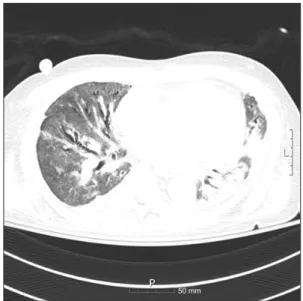
A 58-year-old man was admitted to a local hospital be- cause of fever, dyspnea, and cough with expectoration. His chest radiograph showed bilateral consolidation of the lungs, which was suggestive of pneumonia; therefore, he was started on antibiotic treatment. Since his oxygen satu- ration continued to decrease, he was intubated and kept on a ventilator. After a week of hospitalization, oxygen satura- tion still could not be maintained with mechanical ventila- tion. He was then transferred to Hallym University Sacred Heart Hospital for veno-venous extracorporeal membrane oxygenation (ECMO) treatment. We performed a respira- tory viral panel test, which led to the diagnosis of acute re- spiratory distress syndrome (ARDS) due to influenza A pneumonia. After a few weeks of treatment with awake ve- no-venous ECMO, the levels of inflammatory markers normalized, and his general condition improved. However, he could not be weaned from ECMO due to post-infectious pulmonary sequelae. A chest computed tomography scan
on the 67th day after admission showed extensive ground glass opacities and diffuse bronchiectasis in both lungs (Fig. 1). Bilateral lung transplantation was performed on the 84th day of hospital stay. In the operation, an arterial cannula was inserted into the ascending aorta and 2 can- nulas of veno-venous ECMO were connected and used as a venous cannula to convert the setup to central veno-arteri- al ECMO. The ischemic time of the right donor lung was 3 hours and that of the left donor lung was 4 hours and 56 minutes. After anastomosis finished, weaning from ECMO was attempted, but the patient’s blood pressure was not maintained even with sufficient inotropes and fluids due to decreased heart function. Eventually, a single arterial cannula was inserted into the femoral artery to change central veno-arterial ECMO into peripheral ECMO, and the operation was completed. Induction therapy with methyl- prednisolone (500 mg) was administered intravenously. An immunosuppressive regimen consisting of tacrolimus (tar- get therapeutic range, 5–14 μg/mL) and mycophenolate mofetil (1,000 mg/day) was started after trans plantation.
https://doi.org/10.5090/jcs.20.074
pISSN: 2765-1606 eISSN: 2765-1614
J Chest Surg. 2021;54(5):396-399
397
Byung Mo Gu, et al. Guillain-Barré Syndrome after Lung Transplantation
http://www.jchestsurg.org
JCS
Due to bleeding owing to coagulopathy after surgery, he was re-operated on days 1 and 2 after transplantation. There- after, sedatives were stopped, and we waited for the patient to regain consciousness. However, even 3 days after the second operation for bleeding control, he was unresponsive and showed no movement of the upper and lower extremi- ties except for spontaneous pupil and eye movement. Deep tendon reflexes of the patient’s limbs were absent, and a nerve conduction study was performed to investigate the cause. The results were suggestive of sensory-motor poly- neuropathy (motor-dominant demyelinating neuropathy) (Table 1). Cerebrospinal fluid testing was not performed due to the patient’s poor general condition and bleeding tendency. No electrolyte abnormalities or other causes were found. Guillain-Barré syndrome (GBS) was diagnosed based on its typical clinical manifestations. We started in- travenous immunoglobulin therapy (IVIG) and plasma- pheresis, which are the conventional treatments for GBS.
Bronchoscopic toileting was performed regularly for effec- tive lung care. As the treatment progressed, mild recovery of facial muscles was seen, but the patient died 24 days af- ter surgery due to progression of ARDS and sepsis.
The patient’s spouse provided written informed consent for the publication of his clinical details and images.
Discussion
GBS is a rare disease characterized by acute areflexic pa- ralysis due to damage to the peripheral nervous system
through an impaired immune response. The incidence of GBS is reported as 0.89 to 1.89 cases per 100,000 person- years, with a male-to-female ratio of 1.78 [1]. Although the immuno-pathogenesis of this disease has not been clearly identified, its basic mechanism is an inappropriate autoim- mune response that damages the myelin sheath of neurons.
The treatments available include IVIG and plasmapheresis.
However, GBS has a poor prognosis, since 20% of the pa- tients have persistent disability and approximately 5% die from medical complications [2]. Infections, vaccination, and surgery are known to increase its incidence.
GBS is difficult to diagnose because there are no bio- markers with high specificity and sensitivity. Therefore, the diagnosis of GBS is based on clinical manifestations and ancillary laboratory investigations and the commonly used criteria presented by the National Institute of Neuro- logical Disorders and Stroke, which consist of features re- quired for diagnosis, features strongly supportive of diag- nosis, and features that rule out diagnosis [3]. In this case, the patient showed progressive motor weakness and are- flexia, which are features required for diagnosis. Several strongly supportive features were also observed: symmetry of symptoms, cranial nerve involvement, autonomic dys- function, and electrodiagnostic features of sensory-motor neuropathy.
Since GBS involves an autoimmune mechanism, it was thought that transplant patients undergoing immunosup- pressive management would be protected from GBS. How- ever, there have been many reports of GBS being associated with transplantation. In case of bone marrow transplanta- tion, the incidence of GBS is reported to be 0.3%–0.7%, which is much higher than that in the general population [4]. A study reported by El-Sabrout et al. [5] regarding GBS cases occurring after solid organ transplantation indicated that cytomegalovirus infection plays an important role in the development of GBS in transplant patients. However, several case reports have also been published wherein GBS was reported to occur after transplantation without any history of infection [6,7]. These reports suggested that tac- rolimus, an immunosuppressive drug classified as a calci- neurin inhibitor and used after solid organ transplantation, may cause GBS. Neurotoxicity related to calcineurin inhib- itors, such as tacrolimus and cyclosporine, is well described in the literature; furthermore, it has been claimed that neurotoxicity associated with these drugs may be reversed by reducing the dosage [8].
In our patient, a history of influenza A infection, major surgery, and administration of tacrolimus were the risk factors for GBS. Unlike other cases in which GBS occurred
Fig. 1. Chest computed tomography image showing extensive ground glass opacities and diffuse bronchiectasis in the bilateral lungs.
50 mm