• 검색 결과가 없습니다.

Regional Bone Loss as a Risk Factor for Distal Radius Fracture in Women under 60 Years Old

N/A
N/A
Protected

Academic year: 2021

Share "Regional Bone Loss as a Risk Factor for Distal Radius Fracture in Women under 60 Years Old"

Copied!
6
0
0

로드 중.... (전체 텍스트 보기)

전체 글

(1)

Regional Bone Loss as a Risk Factor for Distal Radius Fracture in Women under 60 Years Old

Hwa-Jae Jeong, MD, PhD, Jinmyung Lee, MD, Hun Kyu Shin, MD, PhD, Jai Hyung Park, MD, PhD, Se-Jin Park, MD, PhD, Taeg Su Ko, MD, Jong-Hyon Park, MD, Eugene Kim, MD, PhD Department of Orthopedic Surgery, Kangbuk Samsung Hospital, Sungkyunkwan University School of Medicine, Seoul, Korea

Objectives: Some studies have suggested that lumbar spine and hip bone mineral density (BMD) are not associated with distal radius fractures (DRF), and a few studies have investigated regional BMDs at the fracture site, not just the lumbar or hip. We correlated distal radius BMD with DRF in postmenopausal women <60 years old.

Methods: A total of 121 women ≥50 years old with DRF were enrolled in the fracture group, and 72 women without fractures were included as a control group. We measured distal radius BMD in the distal radius contralateral to the fractured bone in the fracture group and that of the lumbar body 5 days after the trauma. BMDs at the distal radius of each group were compared in three age groups (50~59, 60~69, and ≥70 years). Age- and site-specific BMDs were analyzed in each group.

Results: No significant differences in the rate of osteoporosis at the distal radius or lumbar spine were observed in patients ≥60 years old. However, BMD and T-score values of the distal radius in female patients were lower than those in controls <60 years old. BMD and T-score values of the distal radius were lower than those of the lumbar spine in the fracture group <60 years old. BMD of the distal radius also carried a higher relative risk.

Conclusions: Low BMD of the distal radius was an indicator of regional BMD and could be a sensitive risk factor for DRF in women <60 years.

Key Words: Bone mineral density, Regional bone loss, Distal radius, Fracture, Osteoporosis

Received: August 26, 2014 Revised: December 15, 2014 Accepted: December 25, 2014

Corresponding Author: Eugene Kim, Department of Orthopedic Surgery, Kangbuk Samsung Hospital, 29 Saemunan-ro, Jongno-gu, Seoul 110-746, Korea

Tel: +82-2-2001-2168, Fax: +82-2-2001-2176, E-mail: dreugene0809@gmail.com

Distal radius fractures (DRF) have a high frequency rate largely among females, and postmenopausal osteoporosis is thought to be one of the causes of this low energy trauma fracture.

1,2

According to 2010 data from The Korean National Health Insurance Corpo- ration (NHIC), DRF occurred in 12,355 patients in 2010, and the aggregate medical costs were $86,643,433 (USD) (NHIC 2010).

However, almost all patients with DRF suffer from continuous pain and functional abnormalities even after

recovery.

3

Thus, many studies have been conducted on the risk factors for DRF in pre and postmenopausal women, particularly based on the change in bone mineral density (BMD). Thus, some authors have tried to predict bone strength for prevention and treatment using BMD measurements taken from the lumbar spine and hip in indicated patients, based on the recommen- dation of the International Society for Clinical Densito- metry.

4,5

Although some studies show that these BMDs are

(2)

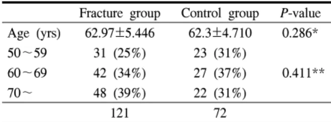
Table 1. Distribution of age in the control and frac- ture subjects

Fracture group Control group P-value Age (yrs) 62.97±5.446 62.3±4.710 0.286*

50~59 31 (25%) 23 (31%)

0.411**

60~69 42 (34%) 27 (37%)

70~ 48 (39%) 22 (31%)

121 72

*data are expressed mean±SD standard deviation) by using independent t-test

**data are expressed n(%) by using chi-square test related with lumbar or hip fractures, age <60 years is

less often associated with hip or spinal fractures,

6

and DRF usually occurs earlier than a hip fracture by an average of 15 years.

7

Thus, other studies have suggested that lumbar spine and hip BMDs are not associated with DRF. A few studies have investigated regional BMDs at the fracture site, not just that of the lumbar or hip.

However, distal radius BMD measurements are uncommon, and their association with other fractures has not been investigated. Thus, we focused on bone mass at the distal radius, which could be related to DRF. We measured and analyzed bone mass at the distal radius where the fractures actually occurred. We studied patients aged ≥50 years, who had experienced DRF, and measured distal radius BMDs. We were interested in determining whether there was a corre- lation between DRF and BMDs, as well as the clinical value of the correlation.

MATERIALS AND METHODS

1. Subject demographics

In total, 121 consecutive female patients ≥50 years (mean age, 62.97 years), who were admitted to our hospital for DRF from March 2008 through December 2010, were included. Patients who had no associated diseases (such as a distal radius disorder, trauma history, bone metabolic disease, or renal osteo- dystrophy) and those who had a low-energy trauma fracture, such as a fall from a standing height, were assessed.

8

The control group included 72 subjects ≥50 years, who had no particular medical history, and who had undergone distal radius and lumbar spine BMD measurements at the Health Screening Center of the hospital from January 2006 through June 2010 (Table 1). All tests and results of the fracture and control groups were used after the appropriate review processes had been completed and were in compliance with the

clinical test management standards of clinical studies established by the institutional review board of our hospital. All procedures complied with the international clinical trial unified guidelines.

2. BMD measurements

We measured distal radius BMDs of the fracture group in the distal radius contralateral to the fractured bone and that of the lumbar body 5 days after the trauma. The BMD measurements were carried out at the ultra-distal area of the radius, and the T-score (the difference from normal maximum) and BMD (g/cm

2

), were obtained. L1–L4 lumbar spine BMD values were also measured. Every measurement was taken by dual-energy x-ray absorptiometry scans using the LUNAR BX-1L system (GE Medical Systems, Wausheka, WI, USA).

3. Statistical analysis

Each group was subdivided by age (50~59, 60~69, and ≥70 years). SPSS 17.0 (SPSS Inc., Chicago, IL, USA) was used for all comparative analyses by age.

The Shapiro–Wilk test was used to verify statistical normality of each group. The independent t-test and chi-square test were used to assess statistical differences by age in the distribution between the fracture and control groups (Table 1).

We compared lumbar spine and distal radius BMDs

in the fracture group with those in the control group by

(3)

Table 2. Proportion of Osteoporosis in fracture and control group

Distal radius   L1-4  

Age

(yrs) Fracture group Control group P-value Fracture group Control group P-value

50~59 8 (25.8%) 3 (13.0%) <0.001 6 (19.3%) 3 (13.0%) 0.017

60~69 13 (30.9%) 9 (30.0%) 0.142 10 (23.8%) 7 (25.9%) 0.062

70~ 41 (85.4%) 19 (86.2%) 0.082 36 (75.0%) 16 (72.7%) 0.124

Table 3. Age- and site related “Inter-Group” comparison in fracture and control group

    Distal radius   L-spine  

    Fracture group Control group P-value Fracture group Control group P-value

50~59 BMD

0.360±0.107 0.741±0.078 0.035 0.914±0.198 1.141±0.161 0.026

T-score -1.079±1.779 -0.361±1.384 0.036 -0.642±1.523 -0.232±1.338 0.012

60~69 BMD 0.294±0.074 0.543±0.101 0.254 0.859±0.169 0.890±0.217 0.350

T-score -2.335±1.553 -1.998±1.742 0.317 -1.723±1.437 -1.664±1.778 0.476

70 over BMD 0.237±0.071 0.255±0.094 0.057 0.584±0.189 0.537±0.156 0.283

T-score -3.120±1.165 -2.968±1.756 0.094 -2.860±1.341 -2.663±1.256 0.164 BMD

: Bone mineral density (g/cm

2

)

age. The so-called “inter-group comparison”, which compares each site-related BMD in the fracture group with those of controls, was assessed with the inde- pendent t-test. The “inter-regional comparison, which is a comparison of age-related T-score values of the distal radius with those of the lumbar spine, was analyzed in both groups using the independent t-test.

Odds ratios (OR) with 95% confidence intervals were calculated for the distal radius and lumbar spine BMDs using multivariate logistic regression analysis to evaluate the association between DRF and BMD. This analysis was conducted for patients <60 years old and those ≥60 years old.

RESULTS

1. Osteoporosis rate

Patients aged <60 years old with DRF had a larger percentage of distal radius and lumbar spine osteoporosis than that in controls. However, no significant difference in the rate of osteoporosis at the distal radius or lumbar spine was observed in patients

≥60 years old (Table 2).

2. BMD analysis

BMDs and T-scores of the distal radius among patients aged 50~59 years in the fracture group were significantly lower than those in the control group, and BMDs and T-scores of the lumbar spine in patients aged 50~59 years in the fracture group were also significantly lower than those in the control group.

However, BMDs of the distal radius and lumbar spine in women aged ≥60 years with DRF were not significantly different from those of the radius and lumbar spine in controls (Table 3).

BMDs and T-score values of the distal radius in patients aged 50~59 years in the fracture group were significantly lower than those in the lumbar spine, although no significant difference was observed between that of the distal radius and lumbar spine in women 60~69 years and those ≥70 years (Table 4).

3. Relative risk of DRF

The association between DRF and low BMD was

significant at all sites. The OR of the distal radius was

larger than that of the lumbar spine. After adjusting for

(4)

Table 4. Age-related "Inter-Region" comparison in fracture and control group

    Fracture group   Control group  

    Distal radius L-spine P-value Distal radius L-spine P-value

50~59 BMD

0.360±0.107 0.914±0.198 <0.001* 0.741±0.078 1.141±0.161 1.042 T-score -1.079±1.779 -0.642±1.523 <0.001 -0.361±1.384 -0.232±1.338 0.353

60~69 BMD 0.294±0.074 0.859±0.169 0.061 0.543±0.101 0.890±0.217 0.941

T-score -2.335±1.550 -1.723±1.437 0.052 -1.998±1.742 -1.664±1.778 2.224

>70 BMD 0.237±0.071 0.584±0.189 0.421 0.255±0.094 0.537±0.156 5.541

T-score -3.120±1.165 -2.860±1.341 0.306 -2.968±1.756 -2.663±1.256 3.327 BMD

: Bone mineral density (g/cm

2

)

*using Bonferroni’s correction

Table 5. Odd ratiosof L-spine and distal radius for fracture of distal radius

  Odd ratios(95% CI

)

Site Crude Adjusted*

Ultradistal forearm (All subjects)

3.9 (1.5~9.3) 3.5 (1.4~8.6)

<60 6.2 (1.3~12.2) 6.1 (1.2~9.8)

≥60 2.4 (0.7~7.4) 2.6 (0.8~8.2)

Lumbar spine (All subjects)

3.0 (1.4~6.8) 2.8 (1.2~6.3)

<60 3.2 (1.9~11.0) 2.8 (1.7~9.8)

≥60 2.5 (0.8~7.2) 3.0 (0.9~9.6)

*Adjusted for age CI

: Confidence Interval

Analysis of multivariate logistic regression test

age, bone loss in the distal radius was significantly associated with DRF in women <60 years old (Table 5).

DISCUSSION

Many studies have correlated fractures with lower bone mass,

4,9

and many studies have been conducted about DRF in patients with a lower bone mass.

10-13

However, it is doubtful that decreased BMD of the lumbar or hip is a key indicator for DRF. No differences were observed between lumbar spine BMDs in the fracture group and those in the control group.

Earnshaw et al. (1998) and Peel et al. (1994) did not find a casual relationship between lumbar or hip BMDs and DRF. Some studies have reported opposite results,

but one study lacked controls, statistically normality, and used different inclusion criteria,

16

whereas another study obtained BMDs from Ward’s triangle, which may overestimate the prevalence of osteoporosis with no hospital-based control.

17

Several studies have found no differences between patients with DRF and controls.

18,19

These results suggest that bone loss differs in different body locations; BMDs measured in every bone are different even in the same person.

20

Some authors have also studied regional bone mass at fracture sites, and lower hip bone mass seems to be more sensitive than lumbar bone mass for hip fractures.

21

Furthermore, several studies have correlated DRF and lower bone mass at the distal radius.

11,14

Kanterewicz et al. (2002) found that distal radius bone mass is more related with DRF than that at the lumbar and hip.

22

We found that distal radius BMDs in females were significantly lower than those of controls in women ≤60 years with DRF.

We suspect that peripheral bone loss may occur markedly in women. Some authors have confirmed the hypothesis of accelerated perimenopausal bone loss with a preferential peripheral target.

23,24

Hesp et al.

(1984) reported a larger deficit in trabecular bone density at the distal radius that seemed to be associated with type I osteoporosis in early post-menopausal women.

25

It is unknown why female patients with DRF have

early bone loss compared with BMDs of controls.

22

Some studies have suggested that genetic factors have

(5)

an effect.

26,27

Bone loss seems to be related with gradual and age-related bone resorption in older patients.14 Kanterewicz et al. (2002) reported that the prevalence of osteoporosis is highest at the distal radius in patients with DRF and controls.

22

The T-score values of distal radius bone mass was lower than that of the lumbar spine in female patients

<60 years old with DRF, although DRF was correlated with low bone mass at the lumbar spine and lower bone mass at the distal radius in women <60 years old. These findings suggest that regional bone loss may be more associated with DRF as a sensitive criterion for recognizing regional osteoporosis. Additionally, BMD of the distal radius had a higher relative risk compared to that of the lumbar spine, and this result was remarkable in women <60 years old. The pro- portion of patients with osteoporosis at the distal radius was 25.8% (8/31), whereas that at the lumbar spine was 19.3% (6/31). Thus, measuring BMD at the distal radius could be an efficient screening test for detecting early bone loss in women with DRF <60 years old.

Some limitations of our study should be mentioned.

Although our study had several advantages, such as a sufficient control, the number of subjects was relatively small to conduct a subanalysis by age and fracture type. Second, this retrospective investigation also had a drawback of using patient recollection. Third, we did not consider patient lifestyle, such as obesity, smoking, and alcohol use, which could affect bone loss. Finally, BMDs of almost all controls were measured on the dominant-right side, although there was a prevalence of left-sided dominance. Thus, we did not match for arm dominance.

Conflict of Interest: None of the authors has a conflict of interest to disclose.

REFERENCES

1. Itoh S, Ohta T, Samejima H, Shinomiya K. Bone

mineral density in the distal radius in a healthy Japanese population and in relation to fractures of the distal radius. J Hand Surg [Br] 1999;24:334-7.

2. Smith D, Khairi M, Johnston Jr C. The loss of bone mineral with aging and its relationship to risk of fracture. J Clin Invest 1975;56:311.

3. Vogt MT, Cauley JA, Tomaino MM, Stone K, Williams JR, Herndon JH. Distal Radius Fractures in Older Women: A 10‐Year Follow‐Up Study of Descriptive Characteristics and Risk Factors. The Study of Osteoporotic Fractures. J Am Geriatr Soc 2002;50:97-103.

4. Kanis JA. Diagnosis of osteoporosis and assess- ment of fracture risk. The Lancet 2002;359:1929- 36.

5. Lewiecki EM, Kendler DL, Kiebzak GM, Schmeer P, Prince RL, El-Hajj Fuleihan G, Hans D. Special report on the official positions of the International Society for Clinical Densitometry. Osteoporos Int 2004;15:779-84.

6. Burge R, Dawson-Hughes B, Solomon DH, Wong JB, King A, Tosteson A. Incidence and Economic Burden of Osteoporosis‐Related Fractures in the United States, 2005-2025. J Bone Miner Res 2007;

22:465-75.

7. Owen R, MELTON III L, Ilstrup D, Johnson K, Riggs B. Colles' fracture and subsequent hip frac- ture risk. Clin Orthop 1982;171:37-43.

8. Lenart BA, Neviaser AS, Lyman S, Chang CC, Edobor-Osula F, Steele B, et al. Association of low-energy femoral fractures with prolonged bis- phosphonate use: a case control study. Osteoporos Int 2009;20:1353-62.

9. Marshall D, Johnell O, Wedel H. Meta-analysis of how well measures of bone mineral density predict occurrence of osteoporotic fractures. Br Med J 1996;312:1254-9.

10. Hegeman J, Oskam J, Van der Palen J, Ten Duis

H, Vierhout P. The distal radial fracture in elderly

women and the bone mineral density of the lumbar

(6)

spine and hip. J Hand Surg [Br] 2004;29:473-6.

11. Löfman O, Hallberg I, Berglund K, Wahlström O, Kartous L, Rosenqvist AM, et al. Women with low-energy fracture should be investigated for osteoporosis. Acta Orthop 2007;78:813-21.

12. Wigderowitz C, Cunningham T, Rowley D, Mole P, Paterson C. Peripheral bone mineral density in patients with distal radial fractures. J Bone Joint Surg Br 2003;85:423-5.

13. Øyen J, Rohde G, Hochberg M, Johnsen V, Haugeberg G. Low-energy distal radius fractures in middle-aged and elderly women—seasonal varia- tions, prevalence of osteoporosis, and associates with fractures. Osteoporos Int 2010;21:1247-55.

14. Earnshaw S, Cawte S, Worley A, Hosking D.

Colles' fracture of the wrist as an indicator of underlying osteoporosis in postmenopausal women:

a prospective study of bone mineral density and bone turnover rate. Osteoporos Int 1998;8:53-60.

15. Peel N, Barrington N, Smith T, Eastell R. Distal forearm fracture as risk factor for vertebral osteo- porosis. BMJ: British Medical Journal 1994;308:

1543-4.

16. Keen R, Griffiths G, Spector T. Patients who have had fractures of the distal forearm do not lose bone as expected. Br Med J 1996;313:821.

17. Lee JO, Chung MS, Baek GH, Oh JH, Lee YH, Gong HS. Age-and site-related bone mineral densi- ties in Korean women with a distal radius fracture compared with the reference Korean female population. J Hand Surg [Am] 2010;35:1435-41.

18. Nordvall H, Glanberg-Persson G, Lysholm J. Are distal radius fractures due to fragility or to falls?:

A consecutive case-control study of bone mineral density, tendency to fall, risk factors for osteo- porosis, and health-related quality of life. Acta Orthop 2007;78:271-7.

19. Sosa M, Saavedra P, Gómez-Alonso C, Mosquera J, Torrijos A, Muñoz-Torres M, et al. Postmeno- pausal women with Colles' fracture have bone

mineral density values similar to those of controls when measured with calcaneus quantitative ultra- sound. Eur J Intern Med 2005;16:561-6.

20. Melton LJ, Atkinson EJ, O'Connor MK, O'Fallon WM, Riggs BL. Bone density and fracture risk in men. J Bone Miner Res 1998;13:1915-23.

21. Shin HK, Choi JY, Lee J, Jeong HJ, Kim E, Park SJ, et al. Lower Hip Bone Mass and Proximal Femur Fractures in Elderly Patients: More Valuable Than Lumbar Vertebrae Bone Mineral Density.

Orthopedics 2010;33:875.

22. Kanterewicz E, Yanez A, Pérez-Pons A, Codony I, Del Rio L, Diez-Perez A. Association between Colles’ fracture and low bone mass: age-based differences in postmenopausal women. Osteoporos Int 2002;13:824-8.

23. Berntsen GKR, Fønnebø V, Tollan A, Søgaard AJ, Magnus JH. Forearm Bone Mineral Density by Age in 7,620 Men and Women The Tromsø Study, a Population-based Study. Am J Epidemiol 2001;

153:465-73.

24. Eastell R. Forearm fracture. Bone 1996;18:S203- S7.

25. Hesp R, Klenerman L, Page L. Decreased radial bone mass in Codes' fracture. Acta Orthopaedica 1984;55:573-5.

26. Deng HW, Chen WM, Recker S, Stegman MR, Li JL, Davies KM, et al. Genetic determination of Colles' fracture and differential bone mass in women with and without Colles' fracture. J Bone Miner Res 2000;15:1243-52.

27. Weichetová M, Štěpán JJ, Michalská D, Haas T, Pols HAP, Uitterlinden AG. COLIA1 polymor- phism contributes to bone mineral density to assess prevalent wrist fractures. Bone 2000;26:287-90.

28. Lochmüller EM, Müller R, Kuhn V, Lill CA,

Eckstein F. Can novel clinical densitometric techni-

ques replace or improve DXA in predicting bone

strength in osteoporosis at the hip and other

skeletal sites? J Bone Miner Res 2003;18:906-12.

수치

Table  1.  Distribution  of  age  in  the  control  and  frac- frac-ture  subjects
Table  3.  Age-  and  site  related  “Inter-Group”  comparison  in  fracture  and  control  group
Table  4.  Age-related  &#34;Inter-Region&#34;  comparison  in  fracture  and  control  group

참조

관련 문서

: Bone response to unloaded titanium implants with a sandblasted and acid-etched surface : A histomorphometric study in the canine mandible.. : Early

Bone transplants, autogenous bones, allogenic bones, xenogenic bones, and synthetic bone substitutes have all been used as bone graft materials.. However,

Result of patients’indirect markers reflecting bone quantity and quality (CTX : C-terminal cross-linking telopeptide of type 1 collagen, FRAX : Fracture Risk

Bone is a common site of distant metastasis in prostate cancer. Association of bone and metastatic cancer cells are important in this site-specific manifestation

Bone mineral content (BMC) and bone mineral density (BMD) of the lumbar spine, total hip, and proximal femur were measured before and after exercise... Results : 1) Body

In 4-week group, the group filled with bone graft with decortication revealed larger new bone formation area than shown in the group that had a defect area

It might also act in conjunction with a growth factor, such as bone morphogenic protein (BMP), insulin-like growth factor, or platelet-derived growth factor.

High complication rate in locking plate fixation of lower periprosthetic distal femur fractures in patients with total knee arthroplasties.. Management and