In-Seok Song
Department of Oral and Maxillofacial Surgery, Korea University Anam Hospital, 73 Inchon-ro, Seongbuk-gu, Seoul 02841, Korea
TEL: +82-2-920-5423 FAX: +82-2-921-7348 E-mail: [email protected]
ORCID: https://orcid.org/0000-0002-0763-8838 Sang-Ho Jun
Department of Oral and Maxillofacial Surgery, Korea University Anam Hospital, 73 Inchon-ro, Seongbuk-gu, Seoul 02841, Korea
TEL: +82-2-920-5423 FAX: +82-2-921-7348 E-mail: [email protected]
ORCID: https://orcid.org/0000-0002-4243-788X
This is an open-access article distributed under the terms of the Creative Commons Attribution Non-Commercial License (http://creativecommons.org/
licenses/by-nc/4.0/), which permits unrestricted non-commercial use, distribution, and reproduction in any medium, provided the original work is properly cited.
CC
Clinical characteristics and recurrence-related factors of medication-related osteonecrosis of the jaw
Mong-Hun Kang, Dong-Keon Lee, Chang-Woo Kim, In-Seok Song, Sang-Ho Jun Department of Oral and Maxillofacial Surgery, Korea University Anam Hospital, Seoul, Korea
Abstract(J Korean Assoc Oral Maxillofac Surg 2018;44:225-231)
Objectives: The purpose of this study was to investigate the demographic and clinical characteristics of patients with medication-related osteonecrosis of the jaw (MRONJ) and to elucidate factors affecting recurrence in surgical treatment.
Materials and Methods: A total of 51 patients who were diagnosed with MRONJ were analyzed according to demographic and clinical features and treatment results through a retrospective chart review from 2013 to 2017 in the Department of Oral and Maxillofacial Surgery, Korea University Anam Hospital, Seoul in Korea.
Results: Alendronate composed the majority of medication doses (55.6%), followed by ibandronate (20.0%), risedronate (15.6%), and zoledronate (6.7%). Forty patients (88.9%) were given oral medication, and five patients (11.1%) were intravenously treated, and the mean duration of medication use was 61.1±42.9 months. A total of 10 patients (22.2%) had a drug holiday before MRONJ-induced dental treatment lasting an average of 6.8±7.0 months. MRONJ occurred 2.7 times more in the mandible, with 41 cases (73.2%) occurring in the mandible and 15 cases (26.8%) occurring in the maxilla, and the prevalence of affected posterior parts (premolar-molar) was six times greater than that of the anterior parts (incisor-canine) (48 cases vs 8 cases, 85.7% vs 14.3%). The most common dental cause of MRONJ was tooth extraction (69.6%). Regarding recurrence, there was no statistical difference in recurrence rate according to either site or stage. However, recurrence occurred in 4 out of 34 cases (11.8%) in the primary closure group and 9 out of 20 cases (45.0%) in the secondary healing group, and there was a statistical difference with respect to closure technique.
Conclusion: The identified risk factors in patients taking bone resorption inhibitors can aid dental clinicians in ensuring prevention and proper treat- ment of MRONJ.
Key words: Bisphophonate-associated osteonecrosis of the jaw, Operative surgical procedure, Recurrence
[paper submitted 2018. 7. 10 / revised 2018. 8. 14 / accepted 2018. 9. 4]
Copyright © 2018 The Korean Association of Oral and Maxillofacial Surgeons. All
I. Introduction
Bisphosphonate-related osteonecrosis of the jaw (BRONJ)
was renamed to medication-related osteonecrosis of the jaw (MRONJ) as the incidence of jaw necrosis is increasing in relation to other bone resorption inhibitors or angiogenesis inhibitors1. Affiliated drugs are currently being used in vari- ous clinical applications. Oral administration of bisphospho- nates is mainly performed for osteoporosis and osteogenesis2. Intravenous bisphosphonates are used for hypercalcemia as- sociated with malignant disease and management of skeletal complications and osteolytic lesions in osteopathic cancer patients and osteoporosis patients3-5. Another bone resorption inhibitor, denosumab, is administered orally or subcutane- ously to inhibit skeletal complications in bone metastatic lesion and to reduce vertebral and hip fractures in osteopo- rosis patients6,7. Pathophysiologically, the exact mechanism of MRONJ development remains unclear, but it has been reported to potentially be due to excessive inhibition of jaw metabolic processes, infection and inflammation, inhibition of angiogenesis, soft tissue toxicity, immune system abnor-
malities, and accumulation of microfractures8-13.
As the number of MRONJ patients continues to increase, related research is actively being conducted. The American Association of Oral and Maxillofacial Surgeons (AAOMS) in 2014 and the Korean Society for Bone and Mineral Research (KSBMR) and the Korean Association of Oral and Maxillo- facial Surgeons (KAOMS) in 2015 published position papers on MRONJ1,14. According to these studies, MRONJ can be diagnosed if (1) there is a history of using a bone resorption inhibitor or an angiogenesis inhibitor and if (2) there is no history of radiation therapy to the jaw, exposure of the jaw, or oral or extraoral fistula lasting more than eight weeks. In addition, MRONJ can be divided into stages according to progression. In stage 0, there are no clinical symptoms of osteonecrosis but a nonspecific symptom. In stage 1, necrotic bone is exposed, but there is no evidence of symptoms or in- fection. In stage 2, there is osteonecrosis with symptoms and infection. Lastly, stage 3 involves the same stage 2 findings with necrotic bone beyond the alveolar bone (i.e., mandibular inferior border or maxillary sinus), pathological fractures, or extraoral fistula (i.e., orocutaneous fistula or oronasal and oroantral fistula).
The risk factors for MRONJ can be divided into systemic factors and local factors. Systemic factors include the dura- tion of related-medication use, use of steroids, age, diabetes, and genetic factors, while local factors include invasive oral surgery, thin mucosa, and periodontal disease15-17. Depending on the symptoms and progression of the disease, conserva- tive treatment including pain control, antibiotics, antibacterial gaggle, and various surgical treatments for removing necrotic tissue can be performed. Surgical treatment has been reported to yield a higher success rate, although failure may lead to advanced necrosis18,19.
Studies on factors affecting recurrence of MRONJ are rare in the South Korean population. Several investigations have suggested duration of administration of related medications, presence of bacterial infections in necrotic areas, and meth- ods of treatment used as factors in recurrence20,21. Therefore, pathophysiologic causes and risk factors of MRONJ as well as factors affecting recurrence should be examined. The pur- pose of the present study was to investigate the demographic and clinical characteristics of patients with MRONJ and to elucidate factors affecting recurrence following surgical treat- ment.
II. Materials and Methods
This study was approved by the Institutional Review Board of Korea University Anam Hospital (Seoul, Korea). From 2013 to 2017, a total of 51 patients who were diagnosed with MRONJ in the Department of Oral and Maxillofacial Surgery, Korea University Anam Hospital were enrolled in the present study. The diagnosis of MRONJ was based on the AAOMS 2014 position paper1. Demographic, clinical, and treatment outcomes data were analyzed by retrospective chart review. When referring to local hospitals, referrals were also consulted. In the demographic analysis, the sex and age of the patients were examined. In the clinical characteristics analysis, the type of MRONJ-inducing drug used and the method and duration of administration were examined. We also investigated the MRONJ-inducing dental treatment per- formed as well as the site, stage, treatment course, and results of MRONJ and furthermore examined the size of the lesion by way of panoramic radiographic study. The closure method was divided into two categories: primary closure and second- ary healing. Primary closure was performed without tension by incision on the periosteum of the mucoperiosteal flap after removal of the lesion. Secondary healing was performed with antibiotic gauze packing (Furacin gauze) with subsequent gauze and dressing changes performed every two to three days following removal of the lesion. Recurrence of MRONJ was defined as multiple infections and severe pain not re- sponding to surgical treatment. Statistical analysis using the IBM SPSS Statistics (ver. 22; IBM Co., Armonk, NY, USA) was performed to analyze the factors affecting recurrence in patients who received surgical treatment. Fisher’s exact test and the chi-square test were also employed for statistical analysis.
III. Results
1. Demographic analysis
Of the total 51 patients, three patients (5.9%) were male and 48 patients (94.1%) were female, and the mean patient age was 76.1±9.5 years (range, 45-92 years). The prevalence rate of MRONJ increased with age, with four patients (7.8%) being younger than 60 years, eight patients (15.7%) being between 61 years and 70 years, 21 patients (41.2%) being be- tween 71 years and 80 years, and 18 patients (35.3%) being older than 80 years.
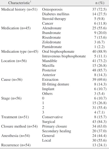
2. Clinical characteristics (Table 1)
1) MRONJ-inducing medications
Of the 51 patients, six patients were taking osteoporosis medication, but the type and duration of the drug were not specified and were excluded. Among the 45 patients, alendro- nate was the most common (25 patients, 55.6%) medication noted, followed by ibandronate (9 patients, 20.0%), rise- dronate (7 patients, 15.6%), zoledronate (3 patients, 6.7%), and pamidronate (1 patient, 2.2%). Forty patients (88.9%) had been given their medications orally, while five patients (11.1%) had undergone intravenous administration. The mean duration of drug use was 61.1±42.9 months (range, 6-240 months). A total of 10 patients had experienced a drug holi- day prior to the dental treatment that induced MRONJ, with a mean length of 6.8±7.0 months.
2) Medical history associated with MRONJ
eases, 37 patients (72.5%) had osteoporosis and 14 patients (27.5%) had diabetes mellitus. Additionally, five patients (9.8%) had been treated with steroids due to lupus, rheuma- tism, or adrenal insufficiency, and six patients (11.8%) had cancer. Patients without associated medical history were tak- ing prophylactic osteoporosis medications.
3) Disease site, cause, stage, and size
Two different disease sites in a single patient were counted as two cases. With regard to site, of the total 56 cases, man- dibular lesions were 2.7 times more prevalent, found in 41 cases (73.2%) versus the maxilla in 15 cases (26.8%). Forty- eight cases (85.7%) of posterior teeth (premolar-molar) made this presentation six times more prevalent than the eight cases (14.3%) of anterior teeth (incisor-canine). Regarding cause, extraction (39 cases, 69.6%) was the most common cause, with ill-fitting denture (8 cases, 14.3%); implant installation (6 cases, 10.7%); and other causes such as periodontal disease, alveoloplasty, and/or trauma (3 cases, 5.4%) also being noted.
With respect to disease stage, stage 2 (31 cases, 55.4%) was the most common, while there were six cases (10.7%) of stage 0, 15 cases (26.8%) of stage 1, and four cases (7.1%) of stage 3. For lesion size, the width and height of both bone de- struction and sclerosis were analyzed on panoramic images.
In 41 cases showing bone destruction, the mean size was 20.0±6.5 mm in width and 12.6±5.2 mm in height.
4) Treatment outcome
Conservative treatment was performed in eight patients (15.7%), and surgical treatment was performed in 43 patients (84.3%). A total of 54 surgical treatments were completed, in- cluding in those who had relapsed and required surgery again.
Of these, general anesthesia was used in 24 cases (44.4%), and local anesthesia was used in 30 cases (55.6%). When classified according to the closure method, 34 cases (63.0%) involved primary suture, and 20 cases (37.0%) involved sec- ondary healing. Recurrence occurred in 13 cases, a rate of 24.1%. The mean follow-up period was 9.6±10.8 months.
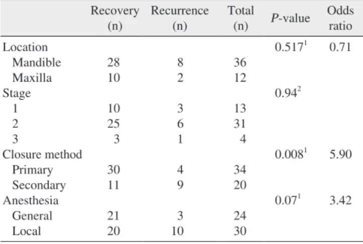
3. Recurrence factors
In the study, we analyzed whether there was a difference in recurrence rate according to site, stage, closure, and anesthe- sia method. Of the 48 MRONJ cases in 43 surgically treated patients, including two cases of multisite disease in a single patient, 8 of the 36 mandible cases (22.2%) and 2 of 12 max-
Table 1. Clinical characteristics of patients suffering from medica- tion-related osteonecrosis of the jaw
Characteristic1 n (%)
Medical history (n=51)
Medication (n=45)
Medication type (n=45) Location (n=56)
Cause (n=56)
Stage (n=56)
Treatment (n=51) Closure method (n=54) Anesthesia (n=54) Recurrence (n=54)
Osteoporosis Diabetes mellitus Steroid therapy Cancer Alendronate Ibandronate Risedronate Zoledronate Pamidronate Oral bisphosphonate Intravenous bisphosphonate Mandible
Maxilla Posterior Anterior Extraction Ill-fitting denture Implant Others 0 1 2 3
Conservative Surgical Primary closure Secondary healing General
Local
37 (72.5) 14 (27.5) 5 (9.8) 6 (11.8) 25 (55.6) 9 (20.0) 7 (15.6) 3 (6.7) 1 (2.2) 40 (88.9)
5 (11.1) 41 (73.2) 15 (26.8) 48 (85.7) 8 (14.3) 39 (69.6)
8 (14.3) 6 (10.7) 3 (5.4) 6 (10.7) 15 (26.8) 31 (55.4) 4 (7.1) 8 (15.7) 43 (84.3) 34 (63.0) 20 (37.0) 24 (44.4) 30 (55.6) 13 (24.1)
1Some patients had more than one disease.
Mong-Hun Kang et al: Clinical characteristics and recurrence-related factors of medi- cation-related osteonecrosis of the jaw. J Korean Assoc Oral Maxillofac Surg 2018
difference in recurrence rate according to site. Additionally, 3 of 13 stage 1 cases (23.1%), 6 of 31 stage 2 cases (19.4%), and 1 of 4 stage 3 cases (25.0%) demonstrated relapse, with no statistical difference in recurrence rate according to stage.
Among 54 cases of surgical treatment, 4 of 34 primary closure cases (11.8%) and 9 of 20 secondary healing cases (45.0%) experienced relapse, and there was a statistically significant difference in recurrence rate according to closure method (P=0.008). Of the 24 cases, 3 general anesthesia cas- es (12.5%) and 10 of 30 local anesthesia cases (33.3%) dem- onstrated relapse. There was a difference in recurrence rate according to anesthesia, although no statistical significance was found.(Table 2)
IV. Discussion
Various systemic and local factors are known to be risk indicators for MRONJ. Previously, the occurrence has dem- onstrated an increase in patients older than 65 years of age22, and similar results were found in the present work. The prev- alence of MRONJ in the patients who received drugs orally was 0.00104% to 0.00169%23-25, whereas higher incidence of MRONJ was noted on the patients with intravenous zole- dronate (0.017%) and denosumab (0.04%), respectively26,27. It has been reported that the risk of MRONJ is increased specifically in diabetic patients, which is due to decreases in bone quality associated with capillary ischemia, vascular endothelial function, osteoblast and bone cell death, immune cell function, and increase in inflammation16. The use of ste- roids may also be a risk factor for MRONJ development due to decreased immune cells, delayed wound healing associated
with steroid use, and worsening oral inflammation28. In an animal study, concurrent use of bisphosphonates and steroids increased the incidence of BRONJ29. MRONJ has addition- ally been reported in patients with cancer such as breast can- cer and multiple myeloma30,31. As a local risk factor, alveolar bone surgery is considered to be the main cause of MRONJ.
In the present study, extraction was the most common cause of MRONJ (70.6%), as in previous studies (70.6%)28,32,33. Other invasive alveolar bone surgeries such as implant instal- lation, endodontic treatment, and periodontal surgery have not been as adequately investigated as a cause of MRONJ, but they are considered to be similar risk factors for extrac- tion. In this study, MRONJ was 2.7 times more common in the mandible than in the maxilla in 73.2% of patients, and the posterior part was more common than the anterior part because the lesion might constitute a more complex vascular network and a more abundant blood supply in the maxilla than in the mandible. Notably, previous studies have reported similar results28.
In a previous study that analyzed panoramic radiographs of patients with MRONJ, it was reported that osseous sclerosis, lamina dura thickening, full-thickness sclerosis, osteolysis, and periapical radiolucency appeared on the panorama34,35. In this study, panorama analysis was performed to estimate the bone destruction size. The mean destruction size was 20.0±6.5 mm in width and 12.6±5.2 mm in height. There have been attempts to classify radiologic features of MRONJ using panoramic radiographs and computed tomographic imaging34,35. However, there exists a lack of clear radiologic criteria for periosteal reaction, cortical hypertrophy, and bone thickness changes to diagnose osteomyelitis in the jaw.
Therefore, more studies are needed to analyze the panoramic radiographs and computed tomographic images of the radio- logic features of MRONJ.
Various studies have been conducted on drug holiday. Ac- cording to the 2011 guidelines of the American Dental Asso- ciation (ADA), patients who received bisphosphonate therapy for less than two years did not require a drug holiday36, and according to the International Osteonecrosis of the Jaw Task Force guidelines, if patients had received bisphosphonate treatment for more than four years or if they had at least one risk factor, a drug holiday is recommended until the bone is completely healed37. The AAOMS recommended a two- month drug holiday based on bone physiology and pharma- cokinetic criteria1,38. Conversely, the KSBMR and KAOMS recommended a two- to four-month drug holiday14. In the present study, many patients underwent invasive procedures
Table 2. Factors affecting recurrence in surgical treatment Recovery
(n) Recurrence
(n) Total
(n) P-value Odds ratio Location
Mandible Maxilla Stage 1 2 3
Closure method Primary Secondary Anesthesia General Local
28 10 10 25 3 30 11 21 20
8 2 3 6 1 4 9 3 10
36 12 13 31 4 34 20 24 30
0.5171 0.942
0.0081 0.071
0.71
5.90 3.42
P-values by 1Fisher’s exact test or 2chi-square test.
Mong-Hun Kang et al: Clinical characteristics and recurrence-related factors of medi- cation-related osteonecrosis of the jaw. J Korean Assoc Oral Maxillofac Surg 2018
without a drug holiday. Therefore, it is predictable that, if a drug holiday was more clearly maintained, the incidence rate would be decreased.
The conservative treatment and surgical treatment are controversial, and evidence is lacking, but stage 1 MRONJ patients are recommended to undergo antibiotic gaggles, systemic antibiotics, and some local surgical procedures39,40. However, in stages 2 and 3, this conservative treatment is often inadequate, and these patients instead require surgical intervention39-41. When considering the failure of conservative treatment in this case, surgical intervention is widely recom- mended. Previous studies have shown that the success rate of surgical treatment was 84.2% to 89%, although there was a slight difference according to surgical method, operative object, and success criteria42-44. Similarly, a success rate of 76% was obtained in this study. Furthermore, various meth- ods such as low level laser therapy and recombinant human bone morphogenetic protein-2 have been used recently for MRONJ treatment45,46.
The success rates were examined according to anesthesia method used in patients who underwent surgical treatment.
There was no statistical significance observed in this regard, though the success rate was significantly different, from 87.5% in the general anesthesia group to 66.7% in the local anesthesia group. Better results were likely obtained with general anesthesia than with local anesthesia because the sur- geon can perform a wider operation with deeper anesthesia47. There was a significant difference in recurrence/reopera- tion rates between the primary closure and secondary healing groups in this study. The primary healing method was supe- rior to the secondary healing method in terms of success rate.
Primary closure enables protection of the bone by soft tissue coverage, provides adequate blood supply, resists traumatic injury, prevents infection, forms strong scar tissue after com- plete healing, and finally enables adequate bone healing44,48,49. The limitations of the present study are the small number of included patients and the short duration of follow-up. Fu- ture research is needed and should include sufficient numbers of patients and longer durations of follow-up. Although, im- portantly, this study tried to analyze local and systemic risk factors thoroughly and clarified the risk indicators associated with recurrence. Thus, clinicians will be able to reduce the risk of MRONJ by knowing these risk factors in patients tak- ing bone resorption inhibitors.
V. Conclusion
In conclusion, MRONJ is more common in the mandible in older women. The potential risk factors at play in a case should be evaluated with consideration of the patient’s medi- cal history, systemic disease, and clinical characteristics, and the incidence of MRONJ may be reduced through appropri- ate drug holiday and/or use of alternative medications. Addi- tionally, the success rate of MRONJ can be improved through extensive surgical treatment and primary closure manner.
ORCID
Mong-Hun Kang, https://orcid.org/0000-0002-6359-7081 Dong-Keon Lee, https://orcid.org/0000-0002-9262-7534 Chang-Woo Kim, https://orcid.org/0000-0003-1967-3267 In-Seok Song, https://orcid.org/0000-0002-0763-8838 Sang-Ho Jun, https://orcid.org/0000-0002-4243-788X
Authors’ Contributions
M.H.K. participated in data collection and wrote the manu- script. C.W.K. and D.K.L. participated in data collection.
I.S.S. participated in the study design and coordination and helped to draft the manuscript. S.H.J. participated in study coordination and helped to draft the manuscript. All authors read and approved the final manuscript.
Ethics Approval and Consent to Participate
This study was approved by the Institutional Review Board of Korea University Anam Hospital (Seoul, Korea).
Conflict of Interest
No potential conflict of interest relevant to this article was reported.
References
1. Ruggiero SL, Dodson TB, Fantasia J, Goodday R, Aghaloo T, Mehrotra B, et al. American Association of Oral and Maxillofacial Surgeons position paper on medication-related osteonecrosis of the jaw--2014 update. J Oral Maxillofac Surg 2014;72:1938-56.
2. Watts NB. Bisphosphonate treatment of osteoporosis. Clin Geriatr Med 2003;19:395-414.
3. Nussbaum SR, Younger J, Vandepol CJ, Gagel RF, Zubler MA, Chapman R, et al. Single-dose intravenous therapy with pamidro- nate for the treatment of hypercalcemia of malignancy: comparison
of 30-, 60-, and 90-mg dosages. Am J Med 1993;95:297-304.
4. Berenson JR, Rosen LS, Howell A, Porter L, Coleman RE, Morley W, et al. Zoledronic acid reduces skeletal-related events in patients with osteolytic metastases. Cancer 2001;91:1191-200.
5. Black DM, Delmas PD, Eastell R, Reid IR, Boonen S, Cauley JA, et al. Once-yearly zoledronic acid for treatment of postmenopausal osteoporosis. N Engl J Med 2007;356:1809-22.
6. Lipton A, Fizazi K, Stopeck AT, Henry DH, Smith MR, Shore N, et al. Effect of denosumab versus zoledronic acid in preventing skeletal-related events in patients with bone metastases by baseline characteristics. Eur J Cancer 2016;53:75-83.
7. Cummings SR, San Martin J, McClung MR, Siris ES, Eastell R, Reid IR, et al. Denosumab for prevention of fractures in postmeno- pausal women with osteoporosis. N Engl J Med 2009;361:756-65.
8. Baron R, Ferrari S, Russell RG. Denosumab and bisphosphonates:
different mechanisms of action and effects. Bone 2011;48:677-92.
9. Ripamonti CI, Maniezzo M, Campa T, Fagnoni E, Brunelli C, Saibene G, et al. Decreased occurrence of osteonecrosis of the jaw after implementation of dental preventive measures in solid tumour patients with bone metastases treated with bisphosphonates. The experience of the National Cancer Institute of Milan. Ann Oncol 2009;20:137-45.
10. Landesberg R, Woo V, Cremers S, Cozin M, Marolt D, Vunjak- Novakovic G, et al. Potential pathophysiological mechanisms in osteonecrosis of the jaw. Ann N Y Acad Sci 2011;1218:62-79.
11. Reid IR, Bolland MJ, Grey AB. Is bisphosphonate-associated osteonecrosis of the jaw caused by soft tissue toxicity? Bone 2007;41:318-20.
12. López-Jornet P, Camacho-Alonso F, Martínez-Canovas A, Moliña- Minano F, Gómez-García F, Vicente-Ortega V. Perioperative antibi- otic regimen in rats treated with pamidronate plus dexamethasone and subjected to dental extraction: a study of the changes in the jaws. J Oral Maxillofac Surg 2011;69:2488-93.
13. Hoefert S, Schmitz I, Tannapfel A, Eufinger H. Importance of mi- crocracks in etiology of bisphosphonate-related osteonecrosis of the jaw: a possible pathogenetic model of symptomatic and non- symptomatic osteonecrosis of the jaw based on scanning electron microscopy findings. Clin Oral Investig 2010;14:271-84.
14. Kim KM, Rhee Y, Kwon YD, Kwon TG, Lee JK, Kim DY. Medi- cation related osteonecrosis of the jaw: 2015 position statement of the Korean Society for Bone and Mineral Research and the Korean Association of Oral and Maxillofacial Surgeons. J Bone Metab 2015;22:151-65.
15. Tsao C, Darby I, Ebeling PR, Walsh K, O'Brien-Simpson N, Reyn- olds E, et al. Oral health risk factors for bisphosphonate-associated jaw osteonecrosis. J Oral Maxillofac Surg 2013;71:1360-6.
16. Peer A, Khamaisi M. Diabetes as a risk factor for medication- related osteonecrosis of the jaw. J Dent Res 2015;94:252-60.
17. McGowan K, McGowan T, Ivanovski S. Risk factors for medica- tion-related osteonecrosis of the jaws: a systematic review. Oral Dis 2018;24:527-36.
18. Stanton DC, Balasanian E. Outcome of surgical management of bisphosphonate-related osteonecrosis of the jaws: review of 33 sur- gical cases. J Oral Maxillofac Surg 2009;67:943-50.
19. Scoletta M, Arduino PG, Dalmasso P, Broccoletti R, Mozzati M.
Treatment outcomes in patients with bisphosphonate-related osteo- necrosis of the jaws: a prospective study. Oral Surg Oral Med Oral Pathol Oral Radiol Endod 2010;110:46-53.
20. Mücke T, Koschinski J, Deppe H, Wagenpfeil S, Pautke C, Mitch- ell DA, et al. Outcome of treatment and parameters influencing recurrence in patients with bisphosphonate-related osteonecrosis of the jaws. J Cancer Res Clin Oncol 2011;137:907-13.
21. Klingelhöffer C, Zeman F, Meier J, Reichert TE, Ettl T. Evaluation of surgical outcome and influencing risk factors in patients with medication-related osteonecrosis of the jaws. J Craniomaxillofac Surg 2016;44:1694-9.
22. Yamazaki T, Yamori M, Ishizaki T, Asai K, Goto K, Takahashi K,
et al. Increased incidence of osteonecrosis of the jaw after tooth extraction in patients treated with bisphosphonates: a cohort study.
Int J Oral Maxillofac Surg 2012;41:1397-403.
23. Etminan M, Aminzadeh K, Matthew IR, Brophy JM. Use of oral bisphosphonates and the risk of aseptic osteonecrosis: a nested case-control study. J Rheumatol 2008;35:691-5.
24. Khan AA, Rios LP, Sándor GK, Khan N, Peters E, Rahman MO, et al. Bisphosphonate-associated osteonecrosis of the jaw in On- tario: a survey of oral and maxillofacial surgeons. J Rheumatol 2011;38:1396-402.
25. Ulmner M, Jarnbring F, Törring O. Osteonecrosis of the jaw in Sweden associated with the oral use of bisphosphonate. J Oral Maxillofac Surg 2014;72:76-82.
26. Grbic JT, Black DM, Lyles KW, Reid DM, Orwoll E, McClung M, et al. The incidence of osteonecrosis of the jaw in patients receiv- ing 5 milligrams of zoledronic acid: data from the health outcomes and reduced incidence with zoledronic acid once yearly clinical tri- als program. J Am Dent Assoc 2010;141:1365-70.
27. Papapoulos S, Chapurlat R, Libanati C, Brandi ML, Brown JP, Czerwiński E, et al. Five years of denosumab exposure in women with postmenopausal osteoporosis: results from the first two years of the FREEDOM extension. J Bone Miner Res 2012;27:694-701.
28. Saad F, Brown JE, Van Poznak C, Ibrahim T, Stemmer SM, Sto- peck AT, et al. Incidence, risk factors, and outcomes of osteone- crosis of the jaw: integrated analysis from three blinded active- controlled phase III trials in cancer patients with bone metastases.
Ann Oncol 2012;23:1341-7.
29. Jang HW, Kim JW, Cha IH. Development of animal model for Bisphosphonates-related osteonecrosis of the jaw (BRONJ). Maxil- lofac Plast Reconstr Surg 2015;37:18.
30. Kim HJ, Park TJ, Ahn KM. Bisphosphonate-related osteonecrosis of the jaw in metastatic breast cancer patients: a review of 25 cases.
Maxillofac Plast Reconstr Surg 2016;38:6.
31. Choi WS, Lee JI, Yoon HJ, Min CK, Lee SH. Medication-related osteonecrosis of the jaw: a preliminary retrospective study of 130 patients with multiple myeloma. Maxillofac Plast Reconstr Surg 2017;39:1.
32. Vahtsevanos K, Kyrgidis A, Verrou E, Katodritou E, Triaridis S, Andreadis CG, et al. Longitudinal cohort study of risk factors in cancer patients of bisphosphonate-related osteonecrosis of the jaw.
J Clin Oncol 2009;27:5356-62.
33. Fehm T, Beck V, Banys M, Lipp HP, Hairass M, Reinert S, et al.
Bisphosphonate-induced osteonecrosis of the jaw (ONJ): incidence and risk factors in patients with breast cancer and gynecological malignancies. Gynecol Oncol 2009;112:605-9.
34. Phal PM, Myall RW, Assael LA, Weissman JL. Imaging findings of bisphosphonate-associated osteonecrosis of the jaws. AJNR Am J Neuroradiol 2007;28:1139-45.
35. Ida M, Watanabe H, Tetsumura A, Kurabayashi T. CT findings as a significant predictive factor for the curability of mandibular osteomyelitis: multivariate analysis. Dentomaxillofac Radiol 2005;34:86-90.
36. Hellstein JW, Adler RA, Edwards B, Jacobsen PL, Kalmar JR, Koka S, et al. Managing the care of patients receiving antiresorp- tive therapy for prevention and treatment of osteoporosis: execu- tive summary of recommendations from the American Dental Association Council on Scientific Affairs. J Am Dent Assoc 2011;142:1243-51.
37. Khan AA, Morrison A, Hanley DA, Felsenberg D, McCauley LK, O'Ryan F, et al. Diagnosis and management of osteonecrosis of the jaw: a systematic review and international consensus. J Bone Miner Res 2015;30:3-23.
38. Damm DD, Jones DM. Bisphosphonate-related osteonecrosis of the jaws: a potential alternative to drug holidays. Gen Dent 2013;61:33-8.
39. Ruggiero SL, Fantasia J, Carlson E. Bisphosphonate-related osteo- necrosis of the jaw: background and guidelines for diagnosis, stag-
ing and management. Oral Surg Oral Med Oral Pathol Oral Radiol Endod 2006;102:433-41.
40. Ruggiero SL. Bisphosphonate-related osteonecrosis of the jaw: an overview. Ann N Y Acad Sci 2011;1218:38-46.
41. Lee HK, Seo MH, Pang KM, Song SI, Lee JK. Comparative study on surgical and conservative management of bisphosphonate-relat- ed osteonecrosis of the jaw (BRONJ) in disease stage 2. J Korean Assoc Maxillofac Plast Reconstr Surg 2013;35:302-9.
42. Lopes RN, Rabelo GD, Rocha AC, Carvalho PA, Alves FA. Surgi- cal therapy for bisphosphonate-related osteonecrosis of the jaw:
six-year experience of a single institution. J Oral Maxillofac Surg 2015;73:1288-95.
43. Stockmann P, Burger M, von Wilmowsky C, Ebker T, Lutz R, Bau- ersachs A, et al. The outcome after surgical therapy of bisphospho- nate-associated osteonecrosis of the jaw--results of a clinical case series with an average follow-up of 20 months. Clin Oral Investig 2014;18:1299-304.
44. Stockmann P, Vairaktaris E, Wehrhan F, Seiss M, Schwarz S, Spriewald B, et al. Osteotomy and primary wound closure in bisphosphonate-associated osteonecrosis of the jaw: a prospective clinical study with 12 months follow-up. Support Care Cancer
2010;18:449-60.
45. Shin SH, Kim KH, Choi NR, Kim IR, Park BS, Kim YD, et al.
Effect of low-level laser therapy on bisphosphonate-treated osteo- blasts. Maxillofac Plast Reconstr Surg 2016;38:48.
46. Kim HC, Song JM, Kim CJ, Yoon SY, Kim IR, Park BS, et al.
Combined effect of bisphosphonate and recombinant human bone morphogenetic protein 2 on bone healing of rat calvarial defects.
Maxillofac Plast Reconstr Surg 2015;37:16.
47. Kim HY, Lee SJ, Kim SM, Myoung H, Hwang SJ, Choi JY, et al.
Extensive surgical procedures result in better treatment outcomes for bisphosphonate-related osteonecrosis of the jaw in patients with osteoporosis. J Oral Maxillofac Surg 2017;75:1404-13.
48. Wilde F, Heufelder M, Winter K, Hendricks J, Frerich B, Schramm A, et al. The role of surgical therapy in the management of intrave- nous bisphosphonates-related osteonecrosis of the jaw. Oral Surg Oral Med Oral Pathol Oral Radiol Endod 2011;111:153-63.
49. Gallego L, Junquera L, Pelaz A, Hernando J, Megías J. The use of pedicled buccal fat pad combined with sequestrectomy in bisphos- phonate-related osteonecrosis of the maxilla. Med Oral Patol Oral Cir Bucal 2012;17:e236-41.