<접수일:2006년 12월 29일, 심사통과일:2007년 2월 16일>
※통신저자:박 성 환
가톨릭대학교 의과대학 강남성모병원 류마티스내과
Tel:02) 590-2712, Fax:02) 599-3589, E-mail:[email protected]
본 연구는 과학기술부/한국과학재단 우수연구센터 육성사업(R11-2002-098-05003-0)과 성의장학연구비(2005)의 지원으로 수행되었음.
베체트 증후군 환자의 말초혈액단핵구에서
종양괴사인자(Tumor Necrosis Factor)- α 의 자극에 의한 Macrophage Migration Inhibitory Factor의 생산 증가
가톨릭대학교 의과대학 류마티스내과학교실, 가톨릭대학교 의과학연구원 류마티스연구센터*
곽승기 ㆍ 박성환 ㆍ 박미경* ㆍ 조미라* ㆍ 서수홍 ㆍ 주지현 ㆍ 윤종현 ㆍ 김호연
= Abstract =
Upregulation of Macrophage Migration Inhibitory Factor (MIF) Production from Peripheral Blood Mononuclear Cells (PBMCs) Stimulated by Tumor
Necrosis Factor (TNF)- α in Patients with Behcet's Syndrome
Seung-Ki Kwok, Sung-Hwan Park, Mi-Kyung Park*, Mi-La Cho*, Soo-Hong Seo, Ji-Hyeon Ju, Chong-Hyeon Yoon, Ho-Youn Kim
Division of Rheumatology, Department of Internal Medicine, College of Medicine, Rheumatism Research Center*, The Catholic University of Korea, Seoul, Korea
Objective: To investigate the effect of tumor necrosis factor (TNF)-α on the production of macrophage migration inhibitory factor (MIF), which might have important roles in the immune mediated inflammatory response of Behcet's syndrome.
Methods: Sixty two patients with Behcet's syndrome and thirty healthy controls were included in this study. The concentrations of TNF-α in sera were determined by enzyme-linked immuno- sorbent assay (ELISA). Peripheral blood mononuclear cells (PBMCs) from eleven patients with Behcet's syndrome were cultured for 48 hours with various concentration of TNF-α. The concentrations of MIF in sera and culture supernatants were determined by ELISA.
Results: Serum levels of TNF-α were significantly higher in patients with Behcet's syndrome than in healthy controls. TNF-α dose-dependently increased MIF production from PBMCs in
patients with Behcet's syndrome. Serum levels of TNF-α tended to correlate with serum levels of MIF, although did not reach statistical significance.
Conclusion: Upregulation of MIF production by increased levels of TNF-α in patients with Behcet's syndrome might be related to the pathogenesis of Behcet's syndrome.
Key Words: Behcet's syndrome, TNF-α, MIF
서 론
베체트 증후군은 원인이 밝혀지지 않은 염증성 질 환으로 구강과 외음부의 재발성 궤양, 포도막염, 피 부병변을 특징으로 하며 관절, 위장관, 중추 신경계 등의 전신을 침범할 수 있다 (1). 이 질환은 만성적 이며 지속적인 염증반응보다는 대부분 급성 염증이 악화와 관해를 반복하는 것을 특징으로 한다. 대부 분의 증상은 경미하고 반복된 안구 침범은 실명을 일으킬 수 있으며 혈관계, 위장관계, 중추 신경계의 침범은 생명을 위협할 수 있다 (2).
베체트 증후군의 정확한 발병기전은 알려져 있지 않으며 혈관 손상, 호중구의 기능 항진, 자가면역반 응 등이 관찰된다 (1). 베체트 증후군 환자의 혈청에 서 염증반응과 관련된 여러 가지 시토카인(cytokine) 의 증가가 관찰되는데 interleukin-2 (IL-2) (3,4), IL-6 (4), IL-8 (3,5), IL-18 (6), 종양괴사인자(TNF)-α (3,4,6), vascular endothelial growth factor (VEGF) (7-10), mon- ocyte chemoattractant protein-1 (MCP-1) (9), macro- phage inflammatory protein (MIP)-1α (11), macro- phage migration inhibitory factor (MIF) (12,13) 등이 베 체트 증후군 환자의 혈청에서 증가되었다는 보고들 이 있었다. 김 등은 MIF가 베체트 증후군 환자의 혈 청에서 정상인에 비하여 증가되어 있고 질병 활성도 와 관련 있다는 것을 보고한 바 있다 (13).
종양괴사인자는 매우 중요한 염증유발성 시토카인 으로 여러 가지 염증성 질환의 발병기전에 중요한 역 할을 하는 것으로 잘 알려져 있다. 그러나, 종양괴사 인자가 베체트 증후군의 발병기전에 어떤 역할을 하 는지는 아직까지 불명확하다. 이에 저자들은 본 연 구에서 종양괴사인자가 베체트 증후군의 질병 활성 도와 연관되고 발병기전에 중요한 역할을 하는 MIF 생산에 어떤 영향을 미치는지를 조사하고자 하였다.
대상 및 방법 1. 대상
강남성모병원에 내원한 International Study Group Criteria for Behcet's disease 진단기준을 (14) 만족시 키는 베체트 증후군 환자 62명(남자 18명, 여자 44 명; 평균 연령 42.24세, range 13∼62세)과 성별과 나 이가 맞춰진 30명(남자 12명, 여자 18명; 평균 연령 43.12세, range 20∼58세)의 정상인을 대조군으로 하 였다. 베체트 증후군 환자의 평균 유병기간은 6.78년 (1∼28)이었다. 환자는 채혈 당시의 임상증상을 기준 으로 활성상태와 비활성상태로 분류하였는데 구강 궤양 혹은 외음부 궤양, 관절증상, 피부병변, 포도막 염, 혈관염, 장염 및 중추신경증상중 두 가지 이상의 증상이 있는 경우를 활성상태로 정의하였다 (5,10,13).
채혈한 혈액은 혈청을 분리한 후 MIF와 TNF-α를 측정하기 위하여 20oC에 보관하였다. 본 연구는 강 남성모병원 임상연구관리 규정과 헬싱키 선언을 준 수하여 시행하였다.
2. 세포 및 세포 배양
베체트 증후군 환자 11명에서 헤파린을 처리한 신 선한 말초혈액을 얻어 Ficoll-Hypaque (Amersham Phar- macia Biotech AB, Sweden)을 이용한 density gradient centrifugation를 통해 말초혈액단핵구를 분리하였다.
분리된 말초혈액단핵구를 24 well plate에 각 well당 1×10⁶개씩 넣어 TNF-α의 자극이 없는 군, 0.1 ng/
mL, 1.0 ng/mL, 5.0 ng/mL, 10.0 ng/mL 농도의 TNF- α로 자극한 군으로 분류하여 배양하였다. 배양액은 serum-free RPMI 1640과 ITSA (insulin transferring selenium A)의 혼합액을 사용하였다. 5% CO2, 37oC 배양기에서 각각 24시간, 48시간 배양한 후 배양액 상층액을 취하여 MIF 농도를 측정하였다.
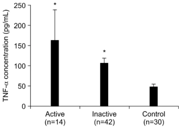
Fig. 1. Serum levels of TNF-α in patients with Behcet's syndrome and healthy controls. Serum levels of TNF-α were significantly higher in patients with Behcet's syndrome than in healthy controls. *p
<0.01 versus healthy controls. Serum levels of TNF-α were higher in patients with active Behcet's syndrome than in patients with inactive Behcet's syndrome, although statistically insig- nificant (p>0.05).
3. MIF, 종양괴사인자의 측정
혈청 MIF와 배양액 상층액에 있는 MIF 농도는 sand- witch ELISA 방법을 이용하였다. Sandwitch ELISA용 96 well plate (NUNC, Denmark)에 단클론성 MIF 항 체(R&D, USA) 4μg/mL로 50μL/well씩 넣고 4oC에 밤새 반응시킨 다음 차단용액(1% BSA/PBST)을 200 μL/well씩 넣고 실온에서 2시간 반응시켰다. Sta- ndard로는 재조합 인형 MIF (R&D, USA)를 이용하 여 5 ng/mL∼7.8 pg/mL 농도를 사용하였다. 표준시 료와 함께 측정할 혈청 및 세포배양 상층액을 50μL/
well씩 넣고 실온에서 2시간 반응시켰다. 반응용기를 세척용액(0.05% Tween 20/Phosphate-Buffered Saline)으 로 4번 세척하고 Biotinylated goat-anti-human MIF an- tibody (R&D, USA)를 200 ng/mL로 희석하여 50μL/
well씩 넣어 실온에서 2시간 반응시킨 후 세척용액 으로 4번 세척하였다. 마지막으로는 Extravidin-Alka- line phosphatase conjugate (SIGMA, USA)를 1:2,000 으로 희석하여 50μL/well씩 넣고 실온에서 2시간 반 응시키고 세척 후 PNPP (Fluka, Phosphate Disodium Salt Hexahydrate)/DEA 용액(Diethanolamine 97 mL, NaN3 0.2 g, MgCl26H2O 0.1 g, 1차 증류수 800 mL) 을 1 mg/mL 농도로 녹여 50μL/well씩 넣어 30분 후 0.2 M NaOH로 반응을 멈추고 405 nm 파장에서 흡 광도를 측정하였다.
TNF-α의 농도는 단클론성 종양괴사인자 항체 (R&D, USA)와 재조합 인형 TNF-α (R&D, USA) Biotinylated goat-anti-human TNF-α antibody (R&D, USA)를 이용하여 제조사의 가이드라인에 따라 sandwitch ELISA를 이용하여 측정하였다.
4. 통계 분석
실험 결과는 평균±평균의 표준오차로 표현하였으 며, SPSS 통계 프로그램(version 10.0)을 사용하였다.
정상인과 환자군의 평균비교는 independent Student's t-test를, 활성상태의 환자군과 비활성 상태의 환자군 간의 평균비교는 Mann-Whitney test를, TNF-α 자극 후 MIF의 분비에 관하여 Mann-Whitney test를, 그리 고 혈청 MIF와 TNF-α 간의 상관관계는 Pearson's correlation coefficient를 이용하였으며, p값이 0.05 이 하일 때 통계적으로 유의하다고 분석하였다.
결 과
1. 베체트 증후군 환자와 정상인의 혈청 TNF-α의 농도
이전에 보고된 논문들과 동일하게(3,4,6) 혈청 TNF- α의 농도는 베체트 증후군 환자에서 118.20± 19.43 pg/mL (평균±평균의 표준오차)였고 정상 대조군은 47.09±7.26 pg/mL로 환자군에서 유의하게 높았다 (p=0.001). 환자군과 정상 대조군 모두에서 성별에 따른 차이는 없었다. 활성상태 환자군의 혈청 TNF- α 농도는 162.65±76.49 pg/mL였고 비활성상태 환자 군은 106.57±12.67 pg/mL로 활성상태 환자군에서 높 은 경향을 보였으나 통계적 유의성은 관찰되지 않았 다. 비활성상태의 환자군도 정상 대조군에 비하여 혈청 TNF-α의 농도가 유의하게 높았다(p=0.001)(그 림 1).
2. TNF-α 자극에 의한 베체트 증후군 환자 말초 혈액단핵구의 MIF 생산증가
11명의 베체트 증후군 환자로부터 말초혈액단핵구
Fig. 2. Effect of TNF-α on macrophage migration inhi- bitory factor (MIF) production by peripheral blood mononuclear cells (PBMCs) in patients with Behcet's syndrome (n=4) and healthy controls (n=10). PBMCs were cultured in duplicate for 48 hours with medium alone or in the presence of TNF-α (0.1 to 1.0 ng/mL). The concentrations of MIF in the culture supernatant were determined by ELISA. TNF-α dose-dependently increased MIF production from PBMCs in patients with Behcet's syndrome not in healthy controls. *p<0.05 versus untreated cells.
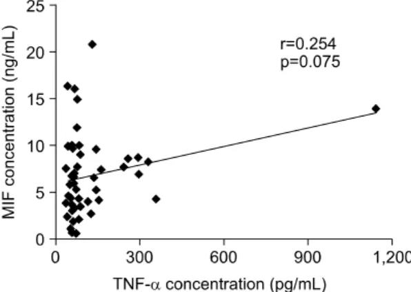
Fig. 3. Correlation of TNF-α concentration with MIF concentration in sera of patients with Behcet's syndrome. Serum TNF-α levels tended to have positive correlation with serum MIF levels, al- though didn't reach statistical significance (Pear- son's correlation coefficient).
를 분리하여 TNF-α의 자극 없이 배양한 군, 0.1 ng/mL, 1.0 ng/mL, 5.0 ng/mL, 10.0 ng/mL 농도의 TNF-α로 자극한 군으로 나누어 24, 48시간 후에 배 양 상층액의 MIF의 농도를 ELISA를 이용하여 측정 하였다. 각각의 환자에 따라 자극조건(자극농도와 배양기간)이 달라 통계분석에 어려움이 있었으나 활 성상태 환자군 4명을 대상으로 0.0 ng/mL, 0.1 ng/
mL, 1.0 ng/mL 농도의 TNF-α로 48시간 자극 후 배 양 상층액의 MIF 농도는 360.66±23.19 pg/mL, 740.60±205.91 pg/mL, 950.70±230.70 pg/mL로 TNF-α 자극에 의한 농도의존적인 생산증가를 보였으며 1.0 ng/mL 농도로 자극한 군의 MIF 생산은 자극없이 배 양한 군보다 통계적으로 유의한 MIF의 생산증가롤 보였다(p<0.05). 이에 반하여 정상 대조군 10명을 대 상으로 동일 조건에서 TNF-α 자극 후 MIF의 농도 를 측정한 결과 농도의존적인 MIF의 생산증가가 관 찰되지 않았다(그림 2).
3. 혈청 TNF-α와 MIF의 상관관계
혈청 TNF-α의 농도와 혈청 MIF 농도를 동시에
측정한 50명의 베체트 증후군 환자로부터 혈청 TNF-α와 MIF의 상관관계를 조사하였다. 혈청 TNF- α와 혈청 MIF의 농도는 양의 상관 관계 경향을 보 였으나 통계적 유의성에는 도달하지 못하였다(p=0.075) (그림 3).
고 찰
베체트 증후군의 정확한 발병기전은 아직 밝혀지 지 않았지만 유전적인 원인으로는 HLA-B51이 질환 의 발생과 관계가 있다는 것이 알려져 있으며, 그 외에도 단핵구, 호중구, T 세포 등이 활성화되어 있 으며 이에 동반된 혈관손상 및 자가면역반응이 질병 의 발생에 중요한 역할을 할 것으로 생각된다 (1,2,15). 특히 염증반응에 관계된 여러 가지 시토카 인, 케모카인(chemokine) (IL-2, IL-6, IL-8, IL-18, TNF, VEGF, MCP-1, MIP-1α, MIF)들이 환자의 혈청 에서 증가되어 있다는 것을 이전의 여러 실험논문에 서 보고하였다 (3-13). 이들 시토카인, 케모카인들 중 IL-8, VEGF, MIF가 질병활성도와 상관관계가 있다 고 알려져 있다 (5,7,13).
MIF는 대식세포의 이동을 억제하는 물질로서 처음 밝혀졌지만 (16) 그 후 많은 연구들로 인하여 MIF의 다양한 기능들이 밝혀지면서 현재는 면역반응 전반
에 중요한 역할을 담당하는 것으로 알려져 있다. 현 재까지 여러가지 염증성 질환들에서 MIF의 역할에 관한 많은 연구들이 있었으며 (17) 특히 류마티스 질환에서는 류마티스관절염의 병인과 관련되어 MIF 의 역할에 관한 많은 연구가 있었다 (17-20). 류마티 스관절염 환자의 혈청 C-반응성 단백질 수치와 활막 MIF 농도가 양의 상관관계를 보였고 치료 전에 활 막 MIF 농도가 증가되어 있다가 치료 후에 감소하 는 경향을 보여 MIF가 류마티스관절염의 임상적 발 현 및 활성화에 중요한 역할을 함을 증명하였다 (19).
베체트 증후군에서 MIF에 관한 연구는 아직 많지 않다. 베체트 증후군환자의 혈청 MIF 농도가 정상인 보다 증가되어 있으며 (12,13) 또한 질환이 활성화 상태인 경우 혈청 MIF 농도가 증가되어 있으며 동 일환자에서 질환이 비활성화 상태로 되면 MIF의 농 도가 감소함이 보고되었다 (13).
TNF-α는 면역반응 전반에 매우 중요한 역할을 하 는 염증 유발성 시토카인으로 많은 자가면역질환에 서 증가되어 있으며 급성 염증반응을 조절하고 지휘 하는 역할을 한다. 최근에 여러 가지 종양괴사인자 길항제가 개발되어 염증성 질환의 치료에 사용되면 서 기존의 치료보다 우월한 치료효과들이 보고되고 있다 (21,22). 베체트 증후군에서 TNF-α에 관한 연 구로 베체트 증후군 환자의 혈청 TNF-α의 농도가 증가되어 있다는 보고들이 있었지만 (3,4,6), TNF-α 가 베체트 증후군의 발병기전에 어떤 역할을 하는지 에 관하여 명확한 해답은 없는 상태이다.
이에 저자들은 본 연구에서 혈청 TNF-α의 농도를 측정하였다. 베체트 증후군 환자는 정상인보다 혈청 TNF-α 농도가 유의하게 높았고 특히 활성상태, 비 활성상태 환자 모두에서 혈청 TNF-α의 농도가 정상 인보다 증가되어 있었다. 또한 TNF-α의 자극에 의 하여 베체트 증후군의 발병기전에 중요한 역할을 하 는 것으로 알려져 있는 MIF 생산이 농도 의존적으 로 증가하는 것을 확인하였다. 이는 TNF-α가 베체 트 증후군의 발병기전에 관여하는 하나의 기전을 제 시한다고 할 수 있다. 그러나, 본 연구에서는 말초혈 액단핵구를 이용하여 실험하였으며, MIF는 단핵구, T 세포, B 세포 모두에서 생산가능하므로 어떤 세포 가 TNF-α의 자극에 의하여 MIF의 생산증가에 기여 하였는지에 관하여 추가연구가 필요할 것이다. 최근
기존의 치료에 반응하지 않는 포도막염, 신경침범, 위장관 침범 등을 동반한 베체트 증후군 환자들을 대상으로 종양괴사인자 길항제를 투여하여 좋은 치 료성적을 나타내는 보고들이 있다 (23-25). 본 연구 결과는 베체트 증후군의 치료에 종양괴사인자 길항 제를 사용하는 이론적 근거로 제시될 수 있을 것이 다. 향후 TNF 자극에 의한 MIF의 생산에 관련된 조 절기전 및 세포 내 신호전달체계 등에 관하여 더 많 은 연구가 필요할 것으로 생각된다.
결 론
본 연구에서 베체트 증후군 환자의 혈청 TNF-α의 농도가 정상인보다 증가되어 있었고 종양괴사인자의 자극에 의하여 말초혈액단핵구의 MIF 생산이 농도 의존적으로 증가하였다. 이는 TNF가 베체트 증후군 의 발병기전에 관여하는 하나의 기전으로 제시될 수 있으며 또한 기존의 치료에 반응이 없는 베체트 증 후군의 치료에 종양괴사인자 길항제를 투여하는 이 론적 근거로 제시될 수 있을 것이다.
REFERENCES
1) Sakane T, Takeno M, Suzuki N, Inaba G. Behcet's disease. N Engl J Med 1999;341:1284-91.
2) Hirohata S, Kikuchi H. Behcet's disease. Arthritis Res Ther 2003;5:139-46.
3) Evereklioglu C, Er H, Turkoz Y, Cekmen M. Serum levels of TNF-alpha, sIL-2R, IL-6, and IL-8 are increased and associated with elevated lipid peroxid- ation in patients with Behcet's disease. Mediators Inflamm 2002;11:87-93.
4) Akdeniz N, Esrefoglu M, Keles MS, Karakuzu A, Atasoy M. Serum interleukin-2, interleukin-6, tumour necrosis factor-alpha and nitric oxide levels in pa- tients with Behcet's disease. Ann Acad Med Singa- pore 2004;33:596-9.
5) Katsantonis J, Adler Y, Orfanos CE, Zouboulis CC.
Adamantiades-Behcet's disease: serum IL-8 is a more reliable marker for disease activity than C-reactive protein and erythrocyte sedimentation rate. Derma- tology 2000;201:37-9.
6) Oztas MO, Onder M, Gurer MA, Bukan N, Sancak B.
Serum interleukin 18 and tumour necrosis factor-alpha
levels are increased in Behcet's disease. Clin Exp Dermatol 2005;30:61-3.
7) Cekmen M, Evereklioglu C, Er H, Inaloz HS, Do- ganay S, Turkoz Y, et al. Vascular endothelial growth factor levels are increased and associated with disease activity in patients with Behcet's syndrome. Int J Dermatol 2003;42:870-5.
8) Erdem F, Gundogdu M, Kiki I, Ali Sari R, Kiziltunc A. Vascular endothelial and basic fibroblast growth factor serum levels in patients with Behcet's disease.
Rheumatol Int 2005;25:599-603.
9) Bozoglu E, Dinc A, Erdem H, Pay S, Simsek I, Kocar IH. Vascular endothelial growth factor and monocyte chemoattractant protein-1 in Behcet's patients with venous thrombosis. Clin Exp Rheumatol 2005;23(4 Suppl 38):S42-8.
10) 홍지현, 김현숙, 김해림, 박미경, 윤종현, 이상헌 등. 베 체트 증후군 환자에서 혈관내피성장인자(Vascular En- dothelial Growth Factor, VEGF)의 상승. 대한류마티 스학회지 2005; 12: 189-96
11) Ozer HT, Erken E, Gunesacar R, Kara O. Serum RANTES, MIP-1 alpha, and MCP-1 levels in Behcet's disease. Rheumatol Int 2005;25:487-8.
12) Kotake S, Kitaichi N, Ohno S. Macrophage migration inhibitory factor in uveitis. Int Ophthalmol Clin 2002;
42:99-103.
13) 김성동, 김상현, 김해림, 박미경, 윤종현, 김완욱 등. 베 체트병 환자에서 혈청 대식세포 유주 억제인자(Ma- crophage Migration Inhibitory Factor, MIF)의 상승.
대한류마티스학회지 2004; 11: 205-11.
14) Criteria for diagnosis of Behcet's disease. International Study Group for Behcet's Disease. Lancet 1990;
335:1078-80.
15) Yurdakul S, Hamuryudan V, Yazici H. Behcet syn- drome. Curr Opin Rheumatol 2004;16:38-42.
16) Nathan CF, Remold HG, David JR. Characterization of a lymphocyte factor which alters macrophage func- tions. J Exp Med 1973;137:275-90.
17) Morand EF. New therapeutic target in inflammatory disease: macrophage migration inhibitory factor. In- tern Med J 2005;35:419-26.
18) Morand EF, Leech M, Bernhagen J. MIF: a new cyto- kine link between rheumatoid arthritis and athero- sclerosis. Nat Rev Drug Discov 2006;5:399-410.
19) Morand EF, Leech M, Weedon H, Metz C, Bucala R, Smith MD. Macrophage migration inhibitory factor in rheumatoid arthritis: clinical correlations. Rheuma- tology (Oxford) 2002;41:558-62.
20) Morand EF, Bucala R, Leech M. Macrophage migra- tion inhibitory factor: an emerging therapeutic target in rheumatoid arthritis. Arthritis Rheum 2003;48:291-9.
21) Anderson PJ. Tumor necrosis factor inhibitors: cli- nical implications of their different immunogenicity profiles. Semin Arthritis Rheum 2005;34:19-22.
22) Chatzantoni K, Mouzaki A. Anti-TNF-alpha antibody therapies in autoimmune diseases. Curr Top Med Chem 2006;6:1707-14.
23) Sarwar H, McGrath H Jr, Espinoza LR. Successful treatment of long-standing neuro-Behcet's disease with infliximab. J Rheumatol 2005;32:181-3.
24) Giansanti F, Barbera ML, Virgili G, Pieri B, Emmi L, Menchini U. Infliximab for the treatment of posterior uveitis with retinal neovascularization in Behcet disease. Eur J Ophthalmol 2004;14:445-8.
25) Ju JH, Kwok SK, Seo SH, Yoon CH, Kim HY, Park SH. Successful treatment of life-threatening intestinal ulcer in Behcet's disease with infliximab: rapid healing of Behcet's ulcer with infliximab. Clin Rheu- matol 2006 Oct 13.