Eagle’s syndrome: a case report
Chang-Sig Moon, Baek-Soo Lee, Yong-Dae Kwon, Byung-Jun Choi, Jung-Woo Lee, Hyun-Woo Lee, Sun-Ung Yun, Joo-Young Ohe
Department of Oral and Maxillofacial Surgery, School of Dentistry, Kyung Hee University, Seoul, Korea
Abstract(J Korean Assoc Oral Maxillofac Surg 2014;40:43-47)
Eagle’s syndrome is a disease caused by an elongated styloid process or calcified stylohyoid ligament. Eagle defined the disorder in 1937 by describ- ing clinical findings related to an elongated styloid process, which is one of the numerous causes of pain in the craniofacial and cervical region. The prevalence of individuals with this anatomic abnormality in the adult population is estimated to be 4% with 0.16% of these individuals reported to be symptomatic. Eagle’s syndrome is usually characterized by neck, throat, or ear pain; pharyngeal foreign body sensation; dysphagia; pain upon head movement; and headache. The diagnosis of Eagle’s syndrome must be made in association with data from the clinical history, physical examination, and imaging studies. Patients with increased symptom severity require surgical excision of the styloid process, which can be performed through an intraoral or an extraoral approach. Here, we report a rare case of stylohyoid ligament bilaterally elongated to more than 60 mm in a 51-year-old female.
We did a surgery by extraoral approach and patient’s symptom was improved.
Key words: Eagle syndrome, Elongated styloid process
[paper submitted 2013. 12. 18 / revised 2014. 2. 3 / accepted 2014. 2. 4]
The carotid artery type of Eagle’s syndrome presents with other symptoms, such as migraines, and neurological symp- toms, caused by irritation of the sympathetic nerve plexus.
If the internal carotid artery is compressed, then ipsilateral headaches can occur. If the external carotid artery is com- pressed, then there can be pain in the temporal and maxillary branch areas4,6.
Here, we report a rare case of the stylohyoid ligament bilat- erally elongated to more than 60 mm in a 51-year-old female.
We did a surgery by extraoral approach and patient’s symp- tom was improved.
II. Case Report
A 51-year-old female patient with throat pain and dizziness was referred to the Department of Oral and Maxillofacial Surgery at Kyung Hee University Dental Hospital in Korea.
Upon turning her head, the patient felt facial swelling, dizzi- ness, as well as neck and shoulder pain. She also had a sense of fullness in her neck. Her past medical history included a thyroid nodulectomy 1 year prior to presentation. During the clinical exam, we could not feel either stylohyoid ligament in the oral cavity. However after imaging, we could see that both ligaments were elongated in the panoramic view.(Fig. 1)
I. Introduction
In 1652, Pietro Marchetti first described an elongated sty- loid process related to an ossifying process of the stylohyoid ligament. Eagle, an otolaryngologist, later defined Eagle’s syndrome in 19371,2. Eagle considered any styloid process longer than 25 mm in an adult to be abnormal. He found that 4% of the population had elongated styloid processes, but only 4% of those with this trait had symptoms3-5. He divided the syndrome into two forms: classic type and carotid artery type.
The classic type of Eagle’s syndrome can develop after tonsillectomy, when scar tissue under the tonsillar fossa com- presses and stretches cranial nerves V, VII, IX, and X. This type includes symptoms such as foreign body sensation, pain referred to the ear, and dysphagia.
Joo-Young Ohe
Department of Oral and Maxillofacial Surgery, Kyung Hee University Dental Hospital, 26, Kyungheedae-ro, Dongdaemun-gu, Seoul 130-701, Korea TEL: +82-2-958-9360 FAX: +82-2-966-4572
E-mail: [email protected]
This is an open-access article distributed under the terms of the Creative Commons Attribution Non-Commercial License (http://creativecommons.org/licenses/by-nc/3.0/), which permits unrestricted non-commercial use, distribution, and reproduction in any medium, provided the original work is properly cited.
CC
Copyright Ⓒ 2014 The Korean Association of Oral and Maxillofacial Surgeons. All
tachments using a rongeur forcep and small osteotome.(Figs. 4, 5) In addition, we extracted all of the wisdom teeth. The day after surgery, we confirmed that most of the elongated stylo- hyoid ligaments were removed using panoramic radiography and CBCT.(Figs. 6, 7) After resection, the patient’s neck and shoulder pain and dizziness upon head movement disap- peared, but she had discomfort with swallowing. She also complained that the skin around the surgical site was loose.
At the 3-month follow-up, we reevaluated the surgical site using panoramic radiography and CBCT. We determined that the patient's symptoms such as pain, dizziness, swollen feel- ing, and odynophagia had resolved. However, the patient still complained of loose skin around the surgical site.
III. Discussion
The styloid process is derived from the second branchial arch of Reichert’s cartilage. This cartilage consists of four components: 1) the tympanohyale, which arises from the periotic capsule of the temporal bone and is a process at- tached to the inferior surface of the petrous part of the tem- poral bone; 2) the stylohyale, which usually forms the greater part of the styloid process proper; 3) the ceratohyale, which forms the stylohyoid ligament; and 4) the hypohyale, which forms the lesser cornu of the hyoid bone7. The styloid pro- cess is an elongated tapered projection that originates in the petrous portion of the temporal bone, lying medially and an- teriorly to the stylomastoid foramen, between the internal and external carotid arteries, and laterally to the tonsillar fossa.
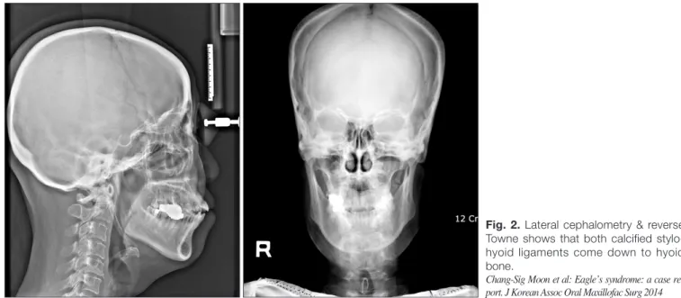
The stylopharyngeal, stylohyoid, and styloglossal muscles are Lateral cephalometry and reverse Towne projection film
showed that the stylohyoid ligaments extended down to the hyoid bone.(Fig. 2) Using cone-beam computed tomography (CBCT), we determined that their lengths were more than 60 mm and their diameters were almost 5 mm at the thickest site, and the ligaments displayed a twig morphology.(Fig. 3)
We diagnosed this patient with Eagle’s syndrome by clini- cal and radiographic exams. We planned for complete remov- al of both elongated stylohyoid ligaments using an extraoral approach under general anesthesia.
A horizontal incision was placed in a skin crease 5 cm in- ferior to the lower border of the mandible. A cervical incision was made from the proximal side of the sternocleidomastoid muscle to the hyoid bone, and the elongated stylohyoid liga- ments were detached and the muscular attachments removed via subperiosteal dissection. We resected the ligaments and at-
Fig. 1. Panoramic view at the first visit shows calcification of both stylohyoid ligaments.
Chang-Sig Moon et al: Eagle’s syndrome: a case report. J Korean Assoc Oral Maxillo- fac Surg 2014
Fig. 2. Lateral cephalometry & reverse Towne shows that both calcified stylo- hyoid ligaments come down to hyoid bone.
Chang-Sig Moon et al: Eagle’s syndrome: a case re- port. J Korean Assoc Oral Maxillofac Surg 2014
to explain the development of Eagle’s syndrome. The first theory is congenital elongation of the styloid process due to the persistence of the cartilaginous precursor, the second is the calcification of the stylohyoid ligament by a mysterious process, and the third is the growth of osseous tissue at the insertion of the stylohyoid ligament. Steinmann proposed attached to the styloid process. This bony process supports
the stylohyoid and stylomandibular ligaments. The stylohy- oid ligament connects the apex of the styloid process and the lesser horn of the hyoid bone, and the stylomandibular liga- ment extends from the styloid process to the parotideomas- seteric fascia between the mastoid process and the mandible8.
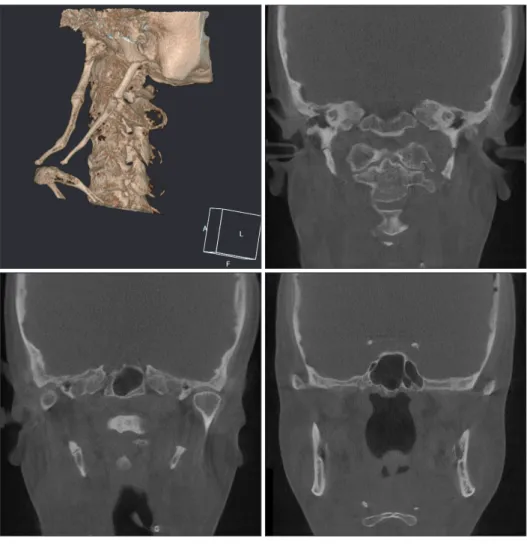
Fig. 3. Cone-beam computed tomogra- phy shows that both calcified stylohyoid ligaments are more longer than 60 mm.
Chang-Sig Moon et al: Eagle’s syndrome: a case re- port. J Korean Assoc Oral Maxillofac Surg 2014
Fig. 4. Clinical photographs were taken during operation. A. Right. B. Left.
Chang-Sig Moon et al: Eagle’s syndrome: a case re- port. J Korean Assoc Oral Maxillofac Surg 2014
ossification of the stylohyoid ligament; and 3) anatomic vari- ance, which occurs without any distinctive trauma10. Camarda et al.11 proposed a fourth mechanism of aging developmental anomaly in the absence of apparent radiographic ossification.
Murtagh et al.9 explained that Eagle’s syndrome symptoms stem from its pathophysiology, which includes: 1) the theory that traumatic fracture of the styloid process with a prolif- eration of granulation tissue is able to apply pressure on the perplasia, when trauma activates the remnants of the original
connective and fibrocartilaginous cells; 2) reactive metapla- sia, or an abnormal healing following a trauma that initiates
Fig. 7. Cone-beam computed tomogra- phy after surgery shows that both calci- fied stylohyoid ligaments were almost removed.
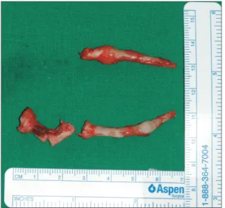
Chang-Sig Moon et al: Eagle’s syndrome: a case re- port. J Korean Assoc Oral Maxillofac Surg 2014 Fig. 5. Resected calcified stylohyoid ligament was more than 60
mm on right side.
Chang-Sig Moon et al: Eagle’s syndrome: a case report. J Korean Assoc Oral Maxillo- fac Surg 2014
Fig. 6. Panoramic view after surgery shows that most of both cal- cified stylohyoid ligaments were removed.
Chang-Sig Moon et al: Eagle’s syndrome: a case report. J Korean Assoc Oral Maxillo- fac Surg 2014
the styloid process. The external approach has the advantage of good anatomic exposure of the styloid process. However, it requires more intervention and results in a visible scar3,12.
In this case, we planned surgical excision because the pa- tient’s symptoms were severe. As both elongated stylohyoid ligaments were quite large, we resected using an external approach. We also extracted 4 wisdom teeth. The resected stylohyoid ligaments measured more than 60 mm long, as in- dicated in the CT scan. After surgery, the patient’s symptoms improved but a scar remained. At the 3-month postoperative follow-up, the patient’s symptoms were further decreased al- though some discomfort remained.
Conflict of Interest
No potential conflict of interest relevant to this article was reported.
References
1. Eagle WW. Elongated styloid processes: report of two cases. Arch Otolaryngol 1937;25:584-7.
2. Dunn-Ryznyk LR, Kelly CW. Eagle syndrome: a rare cause of dysphagia and head and neck pain. JAAPA 2010;23:28, 31-2, 48.
3. Yavuz H, Caylakli F, Erkan AN, Ozluoglu LN. Modified intraoral approach for removal of an elongated styloid process. J Otolaryn- gol Head Neck Surg 2011;40:86-90.
4. Colby CC, Del Gaudio JM. Stylohyoid complex syndrome: a new diagnostic classification. Arch Otolaryngol Head Neck Surg 2011;137:248-52.
5. Johnson GM, Rosdy NM, Horton SJ. Manual therapy assessment findings in patients diagnosed with Eagle's Syndrome: a case series.
Man Ther 2011;16:199-202.
6. Costantinides F, Vidoni G, Bodin C, Di Lenarda R. Eagle's syn- drome: signs and symptoms. Cranio 2013;31:56-60.
7. Ghosh LM, Dubey SP. The syndrome of elongated styloid process.
Auris Nasus Larynx 1999;26:169-75.
8. Valerio CS, Peyneau PD, de Sousa AC, Cardoso FO, de Oliveira DR, Taitson PF, et al. Stylohyoid syndrome: surgical approach. J Craniofac Surg 2012;23:e138-40.
9. Murtagh RD, Caracciolo JT, Fernandez G. CT findings associated with Eagle syndrome. AJNR Am J Neuroradiol 2001;22:1401-2.
10. Steinmann EP. Styloid syndrome in absence of an elongated pro- cess. Acta Otolaryngol 1968;66:347-56.
11. Camarda AJ, Deschamps C, Forest D. I. Stylohyoid chain ossifi- cation: a discussion of etiology. Oral Surg Oral Med Oral Pathol 1989;67:508-14.
12. Leclerc JE. Eagle syndrome: teaching the intraoral surgical ap- proach with a 30 degrees endoscope. J Otolaryngol Head Neck Surg 2008;37:727-9.
surrounding area; 2) compression of the glossopharyngeal nerve, the mandibular branch of the trigeminal, or the chorda tympani; 3) the theory of insertion tendonitis due to inflam- mation of the tendinous part of the stylohyoid insertion; 4) irritation of the pharyngeal mucosa by direct compression or post-tonsillectomy scarring (with involvement of cranial nerves V, VII, IX, and X); and 5) the theory of irritation of the sympathetic nerves surrounding the carotid vessels9.
Diagnosis is guided by the clinical history and physical and radiographic examinations. The physical examination con- sists of palpation of the tonsillar fossa and local infiltration anesthesia. Transpharyngeal palpation demonstrates a bony projection and reproduces the characteristic pain. Symptom relief should follow anesthetic injection in the tonsillar fossa.
Radiologic evaluation, such as panoramic radiography, lateral cephalometry, Towne projection film, or CT, may be used. In the panoramic view, the styloid process is visualized poste- riorly to the external acoustic meatus with a descendant and anterior trajectory. When elongated, it attains over one third of the length of the mandibular ramus. CT allows for the pre- cise measurement of the styloid process length, direction, and anatomic variance, in addition to evaluation of stylohyoid ligament ossification8.
Eagle’s syndrome can be treated nonsurgically and surgi- cally. A pharmacological approach includes transpharyngeal infiltration of steroids or anesthetics into the tonsillar fossa. In addition, there are two surgical approaches, intraoral and ex- traoral. In the intraoral approach, the styloid process is found with palpation of the tonsillar fossa. The overlying mucosa and superior constrictor muscle are incised vertically, and the styloid process are dissected out and resected using a rongeur forcep. If necessary for exposure, a standard tonsillectomy should be performed first. The intraoral approach is good for aesthetic consideration as external scarring is avoided and for shorter operative times. Nevertheless, exposure of the retro- pharyngeal spaces to intraoral contents increases the infection risk. The intraoral approach also has the disadvantages of poor access, as in cases of trismus, and risk of intraoperative injury. An extraoral approach involves making a cervical in- cision from the proximal portion of the sternocleidomastoid muscle to the hyoid bone, and then dissecting and removing