I. 서론
치주질환은 부착소실과 치조골 파괴를 유발하고 치아의 상실을 초래할 수도 있는 만성 염증성 질환 이다. 치주질환의 일차적인 원인요소는 치은열구 내 에 존재하는 몇몇 그람음성 혐기성 세균이다. 치주 질환 병인균주는 단백질 분해효소를 직접 분비하여 숙주세포의 개입없이 치주조직의 파괴를 유발하거 나, 각종 독소, 효소, 그리고 세균내독소 등 병인균주 의 산물들이 숙주세포를 활성화시켜 arachidonic acid 대사산물, 각종 싸이토카인, 그리고 단백질 분 해효소 등의 생물학적 활성물질을 생성하도록 함으 로써 간접적으로 조직파괴를 유발할 수 있다1,2).
Matrix metalloproteinase (MMP)는 숙주에서 유래 하는 일련의 단백질 분해효소로서, 간질성 교원질과 기저막 교원질 뿐 만 아니라, fibronectin, laminin, proteoglycan, 그리고 elastin 등의 세포외 기질 성분 의 대사와 관련이 있다3). 25종 이상의 MMP가 확인 되었으며, 기질 특이성과 구조의 상동성에 의거하여 interstitial collagenase (MMP-1, MMP-8, MMP-13,
MMP-18), gelatinase (MMP-2, MMP-9), stromelysin (MMP-3, MMP-10, MMP-11), matrilysin (MMP-7), metalloelastase (MMP-12), 그리고 membrane-type MMP (MT-MMP) (MMP-14 또는 MT1-MMP, MMP-15, MMP-16, MMP-17) 등의 6개의 하위군으로 분류된다
4-6). MT-MMP를 제외한 대부분의 MMP는 비활성 proenzyme (zymogen) 상태로 분비되며, plasmin, plasma kallikrein, trypsin, neutrophil elastase, 그리고 활성 MMP 등 몇몇 단백질 분해효소들에 의해 propeptide가 분열함으로써 조직 내에서 활성화 된 다7,8). MMP는 zinc와 calcium 의존성을 가지며, zinc 와 calcium 결합 catalytic domain을 가지고 있다.
MMP는 생리적인 개조, 창상 치유, 그리고 치아 발 생 등에 관여한다2,3,9-11). 또한, MMP는 치주염, 류마 토이드 관절염, 그리고 골관절염 등의 다양한 염증성 질환에 있어 중요한 역할을 하는 것으로 알려져 있
다12-14). 섬유아세포, 상피세포, 대식세포, 다형핵 백
혈구, 그리고 혈관내피세포 등 치주조직의 각종 세포 에 의해 MMP-1, MMP-2, MMP-3, MMP-8 그리고 MMP-9 등의 MMP가 발현되고 분비될 수 있으며,
Prevotella intermedia 의 세균내독소가 치은섬유아세포와 치주인대세포에서의 matrix metalloproteinase 및 tissue
inhibitor of metalloproteinase의 발현에 미치는 영향
김성조1ㆍ최은영2ㆍ최인순2ㆍ이주연1ㆍ최점일1ㆍ김종관3
1부산대학교 치과대학 치주과학교실
2신라대학교 자연대학 생명과학과
3연세대학교 치과대학 치주과학교실
대한치주과학회지 : Vol. 35, No. 1, 2005
*이 연구는 한국 보건복지부 (03-PJ1-PG1-CH08-0001) 지원으로 수행되었음.
교신저자: 김성조, 부산광역시 서구 아미동 1-10 부산대학교 치과대학 치주과학교실, 우편번호: 602-739
MMP의 발현은 치주질환과 연관이 있음이 보고된 바 있다15-20).
Prevotella intermedia는 치주질환 주요 병인균주 중의 하나로 성인성 치주염 환자의 치주낭 내에서 우세하게 존재한다21-23). 또한,P. intermedia는 급성 괴사성 궤양성 치은염과 임신성 치은염과도 연관이
있다24,25). 세균내독소 (lipopolysaccharide; LPS)는P.
intermedia와 같은 그람음성 세균의 세포외막의 주 요 성분 중의 하나이다. LPS는 대식세포 등의 숙주세 포를 자극하여 tumor necrosis factor-α, interleukin-1 β, interleukin-6, interleukin-8, 그리고 nitric oxide 등 의 염증매개 물질의 생성과 분비를 유도한다26). 본 연구는 치주질환 주요 병인균주 중의 하나인 P. intermedia의 LPS가 치은섬유아세포와 치주인대세 포에서의 MMP와 tissue inhibitor of metallopro- teinase (TIMP)의 발현에 미치는 영향을 규명하고자 수행되었다.
II. 연구재료 및 방법
1. 균주 및 배양 조건P.intermediaATCC 25611을 통법에 따라 1㎍/ml menadione과 5㎍/ml hemin을 포함하고 있는 enriched trypticase soy agar 또는 GAM broth (Nissui, Tokyo, Japan)를 이용하여 37℃의 혐기성 조 건 (5% H2/5% CO2/90% N2)에서 배양하였다. 액체 배지에서 24시간 배양한 early stationary phase의 균 주를 4℃에서 12,000 x g로 20분 원심분리하여 회수 하고, phosphate buffered saline (PBS, pH 7.2)으로 3회 세척한 후 동결건조 하였다.
2. LPS의 분리
Westphal과 Jann27)의 hot phenol-water 방법에 의 거하여 동결건조한 균주로부터 LPS를 추출하였다.
간략히 소개하면, 균주를 소독된 증류수에 녹인 후 90% phenol을 가하고, 68℃에서 20분간 2회 추출하 여 냉각한 후, 7,000 x g에서 15분간 원심분리하여
aqueous phase를 수집하고, 4℃에서 증류수로 철저 히 투석하였다. 투석 후 105,000 x g에서 3시간 원심 분리하여 동결건조한 LPS를 0.1 M Tris (pH 8.0)에 녹인 DNase (25 μg/ml; Sigma Chemical, St. Louis, Mo, USA)와 RNase (25 μg/ml; Sigma)로 37℃에서 밤새 배양하여 핵산을 제거하였으며, proteinase K (50 μg/ml; Sigma)를 첨가하여 60℃에서 1시간 가열 하고 37℃에서 밤새 배양하여 오염된 단백질을 제거 하였다. 순수분리한 LPS의 단백질 함량은 Markwell 등28)의 방법에 의해 측정한 바에 의하면 0.1 % 미만 이었다. 또한, sodium dodecyl sulfate (SDS)-poly- acrylamide gel에 과량의 분리한 LPS를 가하여 전기 영동한 후 Coomassie blue로 염색한 결과 단백질 밴 드는 보이지 않았다 (자료제시 않함).
3. 치은섬유아세포 및 치주인대세포의 배양
건강한 치은조직과 치근 중앙부의 치주인대를 교 정처치를 위해 발거한 치아로부터 환자 동의 하에 각각 채취하였다. 채취한 조직을 잘게 자른 후, 100 mm 직경의 tissue culture dish (Falcon; Becton Dickinson Labware, Lincoln Park, NJ)에서 10% fetal bovine serum (FBS; Flow Laboratories), 100 U/ml penicillin/streptomycin (Gibco) 그리고 2 mM gluta- mine (Sigma)을 포함하는 alpha minimal essential medium (α-MEM; Flow Laboratories, McLean, VA) 으로 5% CO2하에 37℃에서 배양하였다. Confluent cell monolayer가 형성될 때까지 3일에 1회씩 10-15 일 간 배지를 교환하였다. 0.025% trypsin을 이용하 여 3-4회 subculture 한 후, 섬유아세포와 치주인대 세포의 특징적 형태를 보이는 5-12 subculture level 의 confluent monolayer를 실험에 활용하였다. 100 mm dish 당 1 x 10⁶ 세포를 분주하여 10 % FBS를 포 함하는 α-MEM으로 1-2일 confluence를 이룰 때까 지 배양하여 HBSS로 3회 세척 후, FBS를 포함하지 않는 배지를 가하여 24시간 배양하고, P. intermedia LPS (10 μg/ml)를 포함하는 배지를 가하여 24시간 배양하였다.
4. Reverse Transcriptase-Polymerase Chain Reaction과 PCR product의 분석
배양 후 세포를 PBS로 2회 수세하고, 원심분리하 여 회수하였다. 제조사의 지시에 따라 RNeasy Mini Kit (Qiagen, Valencia, CA, USA)을 활용하여 total RNA를 분리하였다. AccuPower RT/PCR Premix kit (Bioneer, Korea)과 thermal cycler (GeneAmp PCR system 2400; PE Applied Biosystems, USA)를 이용하 여, 추출한 RNA로부터 cDNA를 합성하고, reverse transcription-polymerase chain reaction (RT-PCR)을 수행하여 cDNA를 증폭하였다. Internal control로는 β-actin을 활용하였다. Nonsaturating PCR condition 을 위한 cycle 수는 예비실험을 통해 결정하였다.
PCR 증폭은 95℃에서 1분간, 62℃에서 1분간, 그리 고 72℃에서 1분간, 35 cycle로 수행되었다. 사용된 oligonucleotide primer는 Table 1과 같다. 증폭된 PCR 산물을 ethidium bromide를 포함하고 있는 1.5% agarose gel에서 전기 영동하여 자외선 하에서 관찰하였다. Gel 사진 상의 PCR band의 강도는 den-
sitometry를 이용하여 평가하였다.
III. 결과
1) P. intermedia LPS가 치은섬유아세포에서의 MMP와 TIMP mRNA 발현에 미치는 영향
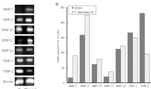
P. intermediaLPS (10 μg/ml)로 24시간 처리한 치 은섬유아세포에서의 MMP-1, -2, -3, -10, -11과 -14, 그리고 TIMP-1과 -2 mRNA의 발현을 RT-PCR을 수행 하여 조사하였다. 이들 MMP 및 TIMP mRNA는 LPS 로 처리하지 않은 치은섬유아세포에서도 발현되었 고, P. intermediaLPS로 처리한 세포에서는 MMP-1, -2, -3, -10과 -14, 그리고 TIMP-1 mRNA의 발현이 증 가되었으며, 이들 중 MMP -1, -3와 -10 mRNA의 증대 가 가장 현저하였다(Figure 1). 반면, MMP-11과 TIMP-2 mRNA의 발현은P. intermediaLPS로 처리한 섬유아세포에서 감소되었으며, 이들 중 TIMP-2 mRNA의 감소가 가장 현저하였다(Figure 1).
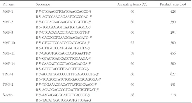
Table 1. Sequences of synthetic oligonucleotide primers for RT-PCR
Primers Sequence Annealing temp (℃) Product size (bp)
MMP-1 F 5'-CTGAAGGTGATGAAGCAGCC-3' 60 428
R 5'-AGTCCAAGAGAATGGCCGAG-3'
MMP-2 F 5'-GCGACAAGAAGTATGGCTTC-3' 60 390
R 5'-TGCCAAGGTCAATGTCAGGA-3'
MMP-3 F 5'-CTCACAGACCTGACTCGGTT-3' 60 294
R 5'-CACGCCTGAAGGAAGAGATG-3'
MMP-10 F 5'-GTCCTTCGATGCCATCAGCA-3' 62 380
R 5'-CTTGCTCCATGGACTGGCTA-3'
MMP-11 F 5'-CAGGTGGCAGCCCATGAATT-3' 58 456
R 5'-GTACTGAGCACCTTGGAAGA-3'
MMP-14 F 5'-CAACACTGCCTACGAGAGGA-3' 60 380
R 5'-GTTCTACCTTCAGCTTCTGG-3'
TIMP-1 F 5'-ACCATGGCCCCCTTTGAGCCCCTG-3' 60 627
R 5'-TCAGGCTATCTGGGACCGCAGGGA-3'
TIMP-2 F 5'-TGGAAACGACATTTATGGCAACC-3' 60 431
R 5'-ACAGGAGCCGTCACTTCTCTTGAT-3'
β-actin F 5'-AAGAGAGGCATCCTCACCCT-3' 66 218
R 5'-TACATGGCTGGGGTGTTGAA-3' Abbreviations: F: forward; R: reverse
Figure 1. Expression of MMP and TIMP mRNAs in human gingival fibroblasts stimulated with P. intermedia LPS. Cells were incubated in the presence of 10 μg/ml of P. intermedia LPS for 24 h. The intensities of the PCR bands on gel photographs were quantified by densitometry. See Materials and methods for further details
MMP-1
MMP-2
MMP-3
MMP-10
MMP-11
MMP-14
TIMP-1
TIMP-2 β-actin
0 10μg/ml
Figure 2. Expression of MMP and TIMP mRNAs in human periodontal ligament fibroblasts stimulated with P.
intermedia LPS. Cells were incubated in the presence of 10 μg/ml of P. intermedia LPS for 24 h. The intensities of the PCR bands on gel photographs were quantified by densitometry. See Materials and methods for further details
MMP-1
A B
A B
MMP-2
MMP-10
MMP-11
MMP-14
TIMP-1
TIMP-2 β-actin
0 10μg/ml
2) P. intermedia LPS가 치주인대세포에서의 MMP와 TIMP mRNA 발현에 미치는 영향
P. intermediaLPS (10 μg/ml)로 24시간 처리한 치 주인대세포에서의 MMP-1, -2, -10, -11과 -14, 그리고 TIMP-1과 -2 mRNA의 발현을 RT-PCR을 수행하여 조 사하였다. 이들 MMP 및 TIMP mRNA는 LPS로 처리 하지 않은 치주인대세포에서도 발현되었고, P. inter- mediaLPS로 처리한 세포에서는 MMP-1, -2, -10, -11 과 -14 mRNA의 발현이 증가되었으며, 이들 중 MMP- 1 mRNA의 증대가 가장 현저하였다(Figure 2). 반면, TIMP-1과 -2 mRNA의 발현은P. intermediaLPS로 처 리한 치주인대세포에서 감소되었으며, 이들 중 TIMP-2 mRNA의 감소가 가장 현저하였다(Figure 2).
IV. 고찰
MMP는 생리적 및 병리적 상황에서 세포외 기질의 개조와 분해에 관여한다2). 치주질환 시의 결체조직 의 파괴는 단핵세포, 대식세포, 임파구, 그리고 다형 핵 백혈구 등의 염증세포와 섬유아세포, 상피세포, 그리고 혈관내피세포 등의 상주세포에 의해 발현되 는 MMP와 그들의 억제제인 TIMP 간의 불균형으로 인한 MMP의 과도한 활성에 부분적으로 기인하는 것 으로 여겨진다12,29).
치주인대 및 치은섬유아세포에 의해 생성되는 Type I 및 III 교원질은 치주조직에 있어 가장 우세한 세포외 기질 성분이다29). 치은 및 치주인대 교원질의 분해는 치주염의 중요한 특징 중의 하나이며, 이러한 분해는 숙주세포에서 유래된 간질성 교원분해 효소 에 의해 수행된다. MMP-1 (collagenase-1)은 섬유아 세포형 간질성 교원분해 효소로, 중성구형 교원분해 효소인 MMP-8과 함께, 치은조직에서의 교원질의 생 리적 및 병적 분해에 관여한다30,31). MMP-1은 섬유아 세포, 상피세포, 그리고 대식세포에 의해 생성된다.
MMP-1은 type I, II 그리고 III 교원질의 helix를 제한 적으로 분열시켜 교원질의 분해를 개시하며, 그 후 gelatinase와 stromelysin에 의해 추가적인 분해가 진 행된다. 치주염 시 MMP-1이 증가되며19,32), MMP-1
mRNA는 치주염에 이환된 치은에 증가되어 있다33). 이는 MMP-1이 치주질환 병소에서의 교원질 분해와 연관이 있다는 것을 의미한다. MMP-1의 활성은 주 로 TIMP-1에 의해 엄격하게 조절된다.
MMP-3 (stromelysin-1)는 type IV와 IX 교원질 뿐 만 아니라 laminin, proteoglycan 그리고 fibronectin 도 분해한다34). MMP-3는 pro-MMP-1, pro-MMP-8 그 리고 pro-MMP-9의 활성화에 있어 중요한 역할을 한 다35). MMP-3는 치주염, 류마토이드 관절염, 그리고 골관절염 등 만성 염증성 질환에서의 결체조직 파괴 에 관여한다29,36). 치주염 이환 부위에서의 치은열구 액과 치은조직 내에 MMP-3 단백질과 MMP-3 mRNA 의 발현이 각각 증가됨이 증명된 바도 있다19,33).
MMP-10 (stromelysin-2)은 MMP-3와 유사하게 aggrecan, type III, IV와 V 교원질과 elastin, fibronectin, gelatin I, casein 그리고 fibrinogen의 분 해에 관여한다6). MMP-11 (stromelysin-3)는 다른 MMP들과는 달리 교원질, laminin, fibronectin, 그리 고 elastin 같은 세포외 기질 성분을 분해하지는 않으 며, α1-proteinase inhibitor, α2-macroglobulin, 그리 고 insulin-like growth factor binding protein 등을 분 해한다37). 또한 대부분의 MMP들이 비활성 zymogen 으로 분비되는데 비해, MMP-11은 45 kDa의 활성 효 소로 분비된다38).
MMP-2 (72 kDa type IV collagenase, gelatinase A) 는 기저막의 개조를 조절한다. MMP-2는 기저막에 존재하는 type IV 교원질, elastin, type V, VII과 X 교 원질 등의 기타 기질 성분을 분해할 수 있다8). 또한, 이들은 간질성 교원 분해효소 (MMP-1, -13, -18)에 의해 교원질 분자가 1/4과 3/4 segments로 분해된 후 gelatin을 분해한다. MMP-2는 섬유아세포, ker- atinocyte, 단핵구, 대식세포, 그리고 골아세포 등 다 양한 세포에서 proenzyme 형태로 분비되나, 다형핵 백혈구에서는 분비되지 않는다. MMP-2는 type IV 교원 분해효소 이므로, 감염부위에서 이들이 부적절 하게 활성화 되는 경우 기저막과 접합상피의 integri- ty에 심각한 영향을 미칠 수 있다.
MT1-MMP라고도 불리우는 MMP-14는 세포막 결 합부위를 가지고 있으며, 세포의 표면에 존재한다.
MMP-14는 types I, II와 III 교원질, fibronectin, type 1 과 5 laminin, vitronectin, fibrin, 그리고 aggrecan 같 은 세포외 기질 성분을 분해한다39,40). MMP-14는 세 포막에 부착되어 있어 여타의 교원 분해효소에 비해 세포표면에서 효율적으로 교원질 분해를 수행할 수 있다41).
TIMP는 MMP 활성을 조절하며, 4종의 TIMP가 보 고된 바 있다42). 이들 중 모든 종류의 MMP에 억제효 과를 발휘하는 TIMP-1과 TIMP-2는 치주질환 병소에 서 발견된다43). TIMP-1은 섬유아세포에서 유래된 MMP에 대해 강력한 억제효과를 발휘하며, TIMP-2 는 다형핵 백혈구에서 유래된 MMP에 강력한 억제효 과를 보인다18). TIMP-1은 28 kDa의 glycoprotein으 로, 활성화된 MMP와 1:1 stoichiometry의 복합체를 형성하며44), MMP를 생성한 세포가 TIMP-1도 생성 분비할 수 있다45). 활성화된 MMP와 TIMP 간의 균형 에 의해 세포외 기질의 개조의 정도가 좌우되며, MMP와 TIMP 간의 균형이 깨지게 되면 세포외 기질 의 병적인 파괴가 일어나 류마토이드 관절염과 혈관 질환 등의 병적인 상황이 초래될 수 있다. TIMP-1은 치은열구액과 치은조직 내에 존재하는 MMP에 대한 주된 억제제이다33,43,46,47). 염증 부위에서의 치은열구 액 내에 TIMP-1 단백질이 증가되어 있으며19,47), 치주 염 이환 치은 조직 내에 MMP-1과 TIMP-1 mRNA가 증가되어 있다33,46).
MMP 유전자의 발현은 싸이토카인을 포함한 다양 한 인자에 의해 조절된다. 본 연구는 치주질환 주요 병인균주 중의 하나인P. intermedia의 LPS가 치은섬 유아세포와 치주인대세포에서의 MMP 및 TIMP 발 현에 미치는 영향을 규명하고자 수행되었다. P. intermedia LPS는 치은섬유아세포와 치주인대세포 에서 MMP-1, -2, -3, -10과 -14, 그리고 TIMP-1 mRNA 와 MMP-1, -2, -10, -11과 -14 mRNA의 발현을 각각 증대시켰다. 반면, MMP-11과 TIMP-2 mRNA 그리고 TIMP-1과 -2 mRNA의 발현은P. intermediaLPS로 처 리한 섬유아세포 및 치주인대세포에서 각각 감소되 었다. P. intermediaLPS는 치은섬유아세포 및 치주 인대세포에서의 MMP 및 TIMP의 발현을 조절함으 로써 치주질환 시의 결체조직 파괴에 있어 중요한
역할을 하는 것으로 사료된다.
V. 결론
MMP는 생리적인 개조, 창상 치유, 그리고 치아 발 생 등에 관여한다. 또한, MMP는 치주염 등의 다양한 염증성 질환에 있어 중요한 역할을 하는 것으로 알 려져 있다. 본 연구는 치주질환 주요 병인균주 중의 하나인P. intermedia의 LPS가 치은섬유아세포와 치 주인대세포에서의 MMP 및 TIMP 발현에 미치는 영 향을 규명하고자 수행되었다. P. intermedia LPS는 치은섬유아세포와 치주인대세포에서 MMP-1, -2, -3, -10과 -14, 그리고 TIMP-1 mRNA와 MMP-1, -2, -10, - 11과 -14 mRNA의 발현을 각각 증대시켰다. 반면, MMP-11과 TIMP-2 mRNA 그리고 TIMP-1과 -2 mRNA의 발현은P. intermediaLPS로 처리한 섬유아 세포 및 치주인대세포에서 각각 감소되었다. P. intermedia LPS는 치은섬유아세포 및 치주인대세포 에서의 MMP 및 TIMP의 발현을 조절함으로써 치주 질환 시의 결체조직 파괴에 있어 중요한 역할을 하 는 것으로 사료된다.
VI. 참고문헌
1. Sorsa T, Ingman T, Suomalainen K et al.
Identification of proteases from periodontopath- ogenic bacteria as activators of latent human neutrophil and fibroblasttype interstitial collage- nases. Infect Immun 1992;60:4491-4795.
2. Birkedal-Hansen H, Moore WGI, Bodden MK et al. Matrix metalloproteinases: a review. Crit Rev Oral Biol Med 1993;4:197-250.
3. Woessner, J. Matrix metalloproteinases and their inhibitors in connective tissue remodeling.
FASEB J 1991;5:2145-2154.
4. Hoekstra R, Eskens FALM , Verweij J. Matrix metalloproteinase inhibitors: current develop- ments and future perspectives. Oncologist . 2001;6:415-427.
5. McCawley LJ, Matrisian LM. Matrix metallopro- teinases: they’re not just for matrix anymore!
Curr Opin Cell Biol 2001;13:534-540.
6. Sternlicht MD, Werb Z. How matrix metallopro- teinases regulate cell behavior. Annu Rev Cell Dev Biol 2001;17:463-516.
7. Nagase H, Okada Y in Textbook of Rheumatology (Kelley WN, Harris ED Jr, Ruddy S, Sledge CB Eds.), 5th ed, pp 323-340, Sanders, 1996, Philadelphia, PA.
8. Nagase H. Activation mechanisms of matrix met- alloproteinases. Biol Chem 1997;378:151-160.
9. Salo T, Mäkelä M, Kylmäniemi M, Autio- Harmainen H, Larjava, H. Expression of matrix metalloproteinase-2 and -9 during early human wound healing. Lab Invest 1994; 70, 176-182.
10. Llano E, Pendas AM, Knauper V et al.
Identification and structural and functional char- acterization of human enamelysin (MMP-20).
Biochemistry 1997;36:15101-15108.
11. Pirilä E, Ramamurthy N, Maisi P et al. Wound healing in ovariectomiced rats. Effects of chemi- cally modified tetracycline (CMT-8) and estrogen on matrix metalloproteinases -8 and -13 and type I collagen expression. Curr Med Chem 2001;8:281-294.
12. Reynolds JJ. Collagenases and tissue inhibitors of metalloproteinases: a functional balance in tis- sue degradation. Oral Dis 1995;2:70-76.
13. Van der Zee E, Everts V, Beertsen W. Cytokines modulate routes of collagen breakdown. J Clin Periodontol 1997;24:297-305.
14. Hanemaaijer R, Sorsa T, Kottinen YT et al.
Matrix metalloproteinase-8 is expressed in rheumatoid synovial fibroblasts and endothelial cells. Regulation by tumor necrosis factor-alpha and doxycycline. J Biol Chem 1997;272:31504- 31509.
15. Tonetti MS, Freiburger K, Lang NP, Bickel M.
Detection of interleukin-8 and matrix metallo- proteinases transcripts in healthy and diseased gingival biopsies by RNA/PCR. J Periodont Res 1993;28:511-513.
16. Sorsa T, Ding YL, Salo T et al. Effects of tetracy- clines on neutrophil, gingival, and salivary colla- genases. A functional and western-blot assess- ment with special reference to their cellular sources in periodontal diseases. Annal New York Acad Sci 1994;732:112-131.
17. Makela M, Salo T, Uitto VJ, Larjava H. Matrix metalloproteinases (MMP-2 and MMP-9) of the oral cavity: cellular origin and relationship to periodontal status. J Dent Res 1994;73:1397- 1406.
18. Aida T, Akeno N, Kawane T, Okamoto H, Horiuchi N. Matrix metalloproteinases-1 and -8 and TIMP-1 mRNA levels in normal and dis- eased human gingivae. EurJ Oral Sci 1996;104:562-569.
19. Ingman T, Tervahartiala T, Ding Y et al. Matrix metallo-proteinases and their inhibitors in gingi- val crevicular fluid and saliva of periodontitis patients. J Clin Periodontol 1996;23:1127-1132.
20. Korostoff JM, Wang JF, Sarment DP et al.
Analysis of in situ protease activity in chronic adult periodontitis patients: Expression of acti- vated MMP-2 and a 40 kDa serine protease. J Periodontol 2000;71:353-360.
21. Tanner ACR, Haffer C, Bratthall GT, Visconti RA, Socransky SS. A study of the bacteria associated with advancing periodontitis in man. J Clin Periodontol 1979;6:278-307.
22. Slots J, Bragd L, Wikstrom M, Dahlen G. The occurrence of Actinobacillus actinomycetem- comitans, Bacteroides gingivalisand Bacteroides intermediusin destructive periodontal disease in adults. J Clin Periodontol 1986;13:570-577.
23. Socransky SS, Haffajee AD. The bacterial etiolo-
gy of destructive periodontal disease: current concepts. J Periodontol 1992;63:322-331.
24. Chung CP, Nisengard RJ, Slots J, Genco RJ.
Bacterial IgG and IgM antibody titers in acute necrotizing ulcerative gingivitis. J Periodontol 1983;54:557-562.
25. Kornman KS, Loesche WJ. The subgingival microbial flora during pregnancy. J Periodont Res 1980;15:111-122.
26. Morrison DC, Ryan JL. Endotoxins and disease mechanisms. Annu Rev Med 1987;38:417-432.
27. Westphal O, Jann K. Bacterial lipopolysaccha- rides: extraction with phenol-water and further applications of the procedure. In: Whistler RL, ed. Methods in carbohydrate chemistry. New York: Academic Press, 1965: 83-91.
28. Markwell MA, Haas SM, Bieber LL, Tolbert NE.
A modification of the Lowry procedure to sim- plify protein determination in membrane and lipoprotein samples. Anal Biochem 1978;87:206- 210.
29. Birkedal-Hansen H. Role of matrix metallopro- teinases in human periodontal diseases. J Periodontol 1993;64:474-484.
30. Goldberg GI, Wilhelm SM, Kronberger A et al.
Human fibroblast collagenases. Complete prima- ry structure and homology to an oncogene transformation-induced rat protein. J Biol Chem 1986; 261:6600-6605.
31. Devarajan P, Mookhtiar K, Wart HV, Berliner N.
Structure and expression of the cDNA encoding human neutrophil collagenase. Blood 1991;77:2731-2738.
32. Sorsa T, Uitto VJ, Suomalainen K, Vauhkonen M, Lindy S. Comparison of interstitial collagenases from human gingiva, sulcular fluid and polymor- phonuclear leukocytes. J Periodont Res 1988;23:386-393.
33. Kubota T, Nomura T, Takahashi T, Hara K.
Expression of mRNA for matrix metallopro- teinases and tissue inhibitors of metallopro- teinases in periodontitis-affected human gingival tissue. Arch Oral Biol 1996;41:253-262.
34. Matrsian LM. The matrix-degrading metallopro- teinases. Bioessays 1992;14:455-463.
35. Ogata Y, Enghild JJ, Nagase H. Matrix metallo- proteinase 3 (stromelysin) activates the precur- sors for human matrix metalloproteinase 9. J Biol Chem 1992;267:3581-3584.
36. McCachren SS. Expression of metalloproteinases and metalloproteinase inhibitor in human arthrit- ic synovium. Arthrit Rheum 1991;34:1085-1093.
37. Manes S, Mira E, Barbacid MM et al.
Identification of insulinlike growth factor-bind- ing protein-1 as a potential physiological sub- strate for human stromelysin-3. J Biol Chem 1997;272:25706-25712.
38. Pei D, Weiss SJ. Furin-dependent intracellular activation of the human stromelysin-3 zymogen.
Nature 1995;375:244-247.
39. d’Ortho MP, Will H, Atkinson S et al. Membrane- type matrix metalloproteinases 1 and 2 exhibit broad-spectrum proteolytic capacities comparable to many matrix metalloproteinases. Eur J Biochem 1997;250:751-757.
40. Ohuchi E, Imai K, Fujii Y et al. Membrane type 1 matrix metalloproteinase digests interstitial colla- gens and other extracellular matrix macromole- cules. J Biol Chem 1997;272:2446-2451.
41. Hotary K, Allen E, Punturieri A, Yana I, Weiss SJ.
Regulation of cell invasion and morphogenesis in a three-dimensional type I collagen matrix by membrane-type matrix metalloproteinases 1, 2, and 3. J Cell Biol 2000;149:1309-1323.
42. Nagase H, Woessner JF. Matrix metallopro- teinases J Biol Chem 1999;274:21491-21494.
43. Kubota T, Matsuki Y, Nomura T, Hara K. In situ hybridization study on tissue inhibitors of metal-
loproteinases (TIMPs) mRNA-expressing cells in human inflamed gingival tissue. J Periodont Res 1997;32:467-472.
44. Stricklin GP, Welgus HG. Human skin fibroblast collagenase inhibitor. Purification and biochemi- cal characterization. J Biol Chem 1983;258:12252- 12258.
45. Welgus HG, Jeffrey JJ, Eisen AZ, Roswit WT, Stricklin GP. Human skin fibroblast collagenase:
interaction with substrate and inhibitor. Coll
Relat Res 1985;5:167-179.
46. Nomura T, Takahashi T, Hara K. Expression of TIMP-1,TIMP-2 and collagenase mRNA in peri- odontitisaffected human gingival tissue. J Periodont Res 1993;28:354-362.
47. Nomura T, Ishii A, Oishi Y, Kohma H, Hara K.
Tissue inhibitors of metalloproteinases level and collagenase activity in gingival crevicular fluid:
the relevance to periodontal diseases. Oral Dis 1998;4:231-240.
-Abstract-
Expression of mRNA for matrix metalloproteinases and tissue inhibitors of metalloproteinases in human gingival
and periodontal ligament fibroblasts treated with lipopolysaccharide from Prevotella intermedia
Sung-Jo Kim1, Eun-Young Choi2, In-Soon Choi2, Ju-Youn Lee1, Jeom-Il Choi1, Chong-Kwan Kim3
11Department of Periodontology, College of Dentistry, Pusan National University
22Department of Life Science, College of Natural Science, Silla University
33Department of Periodontology, College of Dentistry, Yonsei University
Matrix metalloproteinases (MMPs) are a family of host-derived proteolytic enzymes and implicated in the remodeling and degradation of extracellular matrix under both physiological and pathological conditions.
Connective tissue degradation in periodontal diseases is thought to be due to excessive MMP activities over their specific inhibitors. The effects of lipopolysaccharide (LPS) from Prevotella intermedia, one of the major putative pathogens of periodontitis, on the expression of mRNA for MMPs and tissue inhibitors of metallopro- teinases (TIMPs) in human gingival and periodontal ligament fibroblasts were examined by reverse transcrip- tase-polymerase chain reaction (RT-PCR). The expression of mRNAs encoding MMP-1, -2, -3, -10, and -14 was increased in human gingival fibroblasts treated with P. intermediaLPS, whereas MMP-11 and TIMP-2 mRNA expression was decreased in these cells stimulated with LPS. P. intermediaLPS increased the MMP-1, -2, -10, - 11, and -14 mRNA expression and decreased TIMP-1 and -2 mRNA expression in human periodontal ligament fibroblasts. These findings imply that P.intermediaLPS may play an important role in the connective tissue degradation in periodontitis.
Key words : matrix metalloproteinases, tissue inhibitors of metalloproteinases, human gingival fibroblasts, human periodontal ligament fibroblasts, Prevotella intermediaLPS