www.jpis.org
pISSN 2093-2278 eISSN 2093-2286 Copyright © 2010 Korean Academy of PeriodontologyThis is an Open Access article distributed under the terms of the Creative Commons Attribution Non-Commercial License (http://creativecommons.org/licenses/by-nc/3.0/).
Biological effects of a semiconductor diode laser on human periodontal ligament fibroblasts
Eun-Jeong Choi, Ju-Young Yim, Ki-Tae Koo, Yang-Jo Seol, Yong-Moo Lee, Young Ku, In-Chul Rhyu, Chong-Pyoung Chung, Tae-Il Kim* Department of Periodontology and Dental Research Institute, Seoul National University College of Dentistry, Seoul, Korea
Purpose: It has been reported that low-level semiconductor diode lasers could enhance the wound healing process. The peri- odontal ligament is crucial for maintaining the tooth and surrounding tissues in periodontal wound healing. While low-level semiconductor diode lasers have been used in low-level laser therapy, there have been few reports on their effects on periodon- tal ligament fibroblasts (PDLFs). We performed this study to investigate the biological effects of semiconductor diode lasers on human PDLFs.
Methods: Human PDLFs were cultured and irradiated with a gallium-aluminum-arsenate (GaAlAs) semiconductor diode la- ser of which the wavelength was 810 nm. The power output was fixed at 500 mW in the continuous wave mode with various energy fluencies, which were 1.97, 3.94, and 5.91 J/cm2. A culture of PDLFs without laser irradiation was regarded as a control.
Then, cells were additionally incubated in 72 hours for MTS assay and an alkaline phosphatase (ALPase) activity test. At 48 hours post-laser irradiation, western blot analysis was performed to determine extracellular signal-regulated kinase (ERK) activity.
ANOVA was used to assess the significance level of the differences among groups (P<0.05).
Results: At all energy fluencies of laser irradiation, PDLFs proliferation gradually increased for 72 hours without any signifi- cant differences compared with the control over the entire period taken together. However, an increment of cell proliferation significantly greater than in the control occurred between 24 and 48 hours at laser irradiation settings of 1.97 and 3.94 J/cm2 (P<0.05). The highest ALPase activity was found at 48 and 72 hours post-laser irradiation with 3.94 J/cm2 energy fluency (P<0.05).
The phosphorylated ERK level was more prominent at 3.94 J/cm2 energy fluency than in the control.
Conclusions: The present study demonstrated that the GaAlAs semiconductor diode laser promoted proliferation and differ- entiation of human PDLFs.
Keywords: Alkaline phosphatase, Extracellular signal-regulated kinases, Fibroblasts, Periodontal ligament, Semiconductor di- ode lasers.
INTRODUCTION
Lasers have been introduced in the field of periodontology for their adjunctive beneficial effects on conventional peri- odontal treatments [1-3]. Several researchers have reported that lasers show hemostatic and bactericidal effects, which secures good periodontal treatment results [4-6]. Among var-
ious lasers used for periodontal purposes, semiconductor di- ode lasers are mainly applied in subgingival curettage and periodontal pocket disinfection in clinics [7,8]. They can trans- form electric energy to light using the same principle as a light-emitting diode, but with internal reflection capability, thus forming a resonator where a stimulated light can reflect back and forth, allowing only a certain wavelength to be emit-
Received: Mar. 08, 2010; Accepted: Apr. 20, 2010
*Correspondence: Tae-Il Kim
Department of Periodontology, Seoul National University College of Dentistry, 101 Daehang-no, Jongno-gu, Seoul 110-749, Korea Email: [email protected], Tel: +82-2-2072-2642, Fax: +82-2-744-1349
sity, this laser is often used for low-level laser therapy, which accelerates the wound healing process without imparting any thermal effect [9-13].
Periodontal wound healing is a series of interactions among periodontal tissue cells, which includes gingival fibroblasts, osetoblasts, cementoblasts, and periodontal ligament fibro- blasts (PDLFs). In these cell populations, PDLFs have been regarded to be quintessential for maintaining the periodon- tium, as it has been suggested that PDLFs could differentiate into osteoblasts and cementoblasts for restoring lost alveolar bone and cementum [14,15].
While low-level laser therapy has been regarded as an ad- junctive periodontal treatment modality in clinical therapy, there have been few reports on its effects on PDLFs per se [16,17]. Hence, we investigated the biological effects of low- level lasers on PDLFs by using semiconductor diode lasers in this study.
MATERIALS AND METHODS
Cell cultures
Human PDLFs cell lines (ScienCell, San Diego, USA) were maintained in α-MEM (Hyclone, Logan, USA) containing a 10% FBS, and 1% penicillin-streptomycin solution (5,000 units/
mL penicillin and 50 μg/mL streptomycin), and these cultures were incubated at 37°C in a humidified atmosphere in the presence of 5% CO2. PDLFs were allowed to attach for 24 hours before being subject to laser irradiation. Prior to the ir- radiation, the medium was removed and 500 μL of fresh medium was added after the laser irradiation.
Low-level laser irradiation
An 810-nm gallium-aluminum-arsenate (GaAlAs) semicon- ductor diode laser (WhiteStarTM, Creation, Verona, Italy) was used. Laser light was delivered through a 600 μm fiber-optic system with 500 mW power output in the continuous wave mode. The fiber tip was perpendicularly positioned at a 10-cm distance from the cell monolayer (Fig. 1). Laser beam energy was determined by means of the power meter (FieldMasterTM, Coherent Inc., Santa Clara, USA) and irradiation time was 10, 20, or 30 seconds corresponding to energy fluencies of 1.97, 3.94, and 5.91 J/cm2, respectively. The control cultures were treated equally, except for the laser irradiation.
Cell proliferation assay
A 3-(4,5-dimethylthiazol-2-yl)-5-(3-carboxymethoxyphenyl)- 2-(4-sulfophenyl)-2H-tetrazolium (MTS) assay was performed to assess the cell proliferation activity. PDLFs were plated at a density of 1 × 10⁴ cells/well in a 24-well culture plate and after
hours. Then the cell medium was removed and replaced by fresh medium containing MTS reagents (Promega, Madison, USA). After 3 hours of incubation, the absorbance at 490 nm was measured using a spectrophotometer (Molecular Devic- es, Sunnyvale, USA).
Alkaline phosphatase activity test
PDLFs were plated at a density of 1 × 10⁵ cells/well in a 24- well culture plate and all groups were incubated for 24, 48, and 72 hours after the laser irradiation. PDLFs were washed three times with phosphate buffered saline (PBS) and solubilized with alkaline buffer. The lysates with the alkaline phosphatase (ALPase) assay working solution were incubated for 4 min- utes. After incubation, the absorbance of p-nitrophenol was read at 405 nm using a microplate reader (Molecular Devices, Sunnyvale, USA). The total cell protein was measured using a protein assay kit (Invitrogen, Carlsbad, USA) and the results were expressed in μmol/60 minutes/μg.
Western blot analysis for extracellular signal-regulated ki- nase (ERK) activation
Laser-irradiated PDLFs were plated at a density of 1 × 10⁵ cells/well in a 24-well culture plate and incubated for 48 hours,
Figure 1. A gallium-aluminum-arsenate semiconductor diode laser with an 810-nm wavelength was prepared and the fiber tip was per- pendicularly positioned at a 10-cm distance from the cell monolayer.
lysed with RIPA buffer. The protein lysates were centrifuged at 13,000 g for 30 minutes, and the supernatant was collected for protein quantification using the protein assay kit (Invitro- gen, Carlsbad, USA). Total protein in the amount of 50 μg was boiled and separated electrophoretically on SDS-PAGE gel and transferred onto a Hybond-P polyvinylidene difluo- ride (PVDF) membrane (GE Healthcare, Piscataway, USA) by electroblotting. After blocking in TBST buffer (25 mM Tris- HCl pH 7.4, 1.5 M NaCl, 0.5% Tween-20) containing 5% fat- free dry milk for 1 hour, the PVDF membrane sheets were incubated overnight at 4°C with primary antibodies. The pri- mary antibodies used were anti-phosphor-ERK (Cell Signal- ing, Beverly, USA), anti-β-actin (Santa Cruz Biotechnology, Santa Cruz, USA). After washing three times with TBST, the membrane sheets were incubated with horseradish-peroxi- dase conjugated secondary anti-rabbit antibody (Sigma, St.
Louis, USA) for 1 hour. The immunoreactive bands were visu- alized using a luminescent image analysis system (Fujifilm Life Science, Waipahu, USA).
Statistical analysis
The statistical analysis was performed by a statistical soft- ware package (SPSSTM, SPSS Inc., Chicago, USA). The mean values and the standard deviation were calculated for the MTS assay. The data were analyzed by ANOVA with an ad- hoc Tukey’s test to assess the significance level of the differ- ences among the energy doses. The statistical significance was
RESULTS
Cell proliferation assay
After GaAlAs semiconductor diode laser irradiation of hu- man PDLFs for 10, 20, or 30 seconds, increased cell prolifera- tion was recorded in a time-dependent manner. At all levels of applied irradiation, human PDLFs proliferation gradually increased for 72 hours (Fig. 2). While there was no significant difference compared with the control over the entire 72 hours taken together, significant incremental PDLFs proliferation was observed between 24 and 48 hours at both 1.97 and 3.94 J/
cm2 energy fluencies (Fig. 3).
ALPase activity test
The effect of laser irradiation on the ALPase activity of hu- man PDLFs is illustrated in Fig. 4. Compared with the con- trol, significantly increased ALPase activity of laser-irradiated PDLFs was revealed regardless of the amount of laser energy fluency or irradiation time (P<0.05). Among the irradiated groups, the irradiated PDLFs group with 3.94 J/cm2 of laser energy fluency-showed statistically significant ALPase activi- ty at 48 and 72 hours (P<0.05).
Western blot analysis
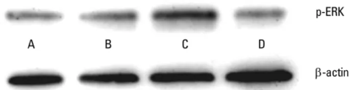
Western blot analysis showed that phosphorylated ERK ac- tivity was prominent in the laser-irradiated PDLFs group at 3.94 J/cm2 (Fig. 5).
Figure 2. Human periodontal ligament fibroblast proliferation af- ter 810-nm semiconductor diode laser irradiation at the energy flu- encies of 1.97 (B), 3.94 (C) and 5.91 (D) J/cm2. Periodontal ligament fi- broblast proliferation was gradually increased up to 72 hours with- out any significant difference compared with the control (A) at all levels of irradiation.
2
1.5
1
0.5
0 A B C D
Absorbance at 490 nm
48 hours 72 hours 24 hours
Groups
Figure 3. Incremental difference in absorbance at 490 nm in hu- man periodontal ligament fibroblasts (PDLFs). PDLFs proliferation between 24 and 48 hours was significant at the energy fluencies of 1.97 (B) and 3.94 (C) J/cm2, but was not in the 5.91 (D) J/cm2 and con- trol group (A) (*P<0.05).
0.7 0.6 0.5 0.4 0.3 0.2 0.1
0 A B C D Incremental absorbance difference
48-24 hours 72-48 hours
Groups
*
*
DISCUSSION
It has been reported that a low-level laser can accelerate wound healing [10-13], enhance bone and collagen formation [18-23] and induce anti-inflammatory effects [18-20]. These findings are supported by in vitro examinations confirming that low-level laser irradiation significantly increases cell pro- liferation [16,21] and collagen deposition [22], and enhances osteogenic differentiation [23].
Studies aiming to confirm the effects of low-level lasers on PDLFs, however, are very few. The responsiveness of PDLFs to low-level laser energy in vitro has been demonstrated by Shimizu et al., who showed that 830 nm GaAlAs laser irradia- tion of stretched human periodontal ligament cells signifi- cantly inhibited the production of prostaglandin E2 and IL-1β [24]. Furthermore, there have seldom been reports regarding the proliferation and differentiation of PDLFs in general.
In the present study, we revealed that an 810 nm GaAlAs semiconductor diode laser increased human PDLFs prolifer- ation, although there were no significant differences among groups. The present result is inconsistent with that of Kreisler et al. [16], who discovered that laser irradiation stimulated the proliferation of PDLFs. In their study, a GaAlAs diode laser with a wavelength of 809 nm significantly increased the meta- bolic activity of PDLFs with a duration of incubation of up to three days. They also reported that laser energy fluencies of 1.96, 3.92, and 7.84 J/cm2 had similar effects on PDLFs. Our current findings are different from theirs. PDLFs proliferation increased in time-dependant manner up to 72 hours irrespec- tive of irradiation regimen. The reasons for the inconsistency
could lie in the differences in power output, fiber distance from the cell monolayer, and irradiation time.
The level of ALPase of periodontal ligament cells is known to be one of the initial differentiation markers of osteoblasts because this level increases in the initial differentiation stage [25]. Hence, we investigated ALPase activity in order to evalu- ate the differentiation of PDLFs after laser irradiation and found that low-level semiconductor diode laser irradiation significantly stimulated PDLFs differentiation at all energy fluencies of 1.97, 3.94, and 5.91 J/cm2. Among the groups, the 3.94 J/cm2 lased group showed significantly greater results compared to the other irradiation groups. This suggests that low-level laser irradiation with a 3.94 J/cm2 energy fluency is suitable for PDLFs differentiation.
The present study found, first, that low-level laser irradia- tion activated ERK. In general, the mitogen-activated protein kinase (MAPK) pathway regulates cellular responses to envi- ronmental changes [26]. ERK, which is a typical MAPK family member, is activated by several growth factors and mitogens to induce cell proliferation and differentiation [27]. We revealed that 3.94 J/cm2 energy fluency more prominently activated ERK of human PDLFs than other energy fluencies and the control. These findings coincide with a previous report which revealed that laser irradiation at 3 and 4 J/cm2 exerted stimu- latory effects, whereas 5 J/cm2 caused inhibitory effects in NIH- 3T3 fibroblasts [22].
Interestingly, the incremental PDLFs proliferation between 24 and 48 hours revealed significantly greater values at 1.97 and 3.94 J/cm2 energy fluencies. In a previous report, the dif- ference in PDLFs proliferation on days 1 and 2 after laser ir- radiation was highly significant and decreased on day 3 [16].
Our data regarding the incremental cell proliferation is com- parable to those of previous studies, which showed peaked molecular and cellular responses up to 2 days after laser irra- diation [16,28]. We can deduct that low-level laser irradiation affects the cellular environment in the early incubation peri- od within 2 days, which requires further investigation.
In conclusion, we demonstrated that the GaAlAs semicon- Figure 4. Effect of semiconductor diode laser irradiation on alka-
line phosphatase (ALPase) activity in human periodontal ligament fibroblasts (PDLFs). All the laser-irradiated groups showed a signifi- cant increase compared to the control. Among the laser-irradiated groups, ALPase activity of PDLFs was significantly greater at 3.94 J/
cm2 of laser energy fluency.
*Statistically significantly greater than the control (P<0.05).
†Statistically significantly greater than other laser-irradiated groups (P<0.05).
20 15 10 5 μmol/60 min/ALPase activity (μg) 0
24 48 72
1.97 J/cm2 5.91 J/cm2
Control 3.94 J/cm
Hours
* * * * *
*
* *
† *
†
p-ERK
β-actin A B C D
Figure 5. Result of western blot analysis of phosphorylated extra- cellular signal-regulated kinase (ERK) at the laser energy fluencies of 1.97 (B), 3.94 (C) and 5.91 (D) J/cm2 compared to the control (A). 3.94 J/cm2 of laser energy-fluency promoted significantly greater ERK activation.
tiation of human PDLFs. Within the limits of this study, the optimal energy fluency for the stimulation of PDLFs differ- entiation was 3.94 J/cm2.
CONFLICT OF INTEREST
No potential conflict of interest relevant to this article was reported.
ACKNOWLEDGEMENTS
This study was supported by the Seoul National University Dental Hospital Research Fund (03-2007-0011).
REFERENCES
Midda M. Lasers in periodontics. Newsl Int Acad Periodon- 1.
tol 1991;1:2-3.
Midda M. Lasers in periodontics. Periodontal Clin Inves- 2.
tig 1992;14:14-20.
Midda M. The use of lasers in periodontology. Curr Opin 3.
Dent 1992;2:104-8.
Ando Y, Aoki A, Watanabe H, Ishikawa I. Bactericidal effect 4.
of erbium YAG laser on periodontopathic bacteria. Lasers Surg Med 1996;19:190-200.
Aoki A, Sasaki KM, Watanabe H, Ishikawa I. Lasers in non- 5.
surgical periodontal therapy. Periodontol 2000 2004;36:
59-97.
Folwaczny M, Mehl A, Aggstaller H, Hickel R. Antimicro- 6.
bial effects of 2.94 microm Er:YAG laser radiation on root surfaces: an in vitro study. J Clin Periodontol 2002;29:73-8.
Moritz A, Schoop U, Goharkhay K, Schauer P, Doertbudak 7.
O, Wernisch J, et al. Treatment of periodontal pockets with a diode laser. Lasers Surg Med 1998;22:302-11.
Kreisler M, Al Haj H, d’Hoedt B. Clinical efficacy of semi- 8.
conductor laser application as an adjunct to conventional scaling and root planing. Lasers Surg Med 2005;37:350-5.
Conlan MJ, Rapley JW, Cobb CM. Biostimulation of wound 9.
healing by low-energy laser irradiation: a review. J Clin Periodontol 1996;23:492-6.
Mester E, Spiry T, Szende B, Tota JG. Effect of laser rays on 10.
wound healing. Am J Surg 1971;122:532-5.
Reddy GK, Stehno-Bittel L, Enwemeka CS. Laser photostim- 11.
ulation accelerates wound healing in diabetic rats. Wound Repair Regen 2001;9:248-55.
Pinfildi CE, Liebano RE, Hochman BS, Ferreira LM. Heli- 12.
um-neon laser in viability of random skin flap in rats. La- sers Surg Med 2005;37:74-7.
Herascu N, Velciu B, Calin M, Savastru D, Talianu C. Low- 13.
Photomed Laser Surg 2005;23:70-3.
Isaka J, Ohazama A, Kobayashi M, Nagashima C, Takigu- 14.
chi T, Kawasaki H, et al. Participation of periodontal liga- ment cells with regeneration of alveolar bone. J Periodon- tol 2001;72:314-23.
Lekic P, McCulloch CA. Periodontal ligament cell popula- 15.
tion: the central role of fibroblasts in creating a unique tis- sue. Anat Rec 1996;245:327-41.
Kreisler M, Christoffers AB, Willershausen B, d’Hoedt B.
16.
Effect of low-level GaAlAs laser irradiation on the prolif- eration rate of human periodontal ligament fibroblasts:
an in vitro study. J Clin Periodontol 2003;30:353-8.
Kreisler M, Meyer C, Stender E, Daublander M, Willershaus- 17.
en-Zonnchen B, d’Hoedt B. Effect of diode laser irradia- tion on the attachment rate of periodontal ligament cells:
an in vitro study. J Periodontol 2001;72:1312-7.
do Nascimento PM, Pinheiro AL, Salgado MA, Ramalho 18.
LM. A preliminary report on the effect of laser therapy on the healing of cutaneous surgical wounds as a consequence of an inversely proportional relationship between wave- length and intensity: histological study in rats. Photomed Laser Surg 2004;22:513-8.
Lopes-Martins RA, Albertini R, Martins PS, Bjordal JM, Far- 19.
ia Neto HC. Spontaneous effects of low-level laser therapy (650 nm) in acute inflammatory mouse pleurisy induced by carrageenan. Photomed Laser Surg 2005;23:377-81.
Correa F, Lopes Martins RA, Correa JC, Iversen VV, Joen- 20.
son J, Bjordal JM. Low-level laser therapy (GaAs lambda = 904 nm) reduces inflammatory cell migration in mice with lipopolysaccharide-induced peritonitis. Photomed Laser Surg 2007;25:245-9.
Kreisler M, Christoffers AB, Al-Haj H, Willershausen B, 21.
d’Hoedt B. Low level 809-nm diode laser-induced in vitro stimulation of the proliferation of human gingival fibro- blasts. Lasers Surg Med 2002;30:365-9.
Pereira AN, Eduardo Cde P, Matson E, Marques MM. Ef- 22.
fect of low-power laser irradiation on cell growth and pro- collagen synthesis of cultured fibroblasts. Lasers Surg Med 2002;31:263-7.
Stein A, Benayahu D, Maltz L, Oron U. Low-level laser ir- 23.
radiation promotes proliferation and differentiation of human osteoblasts in vitro. Photomed Laser Surg 2005;
23:161-6.
Shimizu N, Yamaguchi M, Goseki T, Shibata Y, Takiguchi 24.
H, Iwasawa T, et al. Inhibition of prostaglandin E2 and in- terleukin 1-beta production by low-power laser irradiation in stretched human periodontal ligament cells. J Dent Res 1995;74:1382-8.
Giannopoulou C, Cimasoni G. Functional characteristics 25.
Res 1996;75:895-902.
Gumbiner BM. Cell adhesion: the molecular basis of tis- 26.
sue architecture and morphogenesis. Cell 1996;84:345-57.
Kim TI, Jang JH, Lee YM, Rhyu IC, Chung CP, Han SB, et 27.
al. Biomimetic approach on human periodontal ligament
925-32.
Ozawa Y, Shimizu N, Kariya G, Abiko Y. Low-energy laser 28.
irradiation stimulates bone nodule formation at early stag- es of cell culture in rat calvarial cells. Bone 1998;22:347-54.