Uric Acid Level Has a U-shaped Association with Clinical Outcomes in Patients with Vasospastic Angina
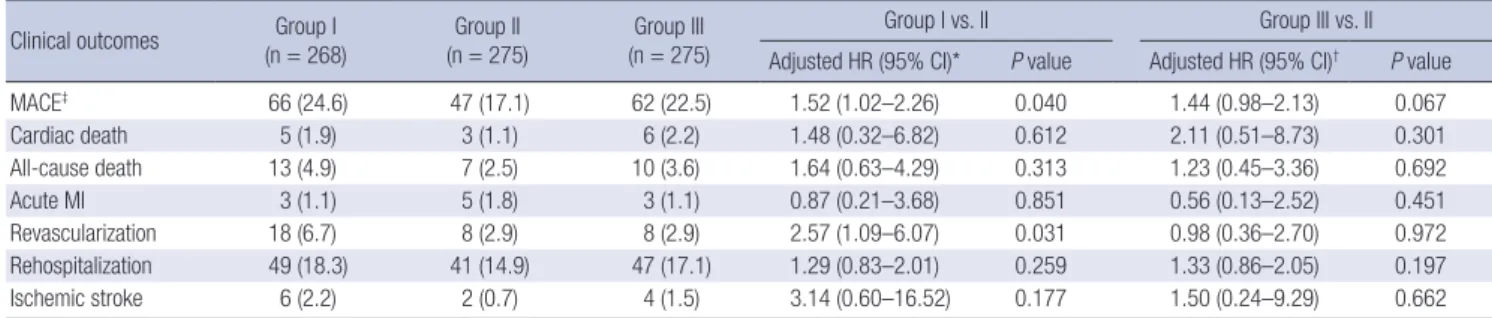
No data are available on the association of serum uric acid and vasospastic angina (VSA) which has endothelial dysfunction as a possible pathophysiologic mechanism. Low uric acid level might cause adverse outcomes in VSA in connection with endothelial dysfunction. We enrolled 818 VSA patients whose uric acid level was measured at admission. Patients were categorized according to tertiles of uric acid level: group I, ≤ 4.8 mg/dL; group II, 4.9–5.9 mg/dL; and group III, ≥ 6.0 mg/dL. Primary outcome was major adverse cardiac events (MACEs), defined as a composite of cardiac death, acute myocardial infarction (MI), ischemic stroke, coronary revascularization, and rehospitalization for angina. Median follow-up duration was 49.2 months. Median uric acid values were 4.1 mg/dL for group I, 5.4 mg/dL for group II, and 6.7 mg/dL for group III. In the overall population, group II had a significantly lower incidence of MACE compared to group I (47 [17.1%] vs. 66 [24.6%]; hazard ratio [HR], 1.52; 95% confidence interval [CI], 1.02–2.26;
P = 0.040) and a tendency of lower incidence of MACEs compared to Group III (47 [17.1%]
vs. 62 [22.5%]; HR, 1.44; 95% CI, 0.98–2.13; P = 0.067). Among group I patients, those who received nitrates had a higher incidence of MACEs than those without nitrate therapy (P < 0.001). Low uric acid level was associated with adverse clinical outcomes, while high uric acid level had a trend toward an increase in it. Use of nitrate in patients with low uric acid level might have adverse effects on clinical outcomes of VSA.
Keywords: Nitrates; Uric Acid; Vasospastic Angina Hye Bin Gwag,1* Jeong Hoon Yang,1,2*
Taek Kyu Park,1 Young Bin Song,1 Joo-Yong Hahn,1 Jin-Ho Choi,1 Sang Hoon Lee,1 Hyeon-Cheol Gwon,1 and Seung-Hyuk Choi1
1Division of Cardiology, Department of Medicine, Samsung Medical Center, Sungkyunkwan University School of Medicine, Seoul, Korea; 2Department of Critical Care Medicine, Samsung Medical Center, Sungkyunkwan University School of Medicine, Seoul, Korea
* Hye Bin Gwag and Jeong Hoon Yang contributed equally to this work.
Received: 7 March 2017 Accepted: 20 May 2017 Address for Correspondence:
Seung-Hyuk Choi, MD, PhD
Division of Cardiology, Department of Medicine, Samsung Medical Center, Sungkyunkwan University School of Medicine, 81 Irwon-ro, Gangnam-gu, Seoul 06351, Korea
E-mail: [email protected]
https://doi.org/10.3346/jkms.2017.32.8.1275 • J Korean Med Sci 2017; 32: 1275-1280
INTRODUCTION
Increased serum uric acid level is proposed to be associated with adverse cardiovascular (CV) events in patients with risk factors such as hypertension, diabetes, heart failure, or meta- bolic syndrome (1-4). However, the causal relationship remains controversial because hyperuricemia can also be caused by conditions like renal dysfunction, gout, alcohol consumption, or diuretic therapy, confounding the association between hy- peruricemia and CV events. Uric acid was reported to have a role as an endogenous antioxidant in previous studies (5,6) and recent studies shows that low uric acid level is associated with endothelial dysfunction (4,5,7). These reports have raised ques- tions about the impact of hypouricemia on CV events. Howev- er, no data are available on the association of serum uric acid level and VSA which has endothelial dysfunction as a possible pathophysiologic mechanism (8-10).
Our hypothesis is that low uric acid level might cause adverse clinical outcomes in vasospastic angina (VSA) in connection with endothelial cell dysfunction. Therefore, we investigated the impact of serum uric acid level on clinical outcomes in VSA patients with or without significant coronary artery stenosis con- firmed by spasm-provocation test.
MATERIALS AND METHODS
The study population was selected from the Samsung Medical Center VSA registry. Between January 2003 and December 2014, 1,199 consecutive patients with VSA were enrolled in a single center registry. VSA was defined by the Guidelines for Diagno- sis and Treatment of Patients with Vasospastic Angina of the Jap- anese Circulation Society (11). Inclusion criteria were: 1) coro- nary spasm proven by coronary angiography (CAG) and spasm- provocation test, and 2) serum uric acid level measured at ad- mission. Enrolled patients were categorized according to the admission uric acid level into group I (≤ 4.8 mg/dL, n = 268), II (4.9–5.9 mg/dL, n = 275), or III (≥ 6.0 mg/dL, n = 275) (Fig. 1).
In addition, we divided patients into pure VSA or mixed angina groups using previous history or the presence of significant fixed stenosis on CAG after intracoronary nitroglycerin injection. Mix- ed angina was defined as: 1) fixed coronary artery stenosis > 50%
in spasm-positive arteries, or 2) previous history of myocardial infarction (MI) or coronary artery revascularization. Patients who did not meet these criteria were assigned to a pure-VSA group (12).
After diagnostic CAG, intracoronary ergonovine was admin- istered for spasm-provocation tests with starting dose of 10 µg ORIGINAL ARTICLE
Cardiovascular Disorders
2017-03-16 https://crossmark-cdn.crossref.org/widget/v2.0/logos/CROSSMARK_Color_square.svg
Fig. 1. Study population.
VSA = vasospastic angina.
Group I Lower uric acid tertile
(n = 268)
Group II Middle uric acid tertile
(n = 275)
Group III Upper uric acid tertile
(n = 275) · Follow-up loss (n = 42)
· No measured uric acid level (n = 339) Samsung Medical Center VSA registry
January 2003-December 2014 (n = 1,199)
Study population (n = 818)
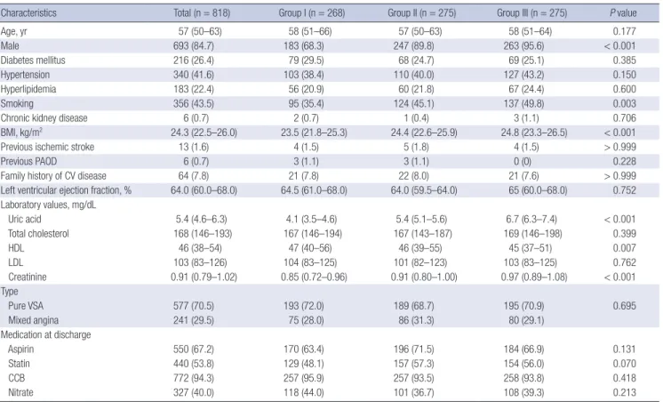
Table 1. Baseline characteristics of patients according to uric acid level
Characteristics Total (n = 818) Group I (n = 268) Group II (n = 275) Group III (n = 275) P value
Age, yr 57 (50–63) 58 (51–66) 57 (50–63) 58 (51–64) 0.177
Male 693 (84.7) 183 (68.3) 247 (89.8) 263 (95.6) < 0.001
Diabetes mellitus 216 (26.4) 79 (29.5) 68 (24.7) 69 (25.1) 0.385
Hypertension 340 (41.6) 103 (38.4) 110 (40.0) 127 (43.2) 0.150
Hyperlipidemia 183 (22.4) 56 (20.9) 60 (21.8) 67 (24.4) 0.600
Smoking 356 (43.5) 95 (35.4) 124 (45.1) 137 (49.8) 0.003
Chronic kidney disease 6 (0.7) 2 (0.7) 1 (0.4) 3 (1.1) 0.706
BMI, kg/m2 24.3 (22.5–26.0) 23.5 (21.8–25.3) 24.4 (22.6–25.9) 24.8 (23.3–26.5) < 0.001
Previous ischemic stroke 13 (1.6) 4 (1.5) 5 (1.8) 4 (1.5) > 0.999
Previous PAOD 6 (0.7) 3 (1.1) 3 (1.1) 0 (0) 0.228
Family history of CV disease 64 (7.8) 21 (7.8) 22 (8.0) 21 (7.6) > 0.999
Left ventricular ejection fraction, % 64.0 (60.0–68.0) 64.5 (61.0–68.0) 64.0 (59.5–64.0) 65 (60.0–68.0) 0.752 Laboratory values, mg/dL
Uric acid 5.4 (4.6–6.3) 4.1 (3.5–4.6) 5.4 (5.1–5.6) 6.7 (6.3–7.4) < 0.001
Total cholesterol 168 (146–193) 167 (146–194) 167 (143–187) 169 (146–198) 0.399
HDL 46 (38–54) 47 (40–56) 46 (39–55) 45 (37–51) 0.007
LDL 103 (83–126) 104 (83–125) 101 (82–123) 103 (83–125) 0.762
Creatinine 0.91 (0.79–1.02) 0.85 (0.72–0.96) 0.91 (0.80–1.00) 0.97 (0.89–1.08) < 0.001
Type
Pure VSA 577 (70.5) 193 (72.0) 189 (68.7) 195 (70.9) 0.695
Mixed angina 241 (29.5) 75 (28.0) 86 (31.3) 80 (29.1)
Medication at discharge
Aspirin 550 (67.2) 170 (63.4) 196 (71.5) 184 (66.9) 0.131
Statin 440 (53.8) 129 (48.1) 157 (57.3) 154 (56.0) 0.070
CCB 772 (94.3) 257 (95.9) 257 (93.5) 258 (93.8) 0.418
Nitrate 327 (40.0) 118 (44.0) 101 (36.7) 108 (39.3) 0.213
Values are expressed as the median (interquartile range) or No. (%). Patients were categorized according to tertiles of uric acid level: group, I ≤ 4.8 mg/dL; group II, 4.9–5.9 mg/dL; and group, III ≥ 6.0 mg/dL.
BMI = body mass index, PAOD = peripheral arterial occlusive disease, CV = cardiovascular, HDL = high-density lipoprotein, LDL = low-density lipoprotein, VSA = vasospastic angina, CCB = calcium channel blocker.
for the right coronary artery (RCA) and 20 µg for the left coro- nary artery (LCA). The dose was doubled in a step-wise manner to a maximum dose of 20 µg for RCA and 80 µg for LCA. When spasm was provoked, intracoronary nitroglycerine was admin- istered and spasm resolution was confirmed by repeated CAG.
The definition of a positive result was: 1) transient, total, or sub- total occlusion of the coronary arteries, and 2) ischemic symp-
toms and/or electrocardiographic (ECG) changes. ECG change was defined as ST-segment elevation, depression (≥ 1 mm), or T-wave inversion in at least 2 consecutive leads (11).
Clinical, laboratory, and outcome data were collected by a trained study coordinator using a standardized case report form and protocol. If necessary, additional information was docu- mented by contacting the principal investigators and/or by re- view of hospital records. The primary outcome was major ad- verse cardiac events (MACEs), which were defined as a com- posite of cardiac death, acute MI, ischemic stroke, coronary re- vascularization, and rehospitalization for angina. Secondary outcomes were each component of MACE and all-cause death.
All deaths were considered to have a cardiac cause unless a def- inite non-cardiac cause could be established.
All values are presented as mean ± standard deviation or me- dian with interquartile range. Comparisons between continu- ous variables were made using a t-test or Wilcoxon rank-sum test when applicable. Categorical data were tested using Fish- er’s exact test, χ2 test, or Kruskal-Wallis test as appropriate. For outcome analysis, event-free survival was estimated by the Ka- plan-Meier method and compared with the log-rank test. Cox proportional hazard model was used to compare relative risk
among uric acid groups and to determine prognostic factors for clinical outcomes. Variables that showed significant differences in baseline characteristics were included for deriving adjusted hazard ratios (HRs) for each clinical outcome. Variables of which the P value was < 0.200 in univariate analysis were included in multivariate analysis for prognostic factors. A P value < 0.050 was considered statistically significant. SPSS version 23 program (IBM Corp., Armonk, NY, USA) was used for statistical analysis.
Ethics statement
The Institutional Review Board at Samsung Medical Center ap- proved the study protocol (IRB No. 2015-07-054). Informed con- sent was waived by the board.
RESULTS
Of the 1,199 patients in our VSA registry, 818 were included in final analysis (Fig. 1). Baseline clinical characteristics are in Ta- ble 1. Median age of the patients was 57 (50–63) years and 693 (84.7%) were men. Most patients (94.3%) were treated with a calcium channel blocker (CCB) and more than half were pre- scribed aspirin or a statin as discharge medication. Three hun- dred ten patients (37.9%) of total population received both CCB
and nitrate, while 479 patients received monotherapy (CCB, n = 462; nitrate, n = 17). Median uric acid values were 4.1 mg/
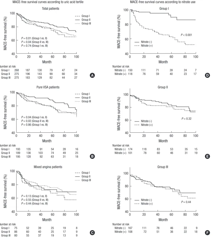
dL for group I, 5.4 mg/dL for group II, and 6.7 mg/dL for group III. Patients were followed for a median 49.2 months (22.7–83.9 months) and no significant difference was observed in the me- dian follow-up duration among the 3 groups (50.1 for group I vs. 49.3 for group II vs. 48.7 months for group III, P = 0.312). Group I had significantly more women and higher high-density lipo- protein (HDL) cholesterol levels than the other 2 groups. The prevalence of smoking, body mass index (BMI), and serum cre- atinine levels were significantly lower in Group I patients than the others (Table 1). No other characteristics showed significant differences among the groups. In the overall population, group II had a significantly lower incidence of MACE compared to group I and a tendency of lower incidence of MACEs compared to Group III (Table 2). Survival curves were generated for VSA patients with or without significant fixed lesions and according to uric acid level. For pure VSA, group II patients showed signif- icantly higher MACE-free survival compared to patients in the other groups (group I, 80.7%, P = 0.037; group II, 91.7%, refer- ence; group III, 80.1%, P = 0.023). In contrast, no significant dif- ferences in MACE-free survival were seen according to uric acid level in patients with mixed angina (P = 0.314) (Fig. 2A-C). MACE-
Table 2. Clinical outcomes of the total population according to uric acid level
Clinical outcomes Group I
(n = 268) Group II
(n = 275) Group III (n = 275)
Group I vs. II Group III vs. II
Adjusted HR (95% CI)* P value Adjusted HR (95% CI)† P value
MACE‡ 66 (24.6) 47 (17.1) 62 (22.5) 1.52 (1.02–2.26) 0.040 1.44 (0.98–2.13) 0.067
Cardiac death 5 (1.9) 3 (1.1) 6 (2.2) 1.48 (0.32–6.82) 0.612 2.11 (0.51–8.73) 0.301
All-cause death 13 (4.9) 7 (2.5) 10 (3.6) 1.64 (0.63–4.29) 0.313 1.23 (0.45–3.36) 0.692
Acute MI 3 (1.1) 5 (1.8) 3 (1.1) 0.87 (0.21–3.68) 0.851 0.56 (0.13–2.52) 0.451
Revascularization 18 (6.7) 8 (2.9) 8 (2.9) 2.57 (1.09–6.07) 0.031 0.98 (0.36–2.70) 0.972
Rehospitalization 49 (18.3) 41 (14.9) 47 (17.1) 1.29 (0.83–2.01) 0.259 1.33 (0.86–2.05) 0.197
Ischemic stroke 6 (2.2) 2 (0.7) 4 (1.5) 3.14 (0.60–16.52) 0.177 1.50 (0.24–9.29) 0.662
Values are expressed as No. (%). Patients were categorized according to tertiles of uric acid level: group, I ≤ 4.8 mg/dL; group II, 4.9–5.9 mg/dL; and group, III ≥ 6.0 mg/dL.
Group II was used as a reference group for analysis.
HR = hazard ratio, CI = confidence interval, MACE = major adverse cardiac events, MI = myocardial infarction, BMI = body mass index.
*Adjusted covariates were sex, smoking, BMI, serum creatinine level, and statin use. †Adjusted covariates were sex and serum creatinine level. ‡MACE was a composite of car- diac death, acute MI, ischemic stroke, revascularization, and rehospitalization for recurrent angina during follow-up.
Table 3. Prognostic factors for MACEs
Factors Univariate analysis Multivariate analysis
HR 95% CI P value HR* 95% CI P value
Age 1.01 1.00–1.03 0.135 1.01 0.99–1.03 0.217
Diabetes mellitus 1.39 1.00–1.92 0.050 1.30 0.93–1.81 0.132
Family history of CV disease 1.72 1.07–2.78 0.026 1.67 1.01–2.75 0.046
Mixed angina 1.64 1.21–2.21 0.001 1.70 1.24–2.34 0.001
Use of nitrate 1.49 1.11–2.01 0.009 1.66 1.21–2.27 0.002
HDL, mg/dL 0.99 0.97–1.00 0.039 0.99 0.97–1.00 0.035
Uric acid group (group II; reference) - - 0.040 - - -
Group I 1.59 1.09–2.31 0.016 1.61 1.10–2.37 0.018
Group III 1.48 1.01–2.17 0.042 1.46 0.99–2.16 0.062
MACE = major adverse cardiac event, HR = hazard ratio, CI = confidence interval, CV = cardiovascular, HDL = high-density lipoprotein.
*Adjusted covariates were age, diabetes mellitus, family history of CV disease, uric acid group, HDL, mixed angina, and use of nitrate.
free survival was also compared according to use of nitrates by uric acid groups (Fig. 2D-F). In group I, patients who received nitrate therapy had a higher incidence of MACE than patients without nitrate therapy (P < 0.001) (Supplementary Table 1).
Cox proportional hazard model analysis identified 6 prog-
nostic factors in univariate analysis. Seven variables were in- cluded in multivariate analysis. Family history of CV disease, lower uric acid tertile, low HDL cholesterol level, mixed angina, and prescription of nitrates at discharge were significant prog- nostic predictors of MACE incidence (Table 3).
Fig. 2. MACE-free survival curves of total (A), pure VSA (B), and mixed angina (C) patients according to uric acid tertile and MACE-free survival curves of group I (D), group II (E), and group III (F) patients according to the use of nitrates.
MACE = major adverse cardiac event, VSA = vasospastic angina.
MACE-free survival curves according to uric acid tertile Total patients
MACE-free survival (%)
Month
0 20 40 60 80 100 100
80
60
40
Number at risk
Group I 268 187 130 79 47 24
Group II 275 196 143 99 66 34
Group III 275 183 129 82 44 27
P = 0.01 (Group I vs. II) P = 0.04 (Group II vs. III) P = 0.74 (Group I vs. III)
Group I Group II Group III
MACE-free survival curves according to nitrate use Group I
MACE-free survival (%)
Month
0 20 40 60 80 100
100
80
60
40
Number at risk
Nitrate (-) 150 111 71 39 24 7
Nitrate (+) 118 76 59 40 23 17
P < 0.001 Nitrate (-)
Nitrate (+)
A D
Pure VSA patients
MACE-free survival (%)
Month
0 20 40 60 80 100 100
80
60
40
Number at risk
Group I 193 135 91 54 28 16
Group II 189 136 103 74 49 25
Group III 195 128 92 63 31 18
P = 0.04 (Group I vs. II) P = 0.02 (Group II vs. III) P = 0.86 (Group I vs. III)
Group I Group II Group III
Group II
MACE-free survival (%)
Month
0 20 40 60 80 100
100
80
60
40
Number at risk
Nitrate (-) 174 118 83 53 35 15
Nitrate (+) 101 78 60 46 31 19
P = 0.32 Nitrate (-)
Nitrate (+)
B E
Mixed angina patients
MACE-free survival (%)
Month
0 20 40 60 80 100 100
80
60
40
Number at risk
Group I 75 52 39 25 19 8
Group II 86 60 40 25 17 9
Group III 80 55 37 19 13 9
P = 0.13 (Group I vs. II) P = 0.55 (Group II vs. III) P = 0.44 (Group I vs. III)
Group I Group II Group III
Group III
MACE-free survival (%)
Month
0 20 40 60 80 100
100
80
60
40
Number at risk
Nitrate (-) 167 111 78 46 22 9
Nitrate (+) 108 72 51 36 22 18
P = 0.44 Nitrate (-)
Nitrate (+)
C F
DISCUSSION
In the present study, we investigated the association of serum uric acid levels and clinical outcomes in VSA patients with or without significant coronary artery stenosis confirmed by spasm- provocation test. The major findings of our study were: 1) pa- tients in group II (middle tertile) showed a higher MACE-free survival than the other 2 groups, 2) this pattern was observed for only pure VSA, not mixed angina, and 3) patients who re- ceived nitrate therapy at discharge had worse clinical outcomes than those without nitrate therapy in group I (lower tertile), but not in group II or III.
It has been reported that hyperuricemia is associated with CV disease as a risk factor or disease consequence. Because the association can be readily confounded by other CV risk factors, several studies have published conflicting results (1-3,13,14) and a number of researchers tried to elucidate the underlying mechanisms of the relationship between hyperuricemia and CV disease. The mechanisms include a compensatory uric acid increase as an antioxidant, stimulated inflammation and vaso- constriction by hyperuricemia, and activation of the renin-an- giotensin system in response to elevated uric acid (2,5,15). On the other hand, a J-shaped relationship between serum uric acid and CV events, which could be explained by hypourice- mia-induced endothelial dysfunction, were reported (4,7); and previous studies reported that uric acid administration restored endothelial function or increased serum antioxidant capacity (16,17).
Unlike the relationship between obstructive coronary artery disease and serum uric acid level, the impact of uric acid on clinical outcomes in VSA has not been evaluated to date (1-3).
We investigated if low or high serum uric acid levels would ad- versely affect clinical outcomes in VSA because endothelial dys- function is known to be the main pathophysiology of the disease.
To the best of our knowledge, this is the first study to analyze the association between uric acid level and clinical outcomes in VSA. Our results showed that patients in group II (middle uric acid tertile) had higher MACE-free survival than the other 2 groups. This pattern was consistent only in patients with pure VSA, not with mixed angina. Differing results for VSA according to the presence of a significant fixed lesion might be explained by the relative contribution of endothelial dysfunction as the main pathogenesis of each type of VSA. It is comprehensible that group III (upper uric acid tertile) showed worse outcomes, taking into account the known relationship between hyperuri- cemia and CV events. Worse clinical outcomes in group I (lower uric acid tertile) patients may be predictable as well consider- ing the link between hypouricemia and endothelial dysfunction (7,16,17). We also performed survival analysis according to use of nitrate for each uric acid group. Interestingly, in the lower uric acid group, patients treated with nitrate had worse clinical out-
comes than patients treated without nitrate; no difference was seen for the middle and upper uric acid groups. A possible ex- planation for this result could come from a study that investi- gated low uric acid level in women with type 1 diabetes in con- ditions of oxidative stress represented by increases in oxidative stress metabolites. The study found that increased oxidative stress was linked to reduced plasma uric acid level that is probably caused by nitric oxide (NO) overproduction (18). Accordingly, we assumed that additional nitrate therapy might harm VSA patients with low uric acid levels by impeding production of the antioxidant uric acid further in a situation in which NO level is already increased. This process could enhance oxidative stress, resulting in poorer clinical outcomes, particularly rehospitaliza- tion due to recurrent angina, in VSA patients with low uric acid level.
This study had several limitations. First, this was a retrospec- tive, observational study with a single-center cohort. There could be missing confounders that we could not assess from retrospec- tive data collection. And the choice of antianginal medication was left to each physician’s discretion. Second, the proportion of women was significantly higher in group I than the other groups.
This might be the consequence of sex differences in baseline uric acid levels, which are usually lower in women (19-21). The lower prevalence of smokers, BMI, and serum creatinine levels in group I might be explained in the same manner. Though we performed multivariate analysis to overcome this limitation, a prospective study is needed to investigate the causal relation- ship between uric acid level and clinical outcomes in VSA. Third, previous studies investigated prognostic role of uric acid in CV disease by only using uric acid level at admission (22,23). Simi- larly, we could not assess the association of follow-up uric acid level with clinical outcomes because routine follow-up of blood test was not mandatory. Finally, our results cannot be simply explained by a single specific mechanism. The pathophysiology of VSA is not fully understood and hypouricemia-induced en- dothelial dysfunction has not been proven in the coronary ar- tery. Despite these limitations, we believe our results have clini- cal implications as the first study suggesting an association be- tween serum uric acid level and clinical outcomes in VSA.
Low or high uric acid level seems to be related with adverse clinical outcomes in pure VSA patients. In particular, patients with low uric acid level had higher incidences of adverse clini- cal outcomes driven by revascularization compared with those with other uric acid levels. Furthermore, use of nitrate was as- sociated with increased rehospitalization rate due to recurrent angina in patients with low uric acid level. Therefore, it requires careful use of nitrate in VSA patients with low uric acid level.
DISCLOSURE
The authors have no potential conflicts of interest to disclose.
AUTHOR CONTRIBUTION
Conceptualization: Gwon HC, Choi SH. Data curation: Gwag HB, Park TK, Song YB, Hahn JY, Choi JH, Lee SH, Gwon HC, Choi SH. Formal analysis: Gwag HB, Yang JH. Investigation: Gwag HB, Yang JH, Park TK, Song YB, Hahn JY, Choi JH, Lee SH. Method- ology: Gwag HB, Yang JH. Project administration: Choi SH. Re- sources: Gwag HB, Yang JH, Park TK, Song YB, Hahn JY, Choi JH, Lee SH, Gwon HC, Choi SH. Software: Gwag HB, Yang JH.
Validation: Yang JH, Choi SH, Gwon HC. Supervision: Gwon HC, Choi SH. Visualization: Gwag HB, Yang JH. Writing - origi- nal draft: Gwag HB, Yang JH. Writing - review & editing: Lee SH, Gwon HC, Choi SH.
ORCID
Hye Bin Gwag https://orcid.org/0000-0001-5610-2872 Jeong Hoon Yang https://orcid.org/0000-0001-8138-1367 Taek Kyu Park https://orcid.org/0000-0003-1440-3583 Young Bin Song https://orcid.org/0000-0002-2581-8891 Joo-Yong Hahn https://orcid.org/0000-0002-4412-377X Jin-Ho Choi https://orcid.org/0000-0002-5421-793X Sang Hoon Lee https://orcid.org/0000-0003-1202-917X Hyeon-Cheol Gwon https://orcid.org/0000-0002-8967-4305 Seung-Hyuk Choi https://orcid.org/0000-0002-0304-6317
REFERENCES
1. Palmer TM, Nordestgaard BG, Benn M, Tybjærg-Hansen A, Davey Smith G, Lawlor DA, Timpson NJ. Association of plasma uric acid with ischaemic heart disease and blood pressure: mendelian randomisation analysis of two large cohorts. BMJ 2013; 347: f4262.
2. Kanbay M, Segal M, Afsar B, Kang DH, Rodriguez-Iturbe B, Johnson RJ.
The role of uric acid in the pathogenesis of human cardiovascular disease.
Heart 2013; 99: 759-66.
3. Brodov Y, Behar S, Boyko V, Chouraqui P. Effect of the metabolic syndrome and hyperuricemia on outcome in patients with coronary artery disease (from the Bezafibrate Infarction Prevention Study). Am J Cardiol 2010;
106: 1717-20.
4. Iso T, Kurabayashi M. Extremely low levels of serum uric acid are associ- ated with endothelial dysfunction in humans. Circ J 2015; 79: 978-80.
5. Nieto FJ, Iribarren C, Gross MD, Comstock GW, Cutler RG. Uric acid and serum antioxidant capacity: a reaction to atherosclerosis? Atherosclerosis 2000; 148: 131-9.
6. Morelli M, Carta AR, Kachroo A, Schwarzschild MA. Pathophysiological roles for purines: adenosine, caffeine and urate. Prog Brain Res 2010; 183:
183-208.
7. Sugihara S, Hisatome I, Kuwabara M, Niwa K, Maharani N, Kato M, Ogino K, Hamada T, Ninomiya H, Higashi Y, et al. Depletion of uric acid due to SLC22A12 (URAT1) loss-of-function mutation causes endothelial dys- function in hypouricemia. Circ J 2015; 79: 1125-32.
8. Kaneda H, Taguchi J, Kuwada Y, Hangaishi M, Aizawa T, Yamakado M, Ogasawara K, Aizawa T, Ohno M. Coronary artery spasm and the poly- morphisms of the endothelial nitric oxide synthase gene. Circ J 2006; 70:
409-13.
9. Yasue H, Nakagawa H, Itoh T, Harada E, Mizuno Y. Coronary artery spasm- -clinical features, diagnosis, pathogenesis, and treatment. J Cardiol 2008;
51: 2-17.
10. Stern S, Bayes de Luna A. Coronary artery spasm: a 2009 update. Circula- tion 2009; 119: 2531-4.
11. JCS Joint Working Group. Guidelines for diagnosis and treatment of pa- tients with vasospastic angina (coronary spastic angina) (JCS 2008): di- gest version. Circ J 2010; 74: 1745-62.
12. Yoo SY, Shin DH, Jeong JI, Yoon J, Ha DC, Cho SW, Cheong SS. Long-term prognosis and clinical characteristics of patients with variant angina. Ko- rean Circ J 2008; 38: 651-8.
13. Culleton BF, Larson MG, Kannel WB, Levy D. Serum uric acid and risk for cardiovascular disease and death: the Framingham Heart Study. Ann In- tern Med 1999; 131: 7-13.
14. De Luca G, Secco GG, Santagostino M, Venegoni L, Iorio S, Cassetti E, Ver- doia M, Coppo L, Di Mario C, Bellomo G, et al. Uric acid does not affect the prevalence and extent of coronary artery disease. Results from a pro- spective study. Nutr Metab Cardiovasc Dis 2012; 22: 426-33.
15. Yu MA, Sánchez-Lozada LG, Johnson RJ, Kang DH. Oxidative stress with an activation of the renin-angiotensin system in human vascular endo- thelial cells as a novel mechanism of uric acid-induced endothelial dys- function. J Hypertens 2010; 28: 1234-42.
16. Waring WS, Webb DJ, Maxwell SR. Systemic uric acid administration in- creases serum antioxidant capacity in healthy volunteers. J Cardiovasc Pharmacol 2001; 38: 365-71.
17. Waring WS, McKnight JA, Webb DJ, Maxwell SR. Uric acid restores endo- thelial function in patients with type 1 diabetes and regular smokers. Di- abetes 2006; 55: 3127-32.
18. Pitocco D, Di Stasio E, Romitelli F, Zaccardi F, Tavazzi B, Manto A, Caputo S, Musella T, Zuppi C, Santini SA, et al. Hypouricemia linked to an over- production of nitric oxide is an early marker of oxidative stress in female subjects with type 1 diabetes. Diabetes Metab Res Rev 2008; 24: 318-23.
19. Antón FM, García Puig J, Ramos T, González P, Ordás J. Sex differences in uric acid metabolism in adults: evidence for a lack of influence of estradi- ol-17 beta (E2) on the renal handling of urate. Metabolism 1986; 35: 343-8.
20. Döring A, Gieger C, Mehta D, Gohlke H, Prokisch H, Coassin S, Fischer G, Henke K, Klopp N, Kronenberg F, et al. SLC2A9 influences uric acid con- centrations with pronounced sex-specific effects. Nat Genet 2008; 40: 430-6.
21. Lim JH, Kim YK, Kim YS, Na SH, Rhee MY, Lee MM. Relationship between serum uric acid levels, metabolic syndrome, and arterial stiffness in Ko- rean. Korean Circ J 2010; 40: 314-20.
22. Akpek M, Kaya MG, Uyarel H, Yarlioglues M, Kalay N, Gunebakmaz O, Dogdu O, Ardic I, Elcik D, Sahin O, et al. The association of serum uric acid levels on coronary flow in patients with STEMI undergoing primary PCI.
Atherosclerosis 2011; 219: 334-41.
23. Lazzeri C, Valente S, Chiostri M, Sori A, Bernardo P, Gensini GF. Uric acid in the acute phase of ST elevation myocardial infarction submitted to pri- mary PCI: its prognostic role and relation with inflammatory markers: a single center experience. Int J Cardiol 2010; 138: 206-9.