서론
신경피부흑색증은 세 개 이상 혹은 크기가 큰(성인의 경우 20 cm 이상, 소인의 경우 6~9 cm 이상) 선천성 멜라닌세포모반과 중추신경계에 멜라닌세포의 증식이 있을 때 진단할 수 있는데, 중추신경계 병소는 다른 곳에서 전이된 것이 아니어야 한다.
증상은 주로 2세 이전에 나타나며 두개내압(intracranial pressure) 상승과 관련하여 수두증(hydrocephalus)과 발작(sei- zure)이 흔한데, 멜라닌세포의 증식과 축적이 뇌척수액 순환을 방해하기 때문이라고 알려져 있다. 드물게 청소년기나 초기 성인 기에 증상을 보이기도 하고 증상이 없는 경우도 있다(1-5).
신경피부흑색증은 Rokitanski가 처음 보고한 이후로 현재까 지 약 100예가 보고되었으나 질환의 영상소견에 관련된 보고 는 드물다(6). 이에 저자들은 영상학적 소견을 중심으로 증상 이 없었던 신경피부흑색증 사례를 보고하겠다.
증례 보고
20세 남자 환자로 태어날 때부터 경부, 몸통 및 사지에 과다 색소침착된 털모반(hyperpigmented hairy nevus)이 있었고 성 장하면서 피부병소(skin lesion)는 상체의 대부분을 덮을 정도
로 커지고 여러 개의 작은 위성모반(satellite nevus)이 사지에 생겼다. 환자에게 특별한 증상은 없었지만 점점 커지고 두꺼워 지는 피부병소의 정확한 진단과 치료에 대해 알아보고자 내원 하였다.
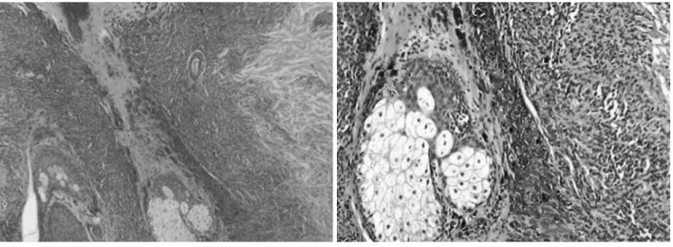
환자의 신체검사에서 과다색소침착된 털모반이 어깨, 등, 목 덜미에 있었고 사지, 가슴, 배에는 위성병변이 있었지만 피부 및 신경학적 증상은 보이지 않았다(Fig. 1). 피부병소의 양상과 진행과정을 고려할 때 신경피부흑색증으로 잠정적인 진단을 하였고 피부생검(skin biopsy)과 중추신경계 자기공명영상 (MRI) 검사를 시행하였다. 피부생검은 왼쪽 넓다리(thigh)와 등에서 시행하였고 조직병리 결과 두 곳 모두에서 멜라닌세포 모반으로 확인되었다(Fig. 2). 뇌 MRI 검사에서는 교뇌(pons) 와 우측 편도(amygdala)에 작은 병변이 있었고 교뇌 병변은 T1 강조영상, T2 강조영상, fluid attenuated inversion recovery (이 하 FLAIR) 영상에서 고신호강도를 보였다. 우측 편도 병변은 T1 강조영상과 FLAIR 영상에서 고신호강도를 보였지만 T2 강조영상에서는 병변확인이 어려웠다. 두 병변 모두 조영증강 T1 강조영상에서 조영증강되지 않았고 척추 MRI 검사에서 특 이소견은 보이지 않았다(Figs. 3, 4).
환자의 피부병소 양상, 피부생검 결과 및 뇌 MRI 소견을 고 려할 때 신경피부흑색증으로 진단할 수 있었다(7).
J Korean Soc Radiol 2011;65(6):537-541
Received June 1, 2011; Accepted September 14, 2011 Corresponding author: Hae Woong Jeong, MD Department of Radiology, Busan Paik Hospital, Inje University College of Medicine, 633-165 Gaegeum- dong, Busanjin-gu, Busan 614-735, Korea.
Tel. 82-51-890-6579 Fax. 82-51-896-1085 E-mail: [email protected]
Copyrights © 2011 The Korean Society of Radiology
Neurocutaneous melanosis is a rare disorder characterized by the presence of a large or multiple congenital melanocytic nevus with proliferation of melanocytes in the central nervous system. The prognosis of neurocutaneous melanosis is extreme- ly poor and its diagnostic approach requires understanding its brain magnetic reso- nance imaging findings. We report a patient with asymptomatic neurocutaneous melanosis and its radiologic findings.
Index terms
Neurocutaneous Syndrome Melanosis
Central Nervous System Magnetic Resonance Imaging
Neurocutaneous Melanosis: A Case Report 신경피부흑색증: 증례 보고
Yoon Nae Seo, MD, Hae Woong Jeong, MD, Hyun Sin In, MD
Department of Radiology, Busan Paik Hospital, Inje University College of Medicine, Busan, Korea
상이 있는 신경피부흑색증이 발생하고 악성 전환에 관계없이 증상이 있는 환자의 90% 이상이 2~3년 내에 사망한다고 한다 (4, 8). 또한 DeDavid 등(9)에 의하면 선천성 멜라닌세포모반 이 두부, 목덜미 척추주변부에 있는 사람이나 20개 이상의 위 성모반을 가진 사람에서 신경피부흑색증이 발생할 위험성이 더 높다고 하였다.
중추신경계 병변을 확인하는 데에는 전산화단층촬영(CT)보 다 MRI가 훨씬 유용한데 대부분의 경우에서 신경피부흑색증
고찰
신경피부흑색증은 선천적, 비유전적 질환이며 악성 전환 가능 성이 있는 양성 질환이다. 정확한 발병경로는 밝혀져 있지 않지 만 neuroectodermal melanocyte precursor cell의 이형성(dys- plasia)이 멜라닌세포의 과다증식(excessive proliferation)과 축 적을 야기한다고 알려져 있다(1, 4).
거대 선천성 멜라닌세포모반을 가진 환자의 6~11%에서 증
Fig. 1. The patient has giant hairy dark nevus covering most of back, shoulder, and posterior neck as well as multiple satellite nevus scattered over the whole body.
Fig. 2. Histopathologic findings show that nevus cells are present near and around appendages and the deep dermis (A. H&E, × 40, B. H&E, × 100).
A B
뇌 실질(parenchyma)이 조영증강되어 보이는 경우, 악성 전환 과 관련하여 종괴 형태로 보이는 경우이다(1, 6).
T1 강조영상에서 고신호강도로 보이는 경우에는 뇌 실질이 나 연수막에 3 cm 미만의 작은 병변이 있지만 조영증강되지 않는다. T1 강조영상에서 고신호강도를 보이는 것은 멜라닌에 이 흑색종으로 전환하지 않으면 CT에서 병변을 확인하기 어렵
고 일부에서 수두증이나 연수막(leptomeninges)의 조영증강을 볼 수 있다(1).
중추신경계 MRI의 소견은 크게 세 가지 형태로 나눌 수 있 는데 T1 강조영상에서 고신호강도를 보이는 경우, 연수막이나
Fig. 3. Neurocutaneous melanosis in a 20-year-old man. (A) T1-weighted MR image and (B) T2-weighted MR image show ill-defined high sig- nal intensity lesion at the ventral part of the left side pons (arrow). (C) Gadolinium-enhanced T1-weighted MR image shows no enhancement in the ill-defined high signal intensity lesion (arrow). (D) FLAIR image shows ill-defined high signal intensity lesion at the ventral part of the left side pons (arrow).
Note.-FLAIR = fluid attenuated inversion recovery
A B C D
D A
E
B C
Fig. 4. Upper level of Fig. 3 in the same patient (A) T1-weighted MR image and (B) FLAIR image show high signal intensity focus at the right amygdala (arrow). (C, D) Gadolinium-enhanced T1-weighted MR image shows no enhancement in the high signal intensity focus (arrow). (E) T2-weighted MR image shows indeterminate signal intensity focus at the right amygdala.
Note.-FLAIR = fluid attenuated inversion recovery
rocutaneous melanosis presenting as chronic partial epi- lepsy. J Clin Neurol 2008;4:134-137
4. Agero AL, Benvenuto-Andrade C, Dusza SW, Halpern AC, Marghoob AA. Asymptomatic neurocutaneous melanocy- tosis in patients with large congenital melanocytic nevi: a study of cases from an Internet-based registry. J Am Acad Dermatol 2005;53:959-965
5. Hsueh CW, Ho CS, Chiu NC, Shen EY. Neurocutaneous melanosis with hydrocephalus: report of one case. Acta Neurol Taiwan 2004;13:29-33
6. Hayashi M, Maeda M, Maji T, Matsubara T, Tsukahara H, Takeda K. Diffuse leptomeningeal hyperintensity on fluid- attenuated inversion recovery MR images in neurocutane- ous melanosis. AJNR Am J Neuroradiol 2004;25:138-141 7. Warakaulle DR, Anslow P. Differential diagnosis of intra-
cranial lesions with high signal on T1 or low signal on T2- weighted MRI. Clin Radiol 2003;58:922-933
8. Bittencourt FV, Marghoob AA, Kopf AW, Koenig KL, Bart RS. Large congenital melanocytic nevi and the risk for de- velopment of malignant melanoma and neurocutaneous melanocytosis. Pediatrics 2000;106:736-741
9. DeDavid M, Orlow SJ, Provost N, Marghoob AA, Rao BK, Wasti Q, et al. Neurocutaneous melanosis: clinical features of large congenital melanocytic nevi in patients with manifest central nervous system melanosis. J Am Acad Dermatol 1996;35:529-538
10. Chu WC, Lee V, Chan YL, Shing MM, Chik KW, Li CK, et al.
Neurocutaneous melanomatosis with a rapidly deteriorat- ing course. AJNR Am J Neuroradiol 2003;24:287-290 하게 반응하여 저신호강도부터 고신호강도까지 다양한 신호강
도를 보인다고 한다. 뇌척수액 내의 높은 단백농도가 FLAIR 영상에서 고신호강도로 보인다는 설명이 있는 한편 FLAIR 영 상에 대한 melanin의 T1 단축효과 때문이라는 보고도 있다.
T2 강조영상에서 고신호강도를 보이는 것은 신경피부흑색증 의 악성전환에 따른 종괴효과, 혈관성 부종(vasogenic ede- ma), 괴사의 결과라고 설명하는 문헌도 있다. 본 증례의 경우 교뇌 병변은 T2 강조영상 FLAIR 영상에서 모두 고신호강도를 보였고 우측 편도의 병변은 FLAIR 영상에서 고신호강도를 보 였다.
연수막이나 뇌 실질의 조영증강되는 경우에는 기저수조(basal cistern), 천막(tentorium), 뇌줄기(brain stem), 하부 소뇌충부 (cerebellar vermis), 소뇌회(cerebellar folia) 등의 뇌 기저부에서 호발하고 뇌 실질보다 연수막이 조영증강되는 경우가 더 많다.
연수막이 결절형(focal nodular)으로 또는 두꺼운 플라크 같이 (thick plaque like) 조영증강되는 경우에는 악성 전환을 의미 한다는 보고가 있다(1, 5).
신경피부흑색증은 나쁜 예후를 보이는 질환이며 적절한 시 기에 진단하는 것이 무엇보다 중요하다. 그러므로 거대 선천성 멜라닌세포모반을 가진 사람에게 신경피부흑색증 발생 여부와 예후를 예측하기 위해서 MRI는 필수적인 검사이며 진단 후 추 적 관찰에 있어서도 중요한 역할을 한다(1, 3, 4).
참고문헌
1. Smith AB, Rushing EJ, Smirniotopoulos JG. Pigmented le- sions of the central nervous system: radiologic-pathologic correlation. Radiographics 2009;29:1503-1524
2. Vanzieleghem BD, Lemmerling MM, Van Coster RN. Neuro- cutaneous melanosis presenting with intracranial amela-
신경피부흑색증: 증례 보고
서윤내 · 정해웅 · 인현신
신경피부흑색증(neurocutaneous melanosis)은 여러 개 혹은 크기가 큰 선천성 멜라닌세포모반(congenital melanocytic nevus)과 중추신경계에 멜라닌세포의 증식을 특징으로 하는 신경피부증후군(neurocutaneous syndrome)이다. 신경피부 흑색증은 드물게 발생하지만 예후가 나쁘기 때문에 진단적 접근을 위한 자기공명영상(MRI) 소견에 대한 이해가 필요하 다. 이에 본 저자들은 영상학적 소견을 중심으로 신경피부흑색증 1예를 보고하고자 한다.
인제대학교 의과대학 부산백병원 영상의학과학교실