Ischemic Stroke in Rats Enhances Bone Resorption in Vitro
We hypothesized that the formation and differentialtion of osteoclasts are accelerated and the potential of bone resorption is increased in the hemiplegic bone marrow in the early stage of stroke. We randomly divided white female Sprague-Dawley (SD) rats (n = 30) into two groups, stroke (n = 15) and sham group (n = 15). On the 7th day after stroke, after cutting away the epiphyses of the femurs and tibias, diaphyseal channels were flushed using α-minimum essential medium (α-MEM) and bone marrow cells were collected. Bone marrow stem cells, which were extracted from the femur and tibia, were cultured on the 7th day after middle cerebral artery occlusion. We then estimated the ratio of non- adherent cells to total bone marrow cells that included osteoclast precursor cells. After culturing these cells separately, cells that tested positive on the tartrate resistant acid phosphatase (TRAP) were counted and bone resorption was evaluated by using the OAASTM plate. In comparison to the control group, the stroke group showed a higher increase of non-adherent cells in the hemiplegic side bone marrow. In addition, after the primary culture, the stroke group showed an increased number of TRAP positive cells and a higher degree of bone resorption estimated by OAASTM plate. As a result, osteoclastogenesis and osteoclast differentiation are accelerated and the potential of bone resorption is increased in the hemiplegic bone marrow and these changes are detected as early as within the first week after middle cerebral artery occlusion in SD rats.
Key Words: Bone Resorption; Osteoclasts; Osteoporosis; Stroke Myung Eun Chung1, Jong In Lee2,
Sun Im3, and Joo Hyun Park2
1Department of Rehabilitation Medicine, St. Paul’s Hospital, College of Medicine, The Catholic University of Korea, Seoul; 2Deparment of Rehabilitation Medicine, Seoul St. Mary’s Hospital, College of Medicine, The Catholic University of Korea, Seoul;
3Deparment of Rehabilitation Medicine, Bucheon St. Mary’s Hospital, College of Medicine, The Catholic University of Korea, Bucheon, Korea
Received: 28 March 2011 Accepted: 4 November 2011 Address for Correspondence:
Joo Hyun Park, MD
Department of Rehabilitation Medicine, Seoul St. Mary’s Hospital, College of Medicine, The Catholic University of Korea, 222 Banpodae-ro, Seocho-gu, Seoul 137-701, Korea Tel: +82.11-9936-3907, Fax: +82.2-2258-2825 E-mail: [email protected]
http://dx.doi.org/10.3346/jkms.2012.27.1.84 • J Korean Med Sci 2012; 27: 84-88 Rehabilitation & Sports Medicine
INTRODUCTION
Osteoporosis is the most common disease among metabolic bone diseases and can be defined clinically as existence of a frac- ture, histomorphologically as reduction in bone matrix per unit volume, and epidemiologically as increased risk of fracture (1).
Osteoporotic fracture increases with age and the frequency of it increases according to aging of society. In the United States, about 1.5 million osteoporotic fracture patients occur every year, resulting in an extremely high social medical expense of about 18 billion dollars (2).
According to recent reports, the bone mineral density of the hemiplegic limb is significantly lower than that of non-hemi- plegic side, and the incidence of hip fracture increases by about 2-4 times after stroke, among which more than 79% occur on the hemiplegic side (3-5). Sato et al. (6, 7) suggested two causes of osteoporosis by stroke. First, bone resorption increases by im- mobilization from paralysis. Second, lack of nutrition and re- duced exposure to sunlight bring deficiency of vitamin D, which induces osteoporosis. In this study, Sato et al. (6) demonstrated increase in bone resorption by increased serum carboxy-termi- nal telopeptide of type I collagen (ICTP; a marker for bone resorp- tion) in patients one week after stroke.
Many studies using serum bone markers have reported on
the induction of osteoporosis on the hemiplegic side since the initial stages of stroke, but there are no studies yet on the effect of stroke on osteoclast differentiation in bone marrow cells. We hypothesized that the formation and differentiation of osteo- clasts are accelerated and the potential of bone resorption is in- creased in the hemiplegic bone marrow in the early stage of stroke. To confirm this, we conducted a study with SD rats by extracting and culturing bone marrow cells in the femur and tibia after one week of stroke.
MATERIALS AND METHODS Animals and treatments
A group of 30 (12 weeks, 220-260 mg) white female Sprague- Dawley rats were bred at a clean animal breeding room of the College of Medicine at the Catholic University of Korea. All ex- perimental procedures were approved by the Animal Experi- mental Committee of the Catholic University of Korea. The feed (Samyang Oil & Feed Co., Ltd., Seoul, Korea) containing 1.15%
calcium and 0.75% phosphorous and water were supplied with- out limits.
Subjects were randomly divided into the control and experi- mental groups, and each group consisted of 15 rats. The experi- mental group received ischemic stroke surgery and the control
group received a sham operation. Both groups were maintained at the same sleep-wake cycle with 12 hr light-dark cycle (light- out 18:00-06:00).
The experimental protocol was approved by the institutional animal care and use committee at the College of Medicine, the Catholic University of Korea.
Preparation of ischemic stroke rats
Ketamine (30 mg/kg) (Yuhan Co., Seoul, Korea) and xylazine hydrochloride (4 mg/kg) (Bayer Korea, Seoul, Korea) were used to anesthetize rats. While maintaining body temperature of 37°C
± 1°C during the surgery, cerebral ischemia was induced using the method of Longa et al. (8). For the method of surgery, the central neck of rats was cut in supine position and the left com- mon carotid artery was dissected. Following the common carotid artery, external carotid artery and internal carotid artery were separated for ligation of the common carotid artery and external carotid artery. The tip of monofilament nylon (4.0) was pushed into the common carotid artery bifurcation to block the middle cerebral artery. The same procedure was performed on the con- trol group until the dissection of the carotid artery, but the rat was sutured without ligation of the artery. After ligation of the middle cerebral artery, permanent local ischemic stroke was confirmed using the following two methods. Three days after operation, neurologic examination was performed on the isch- emic stroke and control rats, following the protocols used by Garcia et al. (9). Seven days after ischemic stroke, the femur and tibia were extracted, and the brain was taken out and placed for 30 min in 2,3,5-Triphenyl-2H-tetrazolium chloride dye (Sigma Diagnostic, St. Louis, MO, USA). After 20 min of culturing at 37°C, it was fixed in 2% paraformaldehyde solution (Sigma Diagnos- tic) to verify ischemic stroke (10).
Culturing of bone marrow
Seven days after ischemic stroke, when all rats in the experimen- tal and control groups were sacrificed, right femurs and tibias of the control group and right femurs and tibias of the experi- mental group were extracted. The epiphyses of the femurs and tibias were cut away, and bone marrow was collected by flusing the diaphyseal channel with α-minimum essential medium (α- MEM). After centrifugation and counting the number of bone marrow cells, they were resuspended on α-MEM containing 15% fetal calf serum. Cells were incubated for three hours un- der 37°C vapor saturated atmosphere with 5% carbon dioxide in a tissue culture flasks (106 cells/cm2). Then, the adherent cells and the non-adherent cells were separated.
Measurement of Tartrate-Resistant Acid Phosphatase (TRAP) positive cells
The ratio of the number of the non-adherent cells to bone mar- row cells was measured and the non-adherent cells were dilut-
ed to a concentration of 1× 10⁶ cells/mL before moving to a cul- ture dish. The cells were cultured on a 24-well plate (1 mL/well) in order to examine the number of TRAP positive cells. After 8 days of culturing, cells were placed in citrate/aceton solution for 30 sec and dyed for one hour in a dark room of 37°C using Sigma Acid Phosphatase Leukocyte Kit (Sigma Diagnostic) to find TRAP positive cells.
Measurement of bone resorption
Among the non-adherent cells, 3× 10⁵ cells were cultured on an OAASTM plate (Osteogenic Core Technology, Cheonan, Korea) (1 mL/well) for evaluation of bone resorption. OAASTM plate is a product covered on the surface by calcium phosphate as a thin layer to allow measurement of bone resorption while culturing the osteoclast. 3× 10⁵ cells per well were cultured for 13 days.
After cell culture, OAASTM plate was washed using distilled wa- ter, dried and observed the percentage of resorbed area. The image was stored using a digital camera under optical micro- scope and the area ratio of bone resorption to total area was eval- uated using an image analysis program called Scion Image.
Statistical analysis
Ratio of the non-adherent cells, the number of TRAP positive cells and the percentage of resorbed area on OAASTM plate were analyzed. Comparison between the control group that received sham operation and the hemiplegic side of the stroke group was analyzed using Mann-Whitney U test. Comparison between the hemiplegic side and the non-hemiplegic side of the experimen- tal group was analyzed using Wilcoxon sign rank test.
RESULTS
Throughout the experimental period, four rats died both in the experimental and control groups. The number of subjects was reduced to 22 rats for analysis of results, each group containing 11 rats.
Body weight
Body weights before sham operation or ischemic stroke surgery and seven days after operations were 230.7 ± 9.5 g and 237.6 ± 4.3 g in the control group and 231.9 ± 9.5 g and 235.4 ± 10.9 g in the experimental group, which showed no significant difference (P = 0.551).
Verification of ischemic stroke
Three days after ischemic stroke surgery, neurologic examina- tion as proposed by Garcia et al. (9) was conducted. Scores of the control group and experimental group were 13.68 ± 0.5 and 17.8 ± 0.4 points. A significant difference (P < 0.001) was shown between the two groups.
Measurement of osteoclastic precursor cell ratio
The ratio of the non-adherent cells containing osteoclastic pre- cursor cells was 47.8% ± 12.5% for the control group, 64.2% ± 13.8% on the hemiplegic side of the experimental group and 48.2% ± 12.8% on the non-hemiplegic side of the experimental group. In comparison to the control group, significantly higher ratio was observed on the hemiplegic side of the experimental group (P = 0.013). There also was a significant difference be- tween the hemiplegic side and the non-hemiplegic side of the experimental group (P = 0.001) (Fig. 1).
Measurement of TRAP positive cells
The number of TRAP positive cells was 38.1 ± 8.2 for the control group, 54.9 ± 11.1 for the hemiplegic side of the experimental group, and 42.6 ± 11.3 for the non-hemiplegic side of the exper- imental group. Compared to the control group, the number was significantly higher (P = 0.001) on the hemiplegic side of the experimental group. In comparison of the hemiplegic and the non-hemiplegic side of the experimental group, significant dif- ference was observed (P = 0.29) (Fig. 2, 3).
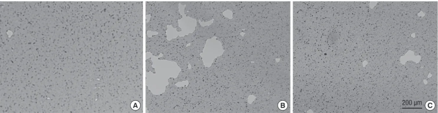
Measurement of bone resorption area
The percentage of resorbed area on an OAASTM plate was mea- sured to be 0.08% ± 0.17% in the control group, 14.8% ± 6.5%
on the hemiplegic side of the experimental group and 1.3% ± 1.4% on the non-hemiplegic side of the experimental group.
Compared to the control group, a significant difference was ob- served on the hemiplegic side of the experimental group (P <
0.001). Significant difference was also shown between hemiple- gic and non-hemiplegic side of the experimental group (P = 0.003) (Fig. 4, 5).
DISCUSSION
Monocytes and macrophage precursor cells in the bone mar- row differentiate to preosteoclast. Then, TRAP gene is gradually expressed, and several cells fuse together to form a polykaryo- cyte. The final form is an activated osteoclast (11).
In this study, the ratio of the non-adherent cells to total bone marrow cells was significantly increased on the hemiplegic side of the experimental group compared to the control group and
% of non-adherent cells
*
CTR S-HS S-NHS
80 70 60 50 40 30 20 10 0
Fig. 1. Percentage of non-adherent cells from bone marrow. CTR, controls; S-HS, hemiplegic side of the stroke group; S-NHS, non-hemiplegic side of the stroke group.
Data are the mean ± SEM. *P < 0.05 control.
TRAP (+) cells/fleld
*
CTR S-HS S-NHS
70 60 50 40 30 20 10 0
Fig. 2. Tartrate resistant acid phosphatase (TRAP) positive cells from bone marrow.
CTR, controls; S-HS, hemiplegic side of the stroke group; S-NHS, non-hemiplegic side of the stroke group. Data are the mean ± SEM. *P < 0.05 control.
A B 50 µm C
Fig. 3. Photomicrographs of osteoclasts in primary culture obtained from bone marrow of rats, TRAP stain, magnification ×100. (A) Control. (B) Hemiplegic side of the stroke rats. (C) Non-hemiplegic side of the stroke rats.
the non-hemiplegic side. Although the total number of bone marrow cells did not differ by large, the ratio of the non-adher- ent cells to total bone marrow cells increased. Since the non-ad- herent cells containing osteoclastic precursors also contained monocytes and macrophage precursor cells, the above results suggest that compositional changes occur with a propensity of increased ratio of osteoclastic precursor cells in bone marrow from the early stages of stroke. The number of TRAP positive cells after osteoclastic precursors culture showed a 40% increase on the hemiplegic side of the MCA occlusion group compared to the control group. We cultured the same number of the non- adherent cells, but a larger number of TRAP positive cells was measured for the hemiplegic side of the MCA occlusion group compared to the control group. This indicates that the formation and differentiation of osteoclasts in bone marrow are accelerat- ed from the early stage of stroke. Bone resorption area measured after culturing the osteoclastic precursors on OAASTM plate was increased by a factor of over 100 in the hemiplegic side of isch- emic stroke compared to the control group. The number of TRAP positive cells was about 40% higher in the experimental group than the control group, but a difference of over 100 times for bone
resorption area measured on OAASTM plate showed an increased potential of osteoclastic activity in the MCA occlusion group.
As such, the formation and differentiation of osteoclasts in the hemiplegic bone marrow are accelerated from the early stages of stroke, thereby increasing the potential of bone resorption and accelerating the development of osteoporosis on the hemi- plegic side.
According to recent studies, excessive bone loss is present in infectious disease. Activated T cells increase the expression of receptor activator of nuclear factor κB ligand (RANKL), and there- by promote osteoclastic activity (12, 13). Acute post-stroke peri- od is vulnerable to infection for humans as well as experimental animals. Therefore, the possible post-stroke infection may be another cause of promoting osteoclastic activity.
A recent study reported that 16.4%-38.5% of hip fracture pa- tients have a past history of stroke (3). In stroke patients with uni- lateral stroke and persisting paresis at the time of fracture, 62.5%
have their fracture on the paretic side. Hip fracture is more com- mon in patients with less severe stroke than in patients with more severe stroke because mobility is better in those with less severe stoke and they have more chance to fall down. This study sug- gests that reduced bone density on the lower limb on the hemi- plegic side as an important cause (3).
Grano et al. (14) reported on the increased bone resorption and formation of osteoclasts in the bone marrow within five days of removal of weight bearing on rat hindlimb. Sato et al. (6) re- ported on the increased biochemical marker which shows bone resorption one week after stroke. In accordance to the results from previous studies, we hypothesize that the increase in osteo- clastogenesis in the bone marrow cells and bone resorption that were detected in this study, were also caused by reduced weight bearing from immobilization of the hemiplegic side. Looking at this, active weight bearing on the hemiplegic side from the ini- tial stages of stroke should be an important therapeutic point in order to reduce the effect of immobilization.
This study has some limitations. Firstly, we demonstrated only the change of osteoclast differentiation and the potential of bone resorption in the early stage of stroke, because we speculated
% of resorbed area
*
CTR S-HS S-NHS
25
20
15
10
5
0
Fig. 4. The percentage of mineral surface resorbed by osteoclasts on OAASTM plate.
CTR, controls; S-HS, hemiplegic side of the stroke group; S-NHS, non-hemiplegic side of the stroke group. Data are the mean ± SEM. *P < 0.05 control.
A B 200 µm C
Fig. 5. Photomicrographs of pits formed by osteoclasts from rats, magnification ×25. (A) Control. (B) Hemiplegic side of the stroke group. (C) Non-hemiplegic side of the stroke group.
that the rate of bone loss in the early stage of stroke was proba- bly accelerated with a disproportionate increase in bone resorp- tion (15). However, osteoporosis is influenced not only by the changes of osteoclast differentiation and bone resorption, but also by the changes of osteoblast differentiation and bone for- mation. Therefore, additional studies about the changes of osteo- blast differentiation and bone formation might be needed. Sec- ondly, the percentage of resorbed area on OAASTM plate was measured to be 0.08% in the control group, but this result was so small that it may be doubted whether the 3× 10⁵ cells cul- tured on the plate were not enough to measure bone resortion area or an OAASTM plate was not adequate for measuring a tiny resorption area. Lastly, this study has another limitation from small sample size.
Since osteoclastogenesis and osteoclast differentiation accel- erate and bone resorption increases at the early stage of stroke, bone resorption inhibitors such as bisphosphonates might have the prevention effect against the osteoporosis on the hemiple- gic limb (16). Poole et al. (17) reported that zoledronate therapy is associated with a reduction in osteoclastsic cell numbers. An- other study showed that treatment with etidronate increases bone mineral density in chronically hospitalized patients with stroke, and may prevent hip fracture (18). Further studies are necessary to measure the effectiveness of prevention of the os- teoporotic fracture on the hemiplegic side by the use of bisphos- phonates in the early stage of stroke. Furthermore, additional studies are needed to estimate the amount of weight bearing time required in order to exert its preventive effect on osteopo- rosis in the early stage of stroke.
In summary, this study demonstrates that osteoclastogenesis and osteoclast differentiation are accelerated and the potential of bone resorption is increased in the hemiplegic bone marrow and these changes are detected as early as within the first week after MCA occlusion in SD rats. After stroke, changes that lead to osteoporosis occur from the early periods of stroke; since hemi- plegic patients have an increased propensity to have an unsta- ble gait or movement pattern, falling down on the hemiplegic side can easily result in hip fractures. The results of this study emphasize the importance of active prevention and manage- ment of osteoporosis from the early stages of stroke.
REFERENCES
1. Kanis JA, Glüer CC. An update on the diagnosis and assessment of osteo- porosis with densitometry. Committee of Scientific Advisors, International Osteoporosis Foundation. Osteoporos Int 2000; 11: 192-202.
2. Gabriel SE, Tosteson AN, Leibson CL, Crowson CS, Pond GR, Hammond CS, Melton LJ 3rd. Direct medical costs attributable to osteoporotic frac- tures. Osteoporos Int 2002; 13: 323-30.
3. Ramnemark A, Nilsson M, Borssén B, Gustafson Y. Stroke, a major and increasing risk factor for femoral neck fracture. Stroke 2000; 31: 1572-7.
4. Ramnemark A, Nyberg L, Lorentzon R, Englund U, Gustafson Y. Progres- sive hemiosteoporosis on the paretic side and increased bone mineral density in the nonparetic arm the first year after severe stroke. Osteopo- ros Int 1999; 9: 269-75.
5. Choi EK. The effects of decreased physical activity to bone mineral densi- ty in hemiparetic stroke patients. Korean J Med 2005; 69: 387-94.
6. Sato Y, Kuno H, Kaji M, Etoh K, Oizumi K. Influence of immobilization upon calcium metabolism in the week following hemiplegic stroke. J Neu- rol Sci 2000; 175: 135-9.
7. Sato Y, Maruoka H, Oizumi K, Kikuyama M. Vitamin D deficiency and osteopenia in the hemiplegic limbs of stroke patients. Stroke 1996; 27:
2183-7.
8. Longa EZ, Weinstein PR, Carlson S, Cummins R. Reversible middle cere- bral artery occlusion without craniectomy in rats. Stroke 1989; 20: 84-91.
9. Garcia JH, Wagner S, Liu KF, Hu XJ. Neurological deficit and extent of neuronal necrosis attributable to middle cerebral artery occlusion in rats.
Statistical validation. Stroke 1995; 26: 627-34.
10. Mehta SH, Dhandapani KM, De Sevilla LM, Webb RC, Mahesh VB, Brann DW. Tamoxifen, a selective estrogen receptor modulator, reduces ischemic damage caused by middle cerebral artery occlusion in the ovariectomized female rat. Neuroendocrinology 2003; 77: 44-50.
11. Boyle WJ, Simonet WS, Lacey DL. Osteoclast differentiation and activa- tion. Nature 2003; 423: 337-42.
12. Rauner M, Sipos W, Pietschmann P. Osteoimmunology. Int Arch Allergy Immunol 2007; 143: 31-48.
13. Jones D, Glimcher LH, Aliprantis AO. Osteoimmunology at the nexus of arthritis, osteoporosis, cancer, and infection. J Clin Invest 2011; 121:
2534-42.
14. Grano M, Mori G, Minielli V, Barou O, Colucci S, Giannelli G, Alexander C, Zallone AZ, Vico L. Rat hindlimb unloading by tail suspension reduces osteoblast differentiation, induces IL-6 secretion, and increases bone re- sorption in ex vivo cultures. Calcif Tissue Int 2002; 70: 176-85.
15. Sato Y, Fujimatsu Y, Kikuyama M, Kaji M, Oizumic K. Influence of im- mobilization on bone mass and bone metabolism in hemiplegic elderly patients with a long-standing stroke. J Neurol Sci 1998; 156: 205-10.
16. Carano A, Teitelbaum SL, Konsek JD, Schlesinger PH, Blair HC. Bisphos- phonates directly inhibit the bone resorption activity of isolated avian osteoclasts in vitro. J Clin Invest 1990; 85: 456-61.
17. Poole KE, Vedi S, Debiram I, Rose C, Power J, Loveridge N, Warburton EA, Reeve J, Compston J. Bone structure and remodeling in stroke pa- tients: early effects of zoledronate. Bone 2009; 44: 629-33.
18. Sato Y, Iwamoto J, Honda Y. Beneficial effect of etidronate therapy in chronically hospitalized, disabled patients with stroke. J Stroke Cerebro- vasc Dis 2010; 19: 198-203.