INTRODUCTION
Malignant fibrous histiocytoma (MFH) is a soft tissue neoplasm with poor prognosis, representing the most com- mon soft tissue sarcoma of middle and late adulthood (1, 2).
This tumor occurs most frequently in the lower and upper extremities and retroperitoneum, but the occurrence in the breast is very rare (1). MFH is also known as malignant fibrous xanthoma, pleomorphic fibrous histiocytoma, and pleomor- phic fibrous xanthoma (1, 3). It is one of the diverse group of benign and malignant tumors that O’Brien and Stout pro- posed first as having a common origin from the tissue histi- ocyte. The newer concepts of these disease entities have been accepted and refined (4). It is possible that some of the tumors diagnosed in the past as MFH were actually pleomorphic variants of liposarcoma, rhabdomyosarcoma, and leiomyosar- coma, and conversely, some of the tumors diagnosed as the latter were MFH. Recently, the diagnosis of MFH has become more accurate by the development of immunohistochemistry and electron microscopy (1, 5).
In this case report, we present a 48-yr-old woman with MFH involving almost the entire left breast, of which the central portion has rotted due to extensive necrosis.
CASE REPORT
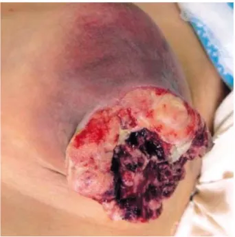
A 48-yr-old woman presented with a huge necrotic mass
in her left breast. She had first noticed the mass 1 yr before, since then it had rapidly grown and become necrotic. She was a mother of two children in good health, and had no family history of breast disease. On physical examination, the mass was seen to involve the entire left breast. The central portion of left breast including the nipple was rotted by severe necro- sis, and the surrounding skin was erythematous. The tumor tissue was very friable with profuse foul-odored discharge (Fig.
1). Several lymph nodes were palpated in the left axilla.
A wide incisional biopsy of the mass was performed. The sarcoma was diagnosed, but the specific type could not be established. Chest computed tomography (CT) demonstrated a hypodense mass, 10×8 cm in size, with central necrosis and numerous enlarged lymph nodes in the left axilla, but there was no abnormal finding in the lung fields. The abdom- inal ultrasonography revealed gallstones, but no abnormalities in the liver parenchyma. The tumor markers were within nor- mal limits, including CA15-3 (5.0 U/mL) and CA125 (7.2 U/mL). All laboratory findings, plain chest radiography film, and bone scintigraphy revealed no abnormalities.
Pseudomonas and Candida were grown on a bacterial cul- ture containing discharge from the necrotic tumor. Although there were no symptoms or signs of generalized infection, systemic antibiotics were administered for fear of dissemi- nating inflammation for several days before operation. At a later date, the patient underwent a radical mastectomy. In the operative field, numerous enlarged lymph nodes were observed in the left axilla. Among these, the largest one was more than
Se Jeong Oh, Kyoung Mee Kim*, Tae Ho Hong, Woo Chan Park, Jeong Soo Kim, Sang Seol Jung
Department of Surgery and Clinical Pathology*, College of Medicine, The Catholic University of Korea, Seoul, Korea
Address for correspondence Se Jeong Oh, M.D.
Department of Surgery, Our Lady of Mercy Hospital, 665 Bupyung-6-dong, Bupyung-gu, Incheon 403-720, Korea
Tel : +82.32-510-5691, Fax : +82.32-510-8615 E-mail : [email protected]
477 J Korean Med Sci 2004; 19: 477-80
ISSN 1011-8934
Copyright � The Korean Academy of Medical Sciences
Giant Cell Malignant Fibrous Histiocytoma of the Breast : A Case Report
A case of primary malignant fibrous histiocytoma of the breast is reported. The patient was a 48-yr-old woman with a huge tumor involving almost the entire left breast. The central portion of her left breast was already rotted by extensive necrosis and inflam- mation. She was treated by radical mastectomy and axillary lymphadenectomy to level I. Pathologic examination supported by an immunohistochemical staining con- firmed the tumor as malignant fibrous histiocytoma of giant cell type. Axillary lymph nodes were free from tumor metastasis. She had not taken any postoperative adju- vant therapy. The metastasis to lungs was found 2 months after the operation, and she died within 6 months.
Key Words : Breast Neoplasms; Sarcoma; Malignant Fibrous Histiocytoma; Myxosarcoma; Histiocytic Disorders, Malignant
Received : 18 April 2003 Accepted : 8 August 2003
478 S.J. Oh, K.M. Kim, T.H. Hong, et al.
2 cm in diameter, and diagnosed benign in the frozen section examination. The axillary node dissection was performed to level I only. Because primary skin closure was impossible, the defect in the left anterior chest wall was covered with the myo- cutaneous flap using the left latissimus dorsi muscle .
During the pathological examination, the breast was re- placed by a 12.5×11×5 cm gray, hard mass occupying almost the whole portion of the left breast, except pectoralis major muscle. On cut section, the mass showed areas of hem-
orrhage and necroses. The nipple and skin also showed nec- roses and involvement of a tumor component. In the micro- scopic examinations, some lobular and ductal structures were well preserved. The mass was composed of spindle-shaped tumor cells having polygonal nuclei and plump eosinophilic cytoplasm. There were some scattered lymphocytes among the tumor cells. Large portions of tumor also showed many bizarre multinucleated giant cells. The nuclei comprising giant cells were hyperchromatic and vesicular, and showed prominent nucleoli with frequent and abnormal mitoses (Fig.
2). On immunohistochemical staining, the cytoplasm of tumor cells were diffusely positive for vimentin and lysozyme, and negative for cytokeratin, epithelial membrane antigen, S-100 protein, and smooth muscle actin. These pathologic findings were compatible with the giant cell type of malignant fibrous histiocytoma.
The patient made an uneventful recovery. While the patient is hesitating to take postoperative adjuvant therapy, she had metastasis to lungs 2 months after the operation. Thereafter, she took only conservative care and died 4 months later.
DISCUSSION
Primary malignancies of the breast stromal elements and sarcomas of mesenchymal origin comprise less than 1% of all breast neoplasms, and are a highly heterogeneous group of tumors. The majority of these are made up of malignant fibrous histiocytomas, fibrosarcomas, liposarcomas, and the less common angiosarcomas; true ‘‘stromal sarcomas’’ of the breast are distinctly rare (6). Stromal sarcoma of the breast was initially defined as all sarcomas of the breast, excluding
Fig. 1.Gross finding of the tumor. The tumor involves almost the entire left breast, which is rotted by extensive necrosis and inflam- mation.
Fig. 2.Microscopic findings of the tumor. Low power (A, H&E, ×100) and high power (B, H&E, ×400) examinations show many multinu- cleated osteoclast-like giant cells and hyperchromatic mononuclear tumor cells.
A B
Malignant Fibrous Histiocytoma of the Breast 479
cystosarcoma phyllodes and angiosarcoma (7); but later it was redefined as rare sarcomas of a nonspecific type that arose in intralobular stroma (8). Since MFH and stromal sarcoma con- sist of spindle cells resembling fibroblasts, the only distin- guishing feature are the primary involvement of extralobular stroma and intralobular stroma, respectively. This distinction is very difficult (2). Jones et al. (9) proposed that MFH and fibrosarcoma have many features in common and thus, clas- sification into one group or another can be arbitrary. There- fore, it is reasonable to group both neoplasms together and divide them into low- and high-grade forms based on nuclear atypia and mitotic activity.
MFH arises most frequently (58-75%) from the deep fas- cia or skeletal muscle of the limbs, and then occurs frequently in the retroperitoneum and abdominal cavity with a frequency of 9-16%. However its primary development in the breast, not in combination with any other malignancies such as cys- tosarcoma phyllodes, or secondary to radiation therapy, is extremely rare; and to our knowledge, only 25 cases have been reported in medical journals written in English (1, 2, 10, 11).
Histologically, the tumor is a pleomorphic sarcoma that con- tains two cell types: fibroblast-like cells and histiocyte-like cells. Each tumor contains varying proportions of the two cell types so that a wide range of histologic features is possible (2).
As a result, MFHs have been characterized into the following subtypes: storiform-pleomorphic, myxoid, giant cell, and inflammatory (12). Most reported cases of primary MFH of the breast were of the storiform-pleomorphic variant (13).
Recently, Ajisaka et al. (2) reported the first case of the myx- oid variant occurring as a primary tumor in the breast. To our knowledge, the giant cell variant has not yet been reported as a primary tumor like the myxoid variant.
For the proper management and prediction of prognosis, differentiating malignant fibrous histiocytoma from other carcinoma or sarcoma is very important. Differential diagno- sis of MFH includes spindle cell (metaplastic) carcinoma, pleomorphic rhabdomyosarcoma, and malignant phyllodes tumor (cystosarcoma phyllodes). Spindle cell carcinoma, the more frequent form of breast carcinoma characterized by a sarcomatous metaplasia of invasive ductal carcinoma, is char- acterized by positive immunohistochemical staining for cytok- eratin, epithelial membrane antigen, and ER (14) while MFH is positive for vimentin and lysozyme. In our case, the tumor cells were positive for vimentin and lysozyme. Although the normal ducts were positive, tumor cells were exclusively neg- ative for cytokeratin and epithelial membrane antigen. Rhab- domyosarcoma can be differentiated for its immunoreactivity for desmin. As MFH can also occurs as a component of cys- tosarcoma phyllodes, it is very important to differentiate them.
To rule out the possibility of cystosarcoma phyllodes com- pletely, many tissue sections for microscopic examination are recommended and immunoreactivity for alph-1-antitrypsin, S-100 protein, and lysozyme should be evaluated.
Metastases of MFH are mainly hematogenous, and lymph
node involvement is very unusual. When the involvement of lymph node is present, it is considered as a sign of a widely disseminated disease (12). For this reason, axillary node dis- section has not generally been recommended in the treatment of this tumor (3). However, clarifying lymph node status by dissection is useful for predicting the prognosis, and helping to determine the indications for the postoperative adjuvant therapy. In addition, van Niekerk et al. (15), who for the first time found metastasis in the axillary lymph nodes, asserted lymphadenectomy to be mandatory. Moreover, axillary lymph node dissection is easy to perform without significant morbid- ity. Consequently, it will be up to the surgeon in case by case matter to decide whether axillary dissection is necessary or not.
Our patient was treated by total mastectomy, including resec- tion of the pectoral muscles and lymphadenectomy was per- formed to level I. There has been no evidence of metastasis to the lymph nodes during the final pathologic examination.
In terms of the postoperative adjuvant therapy, radiation therapy seems to play an important role (16). The effect of chemotherapy, however, is still uncertain, although Clamon et al. (17) reported a remission of more than 42 months by using aggressive combination chemotherapy. In general, prog- nosis of MFH is poor. In our case, the metastases of the lungs were found 2 months during which the patient had hesitated to take the adjuvant therapy, and she died 4 months later.
In spite of its rarity, surgeons should be alert to the existence of this type of soft tissue tumors in the breast. More informa- tion about the biological and pathological behavior of these tumors is necessary in order to expand and improve our ability to treat these patients. So far, surgery is the first step in the treatment. Although it remains doubtful whether radical or modified radical mastectomy is superior to wide local excision, the important point is to secure safe surgical margins of more than 3 cm (5), in which ever procedure that would be carried out.
REFERENCES
1. Tamir G, Nobel M, Hauben DJ, Sandbank J. Malignant fibrous his- tiocytoma of the breast. Eur J Surg Oncol 1995; 21: 210-1.
2. Ajisaka H, Maeda K, Uchiyama A, Miwa A. Myxoid malignant fibrous histiocytoma of the breast. Surgery Today 2002; 32: 887-90.
3. Langham MR Jr, Mills AS, De May RM, O’Dowd GJ, Grathwohl MA, Horsley JS III. Malignant fibrous histiocytoma of the breast. A case report and review of the literature. Cancer 1984; 54: 558-63.
4. Kearney MM, Soule EH, Ivins JC. Malignant fibrous histiocytoma.
A retrospective study of 167 cases. Cancer 1980; 45: 167-78.
5. Iellin A, Waizbard E, Levine T, Behar A. Malignant fibrous histiocy- toma of the breast. Int Surg 1990; 75: 63-6.
6. Freter C. Other cancers of the breast. In: Harris JR, Lippman ME, Morrow M, Osborne CK, editors, Diseases of the breast. 2nd ed.
Philadelphia: Lippincott Williams & Wilkins, 1999; 685.
7. Berg JW, DeCosse JJ, Fracchia AA, Farrow JH. Stromal sarcoma of
the breast. Cancer 1962; 15: 418-24.
8. Callery CD, Rosen PP, Kinne DW. Sarcoma of the breast. A study of 32 patients with reappraisal of classification and therapy. Ann Surg 1985; 201: 527-32.
9. Jones MW, Norris HJ, Wargotz ES, Weiss SW. Fibrosarcoma-Malig- nant fibrous histiocytoma of the breast. A clinicopathological study of 32 cases. Am J Surg Pathol 1992; 16: 667-74.
10. Horii R, Fukuuchi A, Nishi T, Takanashi R. A case of malignant fi- brous histiocytoma after breast conserving therapy for breast cancer.
Breast Cancer 2000; 7: 75-7.
11. De Medici A, Cebrelli CF, Venegoni A, Sirchi M. Malignant fibrous histiocytoma of the breast. A clinical case. Minerva Chir 1997; 52:
1237-41.
12. Weiss SW. Malignant fibrous histiocytoma. A reaffirmation. Am J Surg Pathol 1982; 6: 773-84.
13. Rossen K, Stamp I, Sorensen IM. Primary malignant fibrous histio- cytoma of the breast. A report of four cases and review of the litera- ture. APMIS 1991; 99: 696-702.
14. Tavassoli FA. Mesenchymal lesions. In: Tavassoli FA., editors, Pathol- ogy of the breast. 2nd ed. Hong Kong:McGraw-Hill, 1999; 675-730.
15. van Niekerk JLM, Wobbes T, Holland R, van Haelst UJ. Malignant fibrous histiocytoma of the breast with axillary lymph node involve- ment. J Surg Oncol 1987; 34: 32-5.
16. Belal A, Kandil A, Allam A, Khafaga Y, El-Husseiny G, El-Enbaby A, Memon M, Younge D, Moreau P, Gray A, Schultz H. Malignant fibrous histiocytoma: a retrospective study of 109 cases. Am J Clin Oncol 2002; 25: 16-22.
17. Clamon GH, Robinson RA, Olberding EB. Prolonged remission of metastatic malignant fibrous histiocytoma induced by combination chemotherapy. J Surg Oncol 1984; 26: 113-4.
480 S.J. Oh, K.M. Kim, T.H. Hong, et al.