Relationship between pulmonary
function and bone mineral density
in the Korean National Health and
Nutrition Examination Survey
(KNHANES)
In Seon Lee
Department of Medicine
Relationship between pulmonary
function and bone mineral density
in the Korean National Health and
Nutrition Examination Survey
(KNHANES)
In Seon Lee
Department of Medicine
Relationship between pulmonary
function and bone mineral density
in the Korean National Health and
Nutrition Examination Survey
(KNHANES)
Directed by Professor Young Sam Kim
The Master's Thesis
submitted to the Department of Medicine
the Graduate School of Yonsei University
in partial fulfillment of the requirements for the degree
of Master of Medical Science
In Seon Lee
June 2013
This certifies that the Master's Thesis of
In Seon Lee is approved.
---
Thesis Supervisor : Young Sam Kim
---
Thesis Committee Member#1 : Yumie Rhee
---
Thesis Committee Member#2 : Yoon Ha
The Graduate School
Yonsei University
ACKNOWLEDGEMENTS
First of all, I would like to express my deep and sincere
gratitude to Professor Young Sam Kim, M.D., Ph.D. for his
supervision and encouraging suggestions throughout my
academic life.
I am deeply indebted to Professor Yumie Rhee, M.D., Ph.D.,
who leads academic discussions and encourage me to
broaden my horizons.
I am grateful to Professor Yoon Ha, M.D for his precious and
constructive advices.
I wish to thank my family who has been with me and always
given me lots of support and love.
<TABLE OF CONTENTS>
ABSTRACT ··· 1
I. INTRODUCTION ··· 3
II. MATERIALS AND METHODS ··· 4
1. Study participants ··· 4
2. Blood Sampling ··· 6
3. Body composition and BMD ··· 7
4. Pulmonary Function Test ··· 7
5. Statistical Analysis ··· 8
III. RESULTS ··· 9
IV. DISCUSSION ··· 21
V. CONCLUSION ··· 26
REFERENCES ··· 27
ABSTRACT(IN KOREAN) ··· 29
LIST OF FIGURES
Figure 1. Proportion of osteoporosis and osteopenia according
to severity of airway obstruction. ··· 17
Figure 2. Bone mineral density at the lumbar spine and hip
regions according to severity of airway obstruction ···· 18
LIST OF TABLES
Table 1. Baseline demographics of subjects ··· 10
Table 2. Baseline characteristics of subjects ··· 12
Table 3. Bone mineral density in male & female ··· 13
Table 4. Correlation coefficients between clinical, biochemical
characteristics and BMD ··· 15
Table 5. Multiple regression analysis in male and female ·· 20
ABSTRACT
Relationship between pulmonary function and bone mineral density
in the Korean National Health and Nutrition Examination Survey
(KNHANES)
In Seon Lee
Department of Medicine
The Graduate School, Yonsei University
(Directed by Professor Young Sam Kim)
Background: Osteoporosis is common in chronic obstructive pulmonary
disease (COPD) patients. The relationship between osteoporosis and
COPD has been primarily reported in patients with moderate to severe
COPD, but there is no report in the general population. The aim of this
study is to investigate the relationship between bone mineral density
(BMD) and lung function in the general Korean population.
Methods: This study was based on data acquired from the Fourth and
Fifth Korea National Health and Nutrition Examination Surveys
(KNHANES IV and V), conducted from 2008 to 2010. The study
population included 4,501 subjects (aged ≥50 years) who underwent both
spirometry and BMD. The study populations were divided into two
groups by sex to correct for the effects of gender on BMD.
2
Results: The participants included 2,100 (46.7%) males and 2,401
(53.3%) females. All female participants were postmenopausal women.
For both genders, the BMD of the femur neck, total femur, and L-spine
correlated significantly with age, body mass index (BMI), fat free mass
index (FFMI), body fat percentage, smoking amount, parathyroid
hormone levels(PTH), daily calcium and phosphorus intake,
alkaline phosphataselevels (ALP), and FEV
1/FVC by simple linear analysis. After
adjusting for age, BMI, FFMI, body fat percentage, smoking amount,
serum 25(OH) levels, PTH , daily calcium and phosphorus intake, ALP,
BMD of the femur neck, total femur, and lumbar spine in both males and
females are correlated with age, ALP levels. But pulmonary function is
not associated with BMD in both gender after adjustment.
Conclusion: In the Korean men and women aged over 50, BMD is lower
in the subject with airway obstruction. Osteopenia and osteoporosis are
more common in adult with airflow limitation. But pulmonary function is
not associated with BMD after adjustment. Further studies are needed to
determine the possible role of pulmonary function in BMD.
---
Keywords: pulmonary function test, bone density, osteoporosis
3
Relationship between pulmonary function and bone mineral density
in the Korean National Health and Nutrition Examination Survey
(KNHANES)
In Seon Lee
Department of Medicine
The Graduate School, Yonsei University
(Directed by Professor Young Sam Kim)
I. INTRODUCTION
Chronic obstructive pulmonary disease (COPD) is characterized by a progressive airflow limitation that is not fully reversible1. The degree of airflow limitation can be measured by spirometry and stratified using the Global Initiative for COPD (GOLD) guideline. COPD is a complex disease that affects not only lung function but also many other comorbidities, such as decreased physical activity, right ventricular heart failure, and decreased quality of life2. Osteoporosis is one of the comorbid diseases of COPD, especially in those with advanced airway limitation3. As many as 35% to 72% of patients with COPD are reported to have osteopenia, and 36% to 60% patients have osteoporosis4-6. Risk factors for osteoporosis in COPD patients that have been evaluated by previous studies are a low body mass index (BMI), a low fat-free mass index
4
(FFMI), and severe airflow limitation7. Whereas a large proportion of the COPD population is classified as having mild to moderate airway obstruction, most studies were evaluated in COPD patients with severe to very severe airflow limitation. The risk factor that may contribute to osteoporosis in patients with mild to moderate airway obstruction remains uncertain. Thus, the aim of the present study was to investigate whether bone mineral density (BMD) might correlate with lung function in the Korean population based on the Fourth and Fifth Korean National Health and Nutrition Examination Surveys (KNHANES IV & V).
II. MATERIALS AND METHODS
1. Study participants
A cross-sectional survey representative of the Korean general population (KNHANES IV~V) was carried out from 2007 to 2012 by the Division of Chronic Disease Surveillance, Korea Centers for Disease Control and Prevention. This study was based on data acquired from the second year (2008) of KNHANES IV to the first year (2010) of KNHANES V. The survey consisted of an evaluation of medical history, nutritional status, and health. After an initial interview at home, participants visited mobile centers, where they underwent an extensive physical examination, laboratory testing, BMD measurement, and spirometry.
5
A total of 29,965 adults over 20 years of age participated in the survey. The analysis included 4,501 subjects who were older than 50 years with complete data on both pulmonary function tests and BMD measurements. Regions were categorized as urban and rural. In the adult population, occupations were classified into two groups based on the different levels of sunlight exposure. Outdoor work included primary and secondary industries (agriculture, forestry and fishery, manual labor, engineering, assembling, and technical work), whereas indoor work consisted of tertiary industries, including managers, specialists, clerical workers, salespersons, service personnel, housewives, and students. Unemployed persons and others with unclassifiable work were grouped as indoor work. During the home interview, information was collected on a wide range of variables, including age, gender, smoking history, daily food intake, the frequency of vitamin or mineral supplement intake, and the frequency of undertaking a range of common physical activities. Dietary intakes of calcium (Ca) and phosphorous (P) were estimated using a 24-h dietary recall method. High-risk drinking was defined as ―yes,‖ when men consumed seven cups of alcohol or five cans of beer at one time, or when women consumed five cups of alcohol or three cans of beer at one time. Smoking status was categorized as ―current smoker,‖ ―former smoker,‖ and ―never smoker.‖ Never smokers were defined as those who smoked fewer than five packs during their entire lives. Exercise was indicated as ―yes,‖ when the subject exercised regularly at moderate or vigorous levels (>30 min at a time and >five times per
6
week, in the case of moderate physical activity, and >20 min at a time and >three times per week, in the case of vigorous physical activity). Women who had not menstruated for 1 year were considered postmenopausal. Body weight and anthropometrics were measured with light clothes. BMI was calculated as weight divided by height squared (kg/m2).
2. Blood sampling
Blood samples were collected after an overnight fast and were immediately refrigerated and transported in cold storage to the Central Testing Institute (Neodin Medical Institute, Seoul, South Korea). Samples were analyzed within 24 hours after transportation. The concentrations of plasma glucose, serum calcium, phosphorus, creatinine, alkaline phosphatase, total cholesterol, and parathyroid hormone were measured using standard laboratory techniques. The serum levels of 25(OH) D, which are considered the biomarker for vitamin D metabolic status, were measured using a radioimmunoassay (DISCOVERY-W; Hologic Inc., Waltham, MA, USA) equipped with APEX software (APEX Corporation Software, Pittsburg, PA). The parathyroid hormone (PTH) level was determined by chemiluminescence assay (LIAISON; Diasorin Inc., U.S.A) using N-tact PTH assay (Diasorin Inc., U.S.A).
3. Body composition and BMD
7
a BDR Discovery fan beam densitometer (Hologic, Inc., Bedford, MA, USA) located in the mobile examination centers, and analyses were performed using Hologic Discovery software (version 13.1). BMD was expressed as an absolute value and as a T score (standard deviations from a young, sex-specific reference mean BMD). Osteopenia is defined as a T-score between -1.0 and -2.5. Osteoporosis is defined as a T-score below -2.5. Percentage body fat (fat mass * 100 / total mass) as well as BMD at the lumbar spine (L1–4) and proximal femur (total hip region) were measured using DXA. FFMI was calculated from DXA-measured fat-free mass in kilograms divided by height in meters squared (kg/m2).
4. Pulmonary function test
Spirometry was performed by specially trained technicians according to the 2005 American Thoracic Society and European Respiratory Society recommendations, using a dry rolling seal spirometer (SensorMedics Model 1022; Sensor Medics; Palm Springs, CA). We analyzed only data from subjects with acceptable spirometry performance. Spirometric values were prebronchodilator measurements, and relative values were expressed as the percentage predicted from reference values. Values used in the study were FVC, FEV1, and FEV1 to FVC ratio (FEV1/FVC). We categorized the subjects by
severity of airflow obstruction. Airway obstruction was deemed to be present when the FEV1/FVC was less than 0.7. When the FEV1/FVC was greater than
8
0.7, the subjects were categorized as ―no obstruction.‖ Severity of airway obstruction was determined on the basis of FEV1: ―mild obstruction‖ FEV1
≥80% of predicted value; ―moderate obstruction‖ FEV1 <80% and FEV1
≥50% of predicted value; ―severe obstruction‖ FEV1 < 50% of predicted
value.
5. Statistical analyses
Statistical analyses were performed using SAS version 9.2 (SAS Institute Inc., Cary, NC, USA). To correct the effects of gender on BMD, study participants were divided into two groups by sex and then separately analyzed. BMI was further divided into four categories, which corresponded to the International Classification suggested by the WHO: underweight (BMI <18.5), normal (18.5< BMI <25), overweight (25< BMI <30), and obese (BMI >30). Serum 25(OH)D levels were also divided into quartiles: ≤16.69 ng/mL, 16.79 to 21.44 ng/mL, 21.14 to 26.32 ng/mL, and ≥26.32 ng/mL cutoff values were used for men; ≤13.75 ng/mL, 13.75 to 17.90 ng/mL, 17.90 to 23.42 ng/mL, and ≥23.42 ng/mL cutoff values were used for women. To compare the baseline characteristics between males and females, the Student’s t test or the Chi square test was used for comparisons. Bivariate correlation analysis was used to evaluate the association between possible predictive factors and BMD. Multivariate linear regression analysis was used to evaluate the relationships between BMD and selected covariates that are considered clinically important risk factors for
9
osteopenia. Continuous variables were expressed as mean ± SD. All tests were two-sided, and P < 0.05 was considered statistically significant.
III. RESULTS
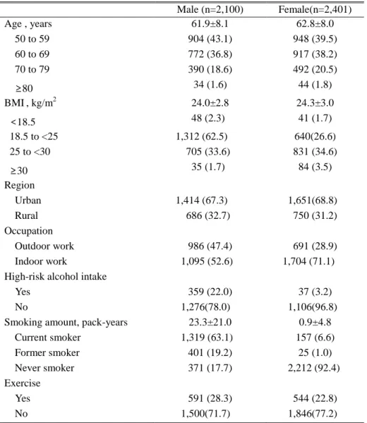
The baseline demographics of the study populations are given in Table 1. The subjects were comprised of 2,100 (46.7%) males and 2,401 (53.3%) females. The mean age of males was 61.9 years, and the mean age of females was 62.8 years. Most participants were in their 50s. The mean BMI was 24.0 kg/m2 in males and 24.3 kg/m2 in females. Most subjects were of normal weight in both genders, followed by being overweight. All female subjects were postmenopausal. In both genders, the proportion living in an urban area was about 68.0%. The proportion of those with high-risk alcohol intake was 22.0% in males and 3.2% in females. Of males, 17.7% were never smokers compared to 92.4 % of female. The proportion of the regular exercise ―yes‖ group was 28.3% in males and 22.8% in females.
10
Table 1. Baseline demographics of subjects
Data are presented as the mean ± SD, or number of subjects (percentage); BMI = body mass index;
Male (n=2,100) Female(n=2,401) Age , years 61.9±8.1 62.8±8.0 50 to 59 904 (43.1) 948 (39.5) 60 to 69 772 (36.8) 917 (38.2) 70 to 79 390 (18.6) 492 (20.5) ≥80 34 (1.6) 44 (1.8) BMI, kg/m2 24.0±2.8 24.3±3.0 <18.5 48 (2.3) 41 (1.7) 18.5 to <25 1,312 (62.5) 640(26.6) 25 to <30 705 (33.6) 831 (34.6) ≥30 35 (1.7) 84 (3.5) Region Urban 1,414 (67.3) 1,651(68.8) Rural 686 (32.7) 750 (31.2) Occupation Outdoor work 986 (47.4) 691 (28.9) Indoor work 1,095 (52.6) 1,704 (71.1)
High-risk alcohol intake
Yes 359 (22.0) 37 (3.2)
No 1,276(78.0) 1,106(96.8)
Smoking amount, pack-years 23.3±21.0 0.9±4.8
Current smoker 1,319 (63.1) 157 (6.6) Former smoker 401 (19.2) 25 (1.0) Never smoker 371 (17.7) 2,212 (92.4) Exercise Yes 591 (28.3) 544 (22.8) No 1,500(71.7) 1,846(77.2)
11
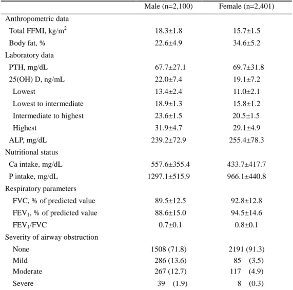
The mean 25(OH) D level was 22.0 ng/mL in males and 19.1 ng/mL in females. The mean FEV1/FVC ratio was 0.7 in males and 0.8 in females. The
mean FEV1 (% of predicted value) was 89.5% in males and 92.8% in females.
While the no airway obstruction group comprised 71.8% of subjects, the mild obstruction group comprised 13.6% and the moderate to severe obstruction group was 14.6% of males; most female participants belonged to the no obstruction group (91.3%), followed by the moderate to severe obstruction group (5.21%) and the mild obstruction group (3.5%) (Table 2).
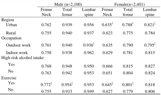
The subjects who lived in rural areas appeared to have lower BMDs than those dwelling in the city, but this trend was statistically significant only in females. The indoor work group was shown to have significantly higher BMDs of the lumbar spine than the outdoor work group in both genders, whereas there was no significant correlation between occupation and BMD of the femur. The regular exercise ―yes‖ group appeared to have lower BMDs than the ―no‖ exercise group in both males and females, except for BMD of the L-spine (Table 3).
12
Table 2. Baseline characteristics of subjects
Male (n=2,100) Female (n=2,401) Anthropometric data Total FFMI, kg/m2 18.3±1.8 15.7±1.5 Body fat, % 22.6±4.9 34.6±5.2 Laboratory data PTH, mg/dL 67.7±27.1 69.7±31.8 25(OH) D, ng/mL 22.0±7.4 19.1±7.2 Lowest 13.4±2.4 11.0±2.1 Lowest to intermediate 18.9±1.3 15.8±1.2 Intermediate to highest 23.6±1.5 20.5±1.5 Highest 31.9±4.7 29.1±4.9 ALP, mg/dL 239.2±72.9 255.4±78.3 Nutritional status Ca intake, mg/dL 557.6±355.4 433.7±417.7 P intake, mg/dL 1297.1±515.9 966.1±440.8 Respiratory parameters FVC, % of predicted value 89.5±12.5 92.8±12.8 FEV1, % of predicted value 88.6±15.0 94.5±14.6
FEV1/FVC 0.7±0.1 0.8±0.1
Severity of airway obstruction
None 1508 (71.8) 2191 (91.3)
Mild 286 (13.6) 85 (3.5)
Moderate 267 (12.7) 117 (4.9)
Severe 39 (1.9) 8 (0.3)
Data are presented as the mean ± SD, or number of subjects (percentage); FFMI = fat-free mass index; PTH = parathyroid hormone; ALP = alkaline phosphatase; FVC = forced vital capacity; FEV1 = forced expiratory volume in
13
Table 3. Bone mineral density in males and females
Male (n=2,100) Female(n=2,401) Femur Neck Total femur Lumbar spine Femur Neck Total femur Lumbar spine Region Urban 0.762 0.939 0.956 0.635† 0.788† 0.821ffi Rural 0.755 0.940 0.937 0.623 0.775 0.784 Occupation Outdoor work 0.761 0.940 0.936† 0.635 0.790 0.795ffi Indoor work 0.758 0.938 0.962 0.629 0.781 0.815 High-risk alcohol intake
Yes 0.768 0.948 0.950 0.666 0.815 0.827 No 0.763 0.942 0.953 0.651 0.804 0.824 Exercise
Yes 0.772† 0.954ffi 0.953 0.645ffi 0.801ffi 0.818
No 0.755 0.933 0.949 0.627 0.779 0.806
Data are presented as the mean.
14
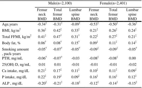
The BMD level was lower with increased age, number of smoking pack-years, and levels of PTH and ALP, whereas it was significantly higher with increased BMI, total FFMI, body fat percentage, daily calcium intake, and daily phosphorus intake in both genders. The levels of 25(OH) D did not correlate with BMDs of the femur neck, total femur, or lumbar spine. The FEV1/FVC ratio was positively correlated with BMDs of all bone regions in
both males and females, except for the BMD of the L-spine in males with FEV1/FVC. In males, FEV1 (% of predicted value) was increased with
increased BMD of total femur and decreased with BMD of the femur neck, whereas the FEV1 (% of predicted value) did not correlate with any region in
females. The FVC (% of predicted value) was lower with increasing BMDs of the lumbar spine in males, whereas the FVC (% of predicted value) was higher with increasing BMDs of the femur neck and total femur in females. BMDs of other regions did not show a significant correlation with FVC (% of predicted value) (Table 4).
15
Table 4. Correlation coefficients between clinical, biochemical characteristics,
and bone mineral density
Male(n=2,100) Female(n=2,401) Femur neck BMD Total femur BMD Lumbar spine BMD Femur neck BMD Total femur BMD Lumbar spine BMD Age,years -0.34ffi -0.31ffi -0.09ffi -0.53ffi -0.50ffi -0.36ffi BMI, kg/m2 0.36ffi 0.42ffi 0.33ffi 0.21ffi 0.26ffi 0.24ffi Total FFMI,kg/m2 0.41ffi 0.47† 0.31† 0.22ffi 0.27ffi 0.21ffi Body fat, % 0.06† 0.08† 0.15† 0.09ffi 0.11ffi 0.14ffi Smoking amount , pack-years -0.05† -0.07† -0.05† -0.09ffi -0.09ffi -0.05† PTH, mg/mL -0.06† -0.07† -0.03 -0.08ffi -0.08ffi 0.00 25(OH) D, ng/mL 0.01 0.01 -0.01 -0.01 -0.01 -0.02 Ca intake, mg/dL 0.21ffi 0.17ffi 0.11ffi 0.10ffi 0.11ffi 0.09ffi P intake, mg/dL 0.22ffi 0.19ffi 0.09ffi 0.16ffi 0.16ffi 0.12ffi ALP , mg/dL -0.20ffi -0.21ffi -0.18ffi -0.12ffi -0.14ffi -0.15ffi †p value <0.05, ffi p value <0.001;
BMI = body mass index; FFMI = fat-free mass index; PTH = parathyroid hormone; ALP = alkaline phosphatase;.
16
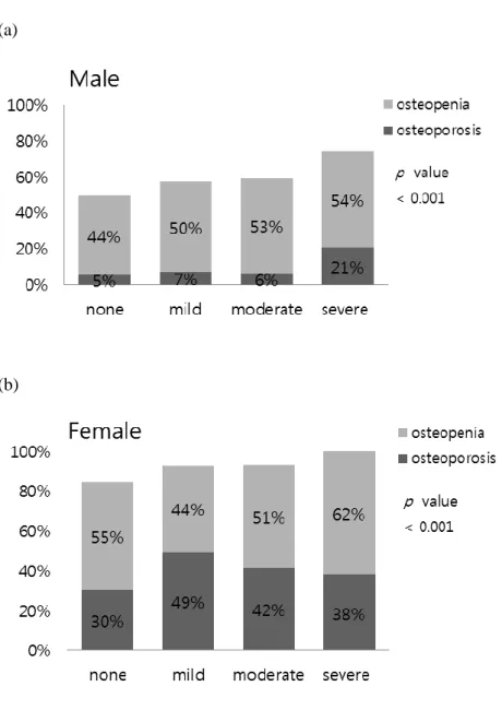
The proportion of osteopenia and osteoporosis was gradually increased according to severity of airway obstruction in both male and female. In the severe obstruction, proportion of osteoporosis were 21% of male and 38% of female. Indeed, female participants in severe airway obstruction group were all osteopenia and osteoporosis status. The proportion of osteopenia and osteoporosis was higher in female than in male at all stage of airway obstruction (Figure1).
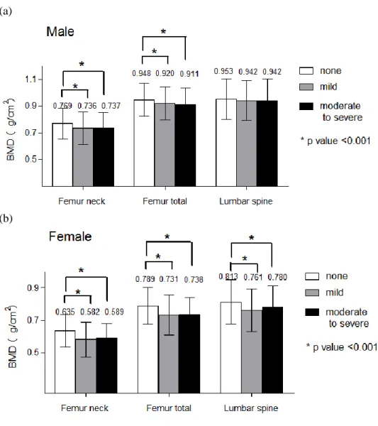
The total BMDs at different regions of the femur and lumbar spine in the ―obstruction‖ group were significantly lower than for the ―no obstruction ― group in both genders, except for the total BMD of the lumbar spine in males. (Figure 2).
17
(a)
(b)
Figure 1. Proportion of osteoporosis and osteopenia according to severity of
airway obstruction.
18
(a)
(b)
Figure 2. Bone mineral density at the lumbar spine and hip regions according
to the severity of airway obstruction. Data are presented as the mean ± SD *Significant difference (p<0.001).
19
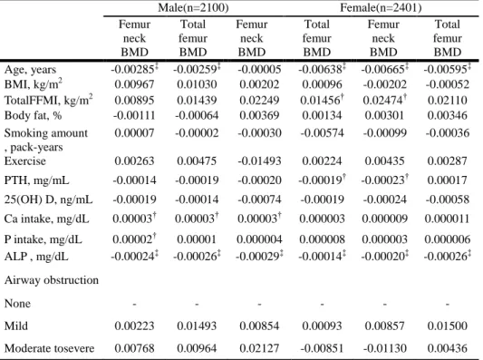
To adjust confounding factors, data were analyzed by multivariate linear regression analysis adjusted for age, smoking amount, BMI, total FFMI, body fat percentage, exercise, 25(OH) D, PTH, daily intake of calcium and phosphorus, and ALP. Results revealed that BMDs of the femur neck, total femur, and lumbar spine in both genders were reduced significantly as age increased. When daily calcium intake increased, BMDs of all regions increased significantly in males. As ALP increased, BMDs of all regions s increased in both males and females. Severity of airway obstruction did not show a significant correlation with BMDs of any region in males or females (Table 5).
20
Table 5. Coefficients between clinical, biochemical characteristics, and bone
mineral density after multiple linear regression analysis in males and females
Male(n=2100) Female(n=2401) Femur neck BMD Total femur BMD Femur neck BMD Total femur BMD Femur neck BMD Total femur BMD Age, years -0.00285ffi -0.00259ffi -0.00005 -0.00638ffi -0.00665ffi -0.00595ffi BMI, kg/m2 0.00967 0.01030 0.00202 0.00096 -0.00202 -0.00052 TotalFFMI, kg/m2 0.00895 0.01439 0.02249 0.01456† 0.02474† 0.02110 Body fat, % -0.00111 -0.00064 0.00369 0.00134 0.00301 0.00346 Smoking amount , pack-years 0.00007 -0.00002 -0.00030 -0.00574 -0.00099 -0.00036 Exercise 0.00263 0.00475 -0.01493 0.00224 0.00435 0.00287 PTH, mg/mL -0.00014 -0.00019 -0.00020 -0.00019† -0.00023† 0.00017 25(OH) D, ng/mL -0.00019 -0.00014 -0.00074 -0.00019 -0.00024 -0.00058 Ca intake, mg/dL 0.00003† 0.00003† 0.00003† 0.000003 0.000009 0.000011 P intake, mg/dL 0.00002† 0.00001 0.000004 0.000008 0.000003 0.000006 ALP , mg/dL -0.00024ffi -0.00026ffi -0.00029ffi -0.00014ffi -0.00020ffi -0.00026ffi Airway obstruction None - - - - Mild 0.00223 0.01493 0.00854 0.00093 0.00857 0.01500 Moderate tosevere 0.00768 0.00964 0.02127 -0.00851 -0.01130 0.00436 † p value <0.05, ffi p value <0.001;
BMI = body mass index; FFMI = fat-free mass index; ALP = alkaline phosphatase;
Regression models were adjusted for age, BMI, FFMI, body fat percentage, smoking amount, serum 25(OH) D, PTH, ALP, daily calcium intake, daily phosphorus intake, exercise, and severity of airway obstruction.
21
IV. DISCUSSION
In this population based, cross-sectional study, osteopenia and osteoporosis are more common as more severe airway obstruction in Korean elderly people. But after adjustment, the deficit of pulmonary function is not associated with BMD. Associations between pulmonary function and BMD have been conflicting in different studies7-9. A meta-analysis reported that the prevalence of osteoporosis was higher in COPD patients than in healthy subjects7. Some researchers have reported that the BMD of the total hip and femoral neck significantly decreased with impairment of lung function8. As airway obstruction becomes more severe, the proportion of patients with osteoporosis increases9. Indeed, one study proposed that the presence of COPD is one of the parameters for increasing risk of osteoporosis in males, but many studies investigated COPD patients who came to the hospital for treatment, so study populations have consisted of moderate to severe airway obstruction. Angela et al. used data from the National Health and Nutrition Examination Survey III of the U.S. population. They validated the Male Osteoporosis Risk Estimation Score (MORES) that included three variables—age, weight, and history of COPD all showed excellent predictive value for developing osteoporosis10. Lower FEV1 showed a correlation with the degree of osteopenia regardless of
glucocorticoid use, although for comparable degrees of pulmonary impairment, the subjects treated with prednisone had more frequent osteopenia. Indeed, a
22
cross-sectional study investigated healthy women, and the BMDs of the lumbar spine and proximal femur revealed a significant positive correlation with FEV1
in postmenopausal women11. Two general population-based studies suggested that FEV1 become higher with increasing BMD in middle-aged to older people.
Additionally, this association remained significant after adjusting for age, weight, height, respiratory disease, cardiovascular diseases, smoking status, and medical history of steroid use and hormone replacement therapy. However, two studies did not adjust for daily vitamin intake, and it is known that serum 25(OH) D, PTH, and ALP levels can all influence BMD. These particular studies also did not quantify smoking amount or use of corticosteroids, and these variables may have dose-related influences on BMD or pulmonary function. Physical activity, alcohol consumption, and nutritional status were also not explored8,12. Don et al. also analyzed data from Caucasian participants in the Third National Health and Nutrition Examination Survey, conducted in the United States between 1988 and 1994. Airflow obstruction was associated with increased odds of osteoporosis compared with the no airflow obstruction group (OR: 1.9; 95% confidence interval [CI]: 1.4 to 2.5). The risk of osteoporosis increased in a linear ―severity-dependent‖ fashion13
. These authors used quantified smoking status (pack-years) and surveyed medication usage (bronchodilators, inhaled or intranasal corticosteroids, estrogens, diuretics, etc.). BMD levels increased gradually with age, showing a peak in the third decade of life, and gradually decreasing thereafter. Their study populations were older than
23
20 years, and menopause in women was not reflected.
Contrary to many other studies, some researchers have failed to find statistically significant difference between BMD values of COPD and non-COPD groups14,15. Twenty-eight male COPD patients with a moderate degree of airway obstruction and 20 male volunteer subjects with normal lung function participated in the study. There was no difference in baseline characteristics (age, BMI, dietary parameters) and the level of physical activity between the two groups, except for smoking history. There was no statistically significant difference in the prevalence of osteoporosis between COPD patients and the control healthy group15. These discrepancies may be derived from differences in the characteristics of subjects among the studies, such as race, age, gender, menopausal status, history of corticosteroid exposure, and other anthropometric variables. These parameters can contribute to the pathophysiology of osteoporosis in COPD patients15.
Our data from a large population-based sample with or without airflow obstruction are a nationally representative sample, as conducted by KNHANES, compared with other studies in which the data were collected from a local hospital. Thus, a different proportion of patients with severe lung function impairment is one reason for the conflicting results. As we analyzed the age group older than 50 years, we eliminated the influences of premenopause and inconsistencies associated with aging on BMD9. Our data contained more detailed information on the demographic and socioeconomic factors, such as
24
complete smoking history and physical activities, as measured by weekly, quantitative energy consumption, so we could analyze the data more deeply and could adjust for various confounding factors. After adjusting for confounding factors, including age, smoking status, BMI, total FFMI, body fat percentage, 25(OH) vitamin D, PTH, ALP, daily intake of calcium and phosphorus, and exercise, the association between airway obstruction and BMD disappeared. These results suggest that the high prevalence of osteoporosis in COPD patients is associated with other factors and not with an impairment of lung function. The strengths of this study, compared with most previous studies of lung function and BMD, include a larger number of participants and the rigorous centralized quality control of the BMD scans. These factors may account for the discrepancy among studies.
We explored possible contributions of body composition, such as FFMI and body fat percentage. Osteoporosis can be influenced by low body weight, fat mass or fat-free mass, and FFMI, depending on age, sex, and menopausal status. Additionally, body composition changes can cause complications, including impaired respiratory muscle function and loss of skeletal muscle mass with reduced force generation. Many researchers have suggested that FFMI is associated with disease severity, exercise capacity, quality of life, and mortality in patients with COPD21-23. The linkage between loss of FFMI and BMD may be
a causative mechanism that can explain osteoporosis in COPD patients. Previous analyses of data from KNHANES IV to V showed that both BMI and FFMI
25
values decreased significantly in subjects with severe airway obstruction, whereas those subjects with mild to moderate airway obstruction had values equivalent to those of the control group.
Several limitations of this study deserve consideration. We used 25(OH) D levels for analysis, which were found to be associated with lung function from the Third National Health and Nutrition Examination Survey in the U.S.16. A previous analysis of the KNHANES III data showed a relationship between serum concentrations of 25(OH) D and lung function. Contrary to previous studies, our analysis from the KNHANES III and IV data did not show such an association. A possible explanation is that these discordances may be derived from differences in age of subjects, season, region, and exposure to sunlight, which are important determinants of BMD. We did not investigate each subject’s amount of sunlight exposure. We assumed that those who work outdoors would have more sunlight exposure, but our data revealed conflicting results between occupation and BMD in each region. Thus, we could not estimate the amount of sunlight exposure. These differences may influence the relationship between BMD and lung function.
The measurements of BMD using DXA may be confounded by several factors17. In particular, measurements of lumbar spine BMD using DXA becomes less useful with increasing age due to confounding effects, such as vertebral collapse, osteophytes, and aortic calcification, all leading to a ― pseudo-normalizing‖ of the BMD. Different calculating patterns have been
26
used for each DXA scan, and thus, these discrepancies should each be considered 18.
The data did not contain information about current medications, such as
steroid use, dosage, and duration. Although the effects of inhaled corticosteroids on osteoporosis and fractures remain controversial, systemic corticosteroids could affect BMD by decreasing new bone formation while increasing bone loss. Indeed, the rate of bone loss correlates loosely with the daily dose and duration of therapy19. In addition, pharmacological treatments of osteoporosis such as bisphosphonates can influence BMD. Smith et al. suggested that alendronate improves significantly T- and Z-scores for lumbar spine BMD, but not for hip BMD, in patients with asthma or COPD20. Further studies are needed to understand the effects on BMD of corticosteroids and medications for osteoporosis.
V. CONCLUSION
In the Korean general population aged over 50, BMD is lower in the subject with airway obstruction. Osteopenia and osteoporosis are more common in adult with airflow limitation. But the results of this study suggest that low BMD levels are not associated with deficits in lung function after adjustment. To determine the association between impaired lung function and osteoporosis, more prospectively controlled studies are needed.
27
REFERENCES
1. Rabe KF, Hurd S, Anzueto A, Barnes PJ, Buist SA, Calverley P, et al. Global strategy for the diagnosis, management, and prevention of chronic obstructive pulmonary disease: GOLD executive summary. Am J Respir Crit Care Med 2007;176:532-55.
2. Katsura H, Kida K. A comparison of bone mineral density in elderly female patients with COPD and bronchial asthma. Chest 2002;122:1949-55.
3. Yawn BP, Kaplan A. Co-morbidities in people with COPD: a result of multiple diseases, or multiple manifestations of smoking and reactive inflammation? Prim Care Respir J 2008;17:199-205.
4. Iqbal F, Michaelson J, Thaler L, Rubin J, Roman J, Nanes MS. Declining bone mass in men with chronic pulmonary disease: contribution of glucocorticoid treatment, body mass index, and gonadal function. Chest 1999;116:1616-24.
5. Shane E, Silverberg SJ, Donovan D, Papadopoulos A, Staron RB, Addesso V, et al. Osteoporosis in lung transplantation candidates with end-stage pulmonary disease. Am J Med 1996;101:262-9.
6. Incalzi RA, Caradonna P, Ranieri P, Basso S, Fuso L, Pagano F, et al. Correlates of osteoporosis in chronic obstructive pulmonary disease. Respir Med 2000;94:1079-84.
7. Graat-Verboom L, Wouters EF, Smeenk FW, van den Borne BE, Lunde R, Spruit MA. Current status of research on osteoporosis in COPD: a systematic review. Eur Respir J 2009;34:209-18.
Lekamwasam S, Trivedi DP, Khaw KT. An association between respiratory function and bone mineral density in women from the general community: a cross sectional study. Osteoporosis international 2002;13:710-5.
9. Biskobing DM. COPD and osteoporosis. Chest 2002;121:609-20. 10. Bikle DD, Halloran B, Fong L, Steinbach L, Shellito J. Elevated
1,25-dihydroxyvitamin D levels in patients with chronic obstructive pulmonary disease treated with prednisone. Journal of Clinical Endocrinology & Metabolism 1993;76:456-61.
11. Choi JW, Pai SH. Association between respiratory function and osteoporosis in pre- and postmenopausal women. Maturitas 2004;48:253-8.
12. Lekamwasam S, Trivedi DP, Khaw KT. An association between respiratory function and hip bone mineral density in older men: a
28
cross-sectional study. Osteoporosis international 2005;16:204-7.
13. Sin DD, Man JP, Man SF. The risk of osteoporosis in Caucasian men and women with obstructive airways disease. Am J Med 2003;114:10-4.
14. Riancho JA, Gonzalez Macias J, Del Arco C, Amado JA, Freijanes J, Anton MA. Vertebral compression fractures and mineral metabolism in chronic obstructive lung disease. Thorax 1987;42:962-6.
15. Karadag F, Cildag O, Yurekli Y, Gurgey O. Should COPD patients be routinely evaluated for bone mineral density? J Bone Miner Metab 2003;21:242-6.
16. Black PN, Scragg R. Relationship between serum 25-hydroxyvitamin d and pulmonary function in the third national health and nutrition examination survey. Chest 2005;128:3792-8.
17. Bolotin HH. DXA in vivo BMD methodology: an erroneous and misleading research and clinical gauge of bone mineral status, bone fragility, and bone remodelling. Bone 2007;41:138-54.
18. El Maghraoui A, Roux C. DXA scanning in clinical practice. QJM 2008;101:605-17.
19. Ferguson GT, Calverley PM, Anderson JA, Jenkins CR, Jones PW, Willits LR, et al. Prevalence and progression of osteoporosis in patients with COPD: results from the TOwards a Revolution in COPD Health study. Chest 2009;136:1456-65.
20. Smith BJ, Laslett LL, Pile KD, Phillips PJ, Phillipov G, Evans SM, et al. Randomized controlled trial of alendronate in airways disease and low bone mineral density. Chron Respir Dis 2004;1:131-7.
21. Schols AM, Broekhuizen R, Weling-Scheepers CA, Wouters EF. Body composition and mortality in chronic obstructive pulmonary disease. Am J Clin Nutr 2005;82:53-9.
22. Bolton CE, Ionescu AA, Shiels KM, Pettit RJ, Edwards PH, Stone MD, et al. Associated loss of fat-free mass and bone mineral density in chronic obstructive pulmonary disease. Am J Respir Crit Care Med 2004;170:1286-93.
23. Slinde F, Gronberg A, Engstrom CP, Rossander-Hulthen L, Larsson S. Body composition by bioelectrical impedance predicts mortality in chronic obstructive pulmonary disease patients. Respir Med 2005;99:1004-9.
29
ABSTRACT(IN KOREAN)
한국 국민건강영양 조사를 토대로한
폐기능과 골밀도의 상관관계
< 지도교수 김 영 삼 >
연세대학교 대학원 의학과
이 인 선
목 적: 골다공증은 만성폐쇄성폐질환 환자에서 흔히 동반되는 질환이다. 폐기능과 골밀도와의 상관관계에 대해 중등도와 중증 만성폐쇄성폐질환 환자에서 주로 연구되어 왔지만, 일반인을 대상으로 조사한 연구는 없다. 본 연구는 한국의 일반인을 대표하는 자료를 이용하여 폐기능과 골밀도 사이의 상관관계에 대하여 알아보고자 하였다. 방 법: 본 연구는 2008 년부터 2010 년까지 시행된 제 4 기와 5 기 국민건강영양조사를 토대로 하였다. 폐기능 검사와 골밀도 검사를 동시에 시행한 50 세 이상 성인 4,501 명의 자료를 분석하였다. 골밀도에 영향을 미치는 요인들을 보정하기 위해 남녀를 따로 구분하였다. 성 적: 대상자는 남성 2,102 명(46.6%), 여성 2404 명(53.4%)로 구성되었다. 여성 모두 폐경 후 상태였다. 상관분석결과 남성과30
여성에서 대퇴 골두, 대퇴골 전체, 요추 골밀도에 유의한 영향을 미친 인자는 연령, BMI, FFMI, 총체지방률, 흡연량, 부갑상선 호르몬, 일일 칼슘 섭취량 및 인 섭취량, ALP, FEV1/FVC 이었다. 연령, BMI, FFMI,
총체지방률, 흡연량, 25(OH)D, 일일 칼슘 섭취량 및 일일 인 섭취량 과 알카라인 포스파타제에 대하여 보정한 다중회귀분석결과 남성과 여성에서 연령, 알카라인포스파타제가 증가할수록 골밀도가 유의하게 증가하였다. 반면,기관지 폐쇄정도와 골밀도는 상관관계가 없었다. 결 론: 기관지 폐쇄가 있는 한국 50 세 이상 남성과 여성에서 골밀도가 낮다. 골감소증과 골다공증은 기도폐쇄가 있는 성인에서 더 흔하다. 하지만 다른 요인들을 보정한 후에 폐기능과 골밀도간에 관계가 없었다. 골밀도에 있어 폐기능이 미치는 영향에 대한 더 많은 연구가 필요하다. --- 핵심되는 말 : 폐기능 검사, 골밀도, 골다공증