213
"J. Korean Soc. Radiol., Vol. 9, No. 4, June 2015"
The effects of labeling gap and susceptibility artifacts in pCASL perfusion MRI
Seong-Hu Kim
Department of radiology, Gyeongsang National Univ. Hospital
pCASL 관류 영상에서 표지 간격과 자화감수성 인공물이 영상에 미치는 영향
김 성 후 경상대학교병원 영상의학과
Abstract
To report problems found in a patient who has implemented stent implantation and then conducted a perfusion MRI using ASL(Arterial Spin Labeling), in order to suggest a solution to them. The perfusion MRI was conducted, using pCASL among ASL methods. Data from pCASL(Pseudo Continuous Arterial Spin Labeling) was acquired together with the structural image simply by changing position(labeling gap 15 mm, 170 mm) of the labeling pulse to avoid stent. Data was processed through the ASLtbx. When perfusion MRI was acquired using pCASL, it showed that the position of the conventional labeling pulse (labeling gap 24 mm) was overlapped with that of stent, which made signal intensity in right brain tissue appear as if it were void.
When the labeling pulse was positioned (labeling gap 15 mm) to avoid stent, high signal intensity images were acquired. In labeling pulse (labeling gap 170 mm), the signal intensity was more reduced due to relaxation before labeled blood arrived at the imaging slice. pCASL can be stably repeated measurements because it does not use a contrast agent. And it should be selected with the appropriate image acquisition parameters for the high quality image.
Keyword : Magnetic resonance imaging, ASL, Stent, Susceptibility artifact
요 약
스텐트 삽입술을 시행한 환자에게 ASL 방법 중 pCASL을 이용한 관류영상에서 나타난 인공물을 보고하고 이에 대 한 해결방법을 제시하고자 한다. pCASL데이터는 구조적 이미지와 함께 스텐트를 피해 표지 펄스(labeling pulse)의 위 치를 변경하여 획득하였다. 데이터는 ASLtbx를 이용하여 처리하였다. pCASL을 이용하여 관류영상을 획득하였을 때 기존의 표지 펄스(표지 간격(labeling gap) 24 mm)의 위치가 스텐트의 위치와 겹쳐져서 우뇌 조직의 신호강도가 비어 있는 것처럼 나타났다. 스텐트를 피해 표지 펄스(표지 간격 15 mm)를 위치시킬 때 높은 신호강도의 영상을 획득 할 수 있었으며, 표지 펄스(표지 간격 170 mm)에서는 labeled 혈액이 영상절편에 도달하기 전에 이완이 되어 낮은 신호 강도의 영상을 획득 하였다. pCASL은 조영제를 사용하지 않기 때문에 안정적으로 반복측정이 가능하며 양질의 영상 획득을 위해서는 알맞은 영상획득인자와 방법들이 선택되어야 한다.
중심단어: 자기공명영상, 동맥스핀표지, 스텐트, 자화감수성인공물 http://dx.doi.org/10.7742/jksr.2015.9.4.213
Corresponding Author: Seong-Hu Kim E-mail: trufi@naver.com Tel: +82-10-8717-6975
Ⅰ. INTRODUCTION
Based on the development of a variety of functional imaging technology advances and hardware of Magnetic Resonance Imaging (MRI) equipment, clinical application of perfusion MRI has been helpful for diagnosis and treatment[1]. Perfusion MRI can be divided into endogenous and exogenous perfusion MRI. Exogenous perfusion MRI methods, including perfusion CT-scan, single photon emission Computed Tomography (SPECT), Positron Emission Tomography (PET), are an invasive method that require ionizing radiation and contrast medium[2]. Unlike exogenous perfusion MRI, Arterial Spin Labeling(ASL) brain perfusion MRI is one of the endogenous perfusion MRI methods using only blood in the human body. ASL was developed in the early 1990s.
ASL is a method for obtaining an image by changing the longitudinal magnetization of arterial blood using a Radio Frequency (RF) pulse[3-4].
The ASL requires no use of irradiation or injection of exogenous contrast medium, and it allows for repeated measuring of tissue blood flow and arterial transit time, which is the reason why ASL is increasingly adopted in clinical fields. Nevertheless, the ASL perfusion signals only account for about 1-5 % of mean MR signals intensity unless background suppression is used[5]. There are disadvantages such as low Signal-to-Noise-Ratio (SNR), a limited spatial resolution, inability to measure the volume of tissue blood flow, and the noise due to external factors.
ASL also acquires control images and labeling images using a labeling pulse. When metal substance for the treatment or blood products, calcification, or air are around the labeling pulse, susceptibility artifact is represented as signal intensity void[3-6]. Therefore, more researchers are looking for optimized imaging parameters in order to solve this problem, and more attempts to develop more advanced technology are being made.
The purpose of this paper is to report the particular case that a portion of the brain of patient who underwent
solve this in attempts to present a solution through a change in labeling pulse location. The patient took a perfusion MRI using pseudo continuous arterial spin labeling (pCASL) and echo-planar imaging (EPI). After preprocessing using statistical parametric mapping (SPM8) (http://fil.ion.ucl.ac.uk), all data was processed by ASLtbx (https://cfn.upenn.edu). pCASL that has combined advantages of continuous arterial spin labeling(CASL) and pulsed arterial spin labeling(PASL) provide better (signal to noise)SNR and reliable data for estimation of cerebral blood flow(CBF)[3], [7], [8].
Ⅱ. MATERIAL AND METHOD
1. Image acquisition
In a patient (male, age 55) who visited the hospital because of motor disturbance, the results of MRI angiography and perfusion MRI using ASL showed a difference in blood flow between right and left brain.
Stent insertion was implemented because the stenosis was found on the common carotid artery. After stent insertion, perfusion MRI using ASL was re-enforced in order to check the progress. Image acquisition was performed on a Philips ingenia 3.0 Tesla system (Philips medical systems, best, the Netherlands) using 32-channel SENSE head coils.
The structural images were acquired using a EPI sequence with the following parameters: TR = 3000 ms, TE = 35 ms, FOV = 220×220 mm2, slice thickness = 7 mm, voxel size = 2.29×2.29×7 mm3, bandwidth = 2052 Hz/pix, dynamic scans = 11. Pseudo continuous arterial spin labeling was applied with the following parameters:
slice order = ascend, TR = 4000 ms, TE = 15 ms, FOV
= 220×220 mm2, slice thickness = 7 mm, voxel size = 2.29×2.29×7 mm3, bandwidth = 1847 Hz/pix, dynamic scans = 25, label duration = 1650 ms ,post label delay = 1600 ms, labeling gap = 15/24/170 mm.
"J. Korean Soc. Radiol., Vol. 9, No. 4, June 2015"
2. Image processing and analysis
ASLtbx is operated, using MATLAB and SPM. This toolbox provides functions of RAW image quality checking, motion correction, coregistration, spatial smoothing, CBF calculation, and outlier cleaning. All the images were realigned by structure image in order to reduce the noise caused by the patient's movement or breathing and background suppression data of pCASL.
Then, the spatial smoothing to prevent signal fluctuation and to spatial SNR of raw image was performed using the 3D isotropic Gaussian kernel of FWHM = 8 mm and structure image. A CBF map was calculated using the ratio of the intensity of control image and the perfusion difference image obtained through a simple subtraction method with a pair of control and label images.
Ⅲ. RESULT
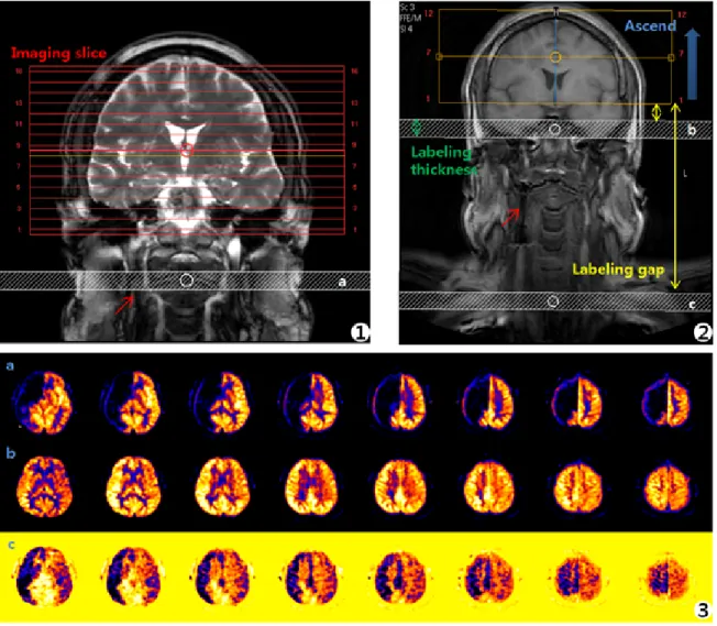
The mean CBF map obtained by 16 slices (labeling gap 24 mm) and 12 slices (labeling gap 15/170 mm) through ASLtbx in the patient who underwent stent insertion was respectively shown in [Fig. 1]. The labeling gap refers to the distance between the labeling pulse and the last slice of the imaging slice which obtained the area of the image.
Each figure's mean labeling gap[Fig. 1-1a : 24 mm, Fig.
1-2b : 15 mm, Fig. 1-2c : 170 mm], and labeling thickness is 20 mm. [Fig. 1-3a, b, c] indicated the mean CBF map at each position of labeling pulse. The overall tissue of the right brain appears as signal intensity void in [Fig. 1-3a], [Fig. 1-3b] appears uniform and high in signal intensity compared to [Fig. 1-3a] and [Fig. 1-3c]. [Fig. 1-3c] appears low in signal intensity as compared to [Fig. 1-3a] and Fig.
1-3b].
Ⅳ. DISCUSSION
In this study, it was confirmed that the tissue of the right brain of the patient who underwent stent insertion at common carotid artery appears as a signal intensity void during clinical application. The position of the labeling
pulse was adjusted by changing the labeling gap and slice number appropriately. The tissue of the right brain appears as signal intensity void in labeling gap 24 mm, and stent located around the labeling pulse results in a susceptibility artifact. The labeling gap 24 mm which is set to the default value in the device, indicated the distance with label offset 90 mm, or label distance, which is the distance from the center of the labeling pulse to the center of the imaging slice. A result of the acquisition of the image by changing labeling gap 15 mm and labeling gap 170 mm in order to avoid the susceptibility artifact by inserted stent, it was possible to obtain the proper image in labeling gap 15 mm. In the case of labeling gap 170 mm, since the time that labeled blood reaches the imaging slice was increased, the signal intensity was reduced by relaxation before labeled blood arrives at the imaging slice. For obtaining high quality images by the ASL from normal patients, several researchers have reported to be able to acquire that high signal intensity when labeling pulse positions labeling gaps 15~20 mm[9] or 74~94 mm[10] from the anterior commissure-posterior commissure line. However, as in the present study, when metal material for the treatment, blood products, calcification, or air are around the labeling pulse, this area should be avoided to acquire images. Also, if the time that labeled blood reaches the imaging slice is increased, the distance between the labeling pulse and imaging slice should be properly set since signal intensity was reduced by time.
The limitations of this study are that the images were not acquired by regular intervals from the inserted stent.
In order to obtain more reliable results, the result images have to be acquired by setting the various positions of labeling pulses. It is also necessary to discuss which has the highest signal intensity by comparing these results.
Ⅴ. CONCLUSION
Development of brain perfusion MRI technologies using the ASL method has been much help to clinical researchers especially for diagnosis and treatment issues.
patients, image parameters and methods of image acquisition not only should be properly selected but also prepared with a countermeasure for artifacts in metal material, blood products, calcification, and air. The brain perfusion MRI using the ASL can be possible with
use a contrast medium. And also, the women of childbearing age and children, patients who have a riskfor radioactive material in a contrast medium, can have the image stably acquired.
Fig. 1. Changes in the results of mean CBF map corresponding to the position of the implanted stent and labeling pulse. The mean CBF map can be changed according to the location of the labeling pulse, the surrounding environment, and the time that labeled
blood reaches the region of interest (ROI). Each letter a, b, and c shows the mean of 2 mm, 15 mm, 170 mm of labeling gap respectively and results of the mean CBF map by location of labeling pulse. The red arrow indicates an inserted stent.
"J. Korean Soc. Radiol., Vol. 9, No. 4, June 2015"
Reference
[1] Detre JA, Alsop DC, "Perfusion magnetic resonance imaging with Continuous arterial spin labeling: methods and clinical applications in the central nervous system," European Journal of Radiology Vol. 30, pp.115-124, 1999.
[2] Mahani NK, Baerends E, Soeter RP, et al, "Pseudocontinuous arterial spin labeling reveals dissociable effects of morphine and alcohol on regional cerebral blood flow," Journal of Cerebral Blood Flow & Metabolism Vol. 30, pp.1321-1333, 2011.
[3] Ferre JC, Bannier E, Raoult H, Mineur G, Nicol BC, Gauvrit JY, "Arterial spin labeling(ASL) perfusion:Technique and clinical use," Diagnostic and Interventional Imaging, Vol. 94,
pp.1211-1223, 2013.
[4] Wang Z, Das SR, Xie SX, Arnold SE, Detre JA, Wolk DA,
"Arterial spin labeled MRI in prodromal Alzheimer's disease: a multi-site study," NeuroImage Clinical, Vol. 2, pp.630-636, 2013.
[5] Wang Z, Aguirre GK, Rao H, et al, "Empirical optimization of ASL data analysis using an ASL data processing toolbox:
ASLtbx," Magn Reson Imaging, Vol. 26, pp.261-269, 2008.
[6] Deibler AR, Pollock JM, Kraft RA, Tan H, Burdette JH, Maldjian JA, "Arterial Spin-Labeling in Routine Clinical Practice, Part 1: Technique and Artifacts," AJNR Am J Neuroradiol, Vol. 29, pp.1228-1234, 2008.
[7] Dai W, Garcia D, Bazelaire CD, Alsop DC, "Continuous Flow Driven Inversion for Arterial Spin Labeling Using Pulsed Radiofrequency and Gradient Fields," Magn Reson Med, Vol.
60, pp.1488-1497, 2008.
[8] Wu WC, Seara MF, Detre JA, Wehrli FW, Wang JJ, "A Theoretical and Experimental Investigation of the Tagging Efficiency of Pseudocontinuous Arterial Spin Labeling,"
Magnetic Resonance in Medicine, Vol. 58, pp.1020-1057, 2007.
[9] Jang GH, Byun JH, Park MH, Kang JY, Lee JW, Lee KW,
"Investigation of Perfusion-weighted Signal Changes on a Pulsed Arterial Spin Labeling Magnetic Resonance Imaging Technique:
Dependence on the Labeling Gap, Delay Time, Labeling Thickness, and Slice Scan Order," Korean Journal of Medical Physics, Vol. 24, pp.108-118, 2013
[10] Aslan S, Xu F, Wang PL, et al, "Estimation of Labeling Efficiency in Pseudocontinuous Arterial Spin Labeling,"
Magnetic Resonance in Medicine, Vol. 63, pp.765-771, 2010.