Corneal Asphericity and Optical Performance after Myopic Laser Refractive Surgery
Jeong-Mee Kim
1, A-Young Lee
1, and Koon-Ja Lee
1,2,*1
Graduate School Dept. of Optometry, Eulji University, Daejeon 301-832, Korea
2
Dept. of Optometry, Eulji University, Sungnam 461-713, Korea (Received April 23, 2013: Revised May 23, 2013: Accepted June 15, 2013)
···
Purpose: To compare corneal asphericity, visual acuity (VA), and ocular and corneal higher-order aberrations (HOAs) between myopic refractive surgery and emmetropia groups. Methods: Twenty three subjects (23.0±2.5 years) who underwent myopic refractive surgery and twenty emmetropia (21.0±2.6 years) were enrolled. The subjects’ criteria were best unaided monocular VA of 20/20 or better in both two groups. High and low contrast log MAR visual acuities were measured under photopic and mesopic conditions. Corneal and ocular HOAs were measured using Wavefront Analyzer (KR-1W, Topcon) for 4 mm and 6 mm pupils. Corneal asphericity was taken by topography in KR-1W. Results: There was no significant difference in VA between two groups under either photopic or mesopic conditions. In ocular aberrations, there were significant differences in total HOAs, fourth- order and spherical aberration (SA) for a 6 mm between two groups (p=0.045, p<0.001, and p<0.001, respectively). In corneal aberrations, there was a significant difference in SA for 4 mm (p=0.001) and 6 mm (p<0.001) pupils between two groups and there were statistically significant differences in total HOAs (p<0.001) and fourth-order aberrations (p<0.001) between two groups for a 6 mm pupil. There was a significant correlation in emmetropia between Q-value and SA in ocular aberrations for 4 mm and 6 mm pupils (r=0.442, p=0.004, and r=0.519, p<0.001) and in corneal aberrations for 4 mm and 6 mm pupils (r=0.358, p=0.023, and r=0.646, p<0.001). No significant correlations were found between Q-value and SA in refractive surgery group.
Conclusions: VA in myopic refractive surgery is better than or similar to emmetropia. Nevertheless, the more increasing pupil size is, the more increasing aberrations are. Thus, it could have an influence on the quality of vision at night.
Key words: Higher-order aberrations, Spherical aberration, Corneal asphericity, Refractive surgery
···
INTRODUCTION
With the advances of wavefront technology and laser tech- nology, refractive surgery is now used as a means for cor- recting monochromatic higher-order aberrations (HOAs) beyond spherical and astigmatic refractive errors in clini- cal practice. New technology of the aberrometer could allow us to measure HOAs, such as coma, trefoil, spheri- cal aberration, and tetrafoil. The aberrometers have led to investigations of how HOAs affect visual performance and give us a better understanding of our visual systems.[1-3]
Ocular aberrations that combine various HOAs in optical systems produce defects in retina image quality.[4] In a nor-
mal, spherical aberration is one of the largest of the HOAs.[5]
Previous studies have shown that HOAs become signifi- cant factors in the quality of vision after correcting lower order aberrations.[6,7] Correcting lower order aberrations by refractive surgery does not always provide for mainte- nance and enhancement of our vision. Furthermore, many factors including an irregular corneal shape or changed corneal shape can induce aberrations. The shape change in cornea after refractive surgery can have an impact on cor- neal asphericity and HOAs, especially in terms of spheri- cal aberration and coma.[8,9] In addition to this, the effect of HOAs on vision depends on pupil size.[10,11]
The purpose of this study was to compare not only the
*Corresponding author: Koon-Ja Lee, TEL: +82-31-740-7182, E-mail: [email protected]
changes of high or low contrast visual acuity in both pho- topic and mesopic conditions but also ocular, corneal, and internal HOAs for 4 mm and 6 mm pupils between emme- tropic eyes and eyes undergone laser in-situ keratomileu- sis (LASIK) or laser sub-epithelial keratectomy (LASEK) for myopia and to analyze the influence of corneal asphe- ricity on spherical aberration in two groups.
METHODS
1. Subjects
Subjects were recruited from the Dept. of Optometry of Eulji University. There were 23 subjects who underwent conventional myopic LASIK or LASEK (except wave- front-guided refractive surgery) and 20 emmetropia sub- jects. The demographics and biometric data of the subjects are listed in Table 1. The subjects who met the criterion that was best unaided monocular VA of 20/20 (including no more than 0.50D of astigmatic refractive errors) or bet- ter in both two groups were selected for this study. None of the subjects had any ocular diseases other than refrac- tive errors or refractive surgery.
2. Visual acuity testing
VA was measured by subjective manifest refractions under photopic and mesopic conditions with a natural pupil, using a high (100%) and low (10%)-contrast ETDRS acuity charts. VA score was recorded by logMAR units. There were five letters for each line in the ETDRS acuity charts and each letter was assigned a score of 0.02 log units. The test was ended when subjects read twice incorrectly. The testing distance was 4 m. The illumination at the eye plane
for high contrast visual acuity (HCVA) and low contrast visual acuity (LCVA) under photopic and mesopic condi- tions was 340 lux and 20 lux, respectively (Digital Light Meter, TES-1330A, Taiwan).
3. Wavefront aberration measurement
Ocular wavefront aberrations were measured by Wave- front Analyzer (KR-1W, Topcon, Japan) with a Hartmann- shack aberrometry. In this study, ocular, corneal and inter- nal higher-order aberrations for 4 mm and 6 mm pupils were measured. The root mean square (RMS) of the third-order Zernike coefficients was used to represent third-order aberrations and the RMS of the fourth-order coefficient was used to represent fourth-order aberra- tions. Total HOAs were calculated as the RMS of the third-order and fourth-order coefficients. In order to max- imize the influence of pupil size, ocular aberration mea- surement was performed in a dark room and taken on the unaided eye. All measurements were carried out monocu- larly with an undilated pupil. Pupil diameter was taken under a dark room with a natural pupil using KR-1W, where a pupillometry function is incorporated within the Wavefront Analyzer.
4. Corneal topography measurement
Corneal asphericity, eccentricity and sim K were mea- sured by KR-1W, where corneal topography function is incorporated within the Wavefront Analyzer. The corneal asphericity coefficient Q describes the rate of variation in the curvature of the cornea from its center to the periph- ery. [12,13]
5. Statistical analysis
VA was compared between emmetropic and myopic refractive surgery groups by performing the independent t- test. For root mean square (RMS) values of HOAs, com- parisons between the two groups were also analyzed by the independent t-test. The results were expressed as mean
±SD. Pearson correlation test was analyzed for correla- tion between spherical aberration and corneal asphericity (Q-value) in emmetropia or conventional refractive sur- gery eyes. Origin 8.0 program (OriginLab Co., Northamp- ton, USA) was used for statistical analysis throughout this study. For statistical significance, p value of < 0.05 was used.
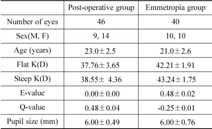
Table 1. Subjects’ demographics and biometric data Post-operative group Emmetropia group
Number of eyes 46 40
Sex(M, F) 9, 14 10, 10
Age (years) 23.0±2.5. 21.0±2.6 Flat K(D) 37.76±3.65. 42.21±1.91 Steep K(D) 38.55± 4.36 .43.24±1.75
E-value .0.00±0.00 00.48±0.02
Q-value 0.48±0.04 .-0.25±0.01 Pupil size (mm) .6.00±0.49 06.00±0.76 E, eccentricity; K, keratometry; Q-value, corneal asphericity
RESULTS
Twenty-three subjects who underwent conventional myo- pic LASIK or LASEK and 20 emmetropia subjects were participated in this study. The mean age of the 23 laser refractive surgical subjects was 23.0±2.5 years (range 20 to 27), and the period after refractive surgery was 16.0±
12.1 months. The mean age of emmetropia subjects was 21.0±2.6 years (range 19 to 24).
1. Visual acuity
Visual acuity under photopic and mesopic lighting condi- tions is summarized in Table 2.
In emmetropia group, HCVA and LCVA under photopic conditions were 0.003±0.005 and 0.172±0.008, respec- tively. In myopic refractive surgery group, Log MAR VA of HCVA and LCVA under photopic conditions were 0.013±0.003 and 0.156±0.005, respectively. There was no statistically significant difference in HCVA or LCVA between two groups under photopic conditions.
In mesopic conditions, log MAR VA of HCVA and LCVA for emmetropia group were 0.151±0.004 and 0.431±
0.006. Log MAR VA of HCVA and LCVA for myopic refrac- tive surgery group were 0.146±0.003 and 0.453±0.006, respectively. There was no statistically significant differ- ence in HCVA or LCVA between emmetropia and myo- pic refractive surgery groups under mesopic conditions.
2. Wavefront higher-order aberrations
Ocular aberrations of total HOA, third-order aberrations, fourth-order aberrations, coma and spherical aberration for 4 mm and 6 mm pupils are shown in Table 3 and Fig. 1.
For a 6 mm pupil size, in emmetropic eyes with ocular aberrations, the RMS values of total HOA, third-order and fourth-order aberrations were 0.547±0.045 µm, 0.435±
0.045 µm and 0.276±0.012 µm respectively, and coma and spherical aberrations were 0.342±0.042 µm and 0.224±
0.010 µm. In myopic refractive surgery eyes, the RMS values of total HOA, third-order and fourth-order aberrations were 0.649±0.06 µm, 0.406±0.039 µm and 0.443±0.051 µm
Table 2. Log MAR visual acuity measured under photopic and mesopic lighting conditions
Photopic Condition Mesopic Condition
HCVA(100%) LCVA(10%) HCVA(100%) LCVA(10%)
Post-operative 0.013±0.003a 0.156±0.0 0.146±0.003 0.453±0.006
Emmetropia 0.003±0.005 0.172±0.008 0.151±0.004 0.431±0.006
p - valueb p = 0.518 p = 0.360 p = 0.683 p = 0.205
aMean±standard deviation; HCVA, high contrast visual acuity (log MAR); LCVA, low contrast visual acuity (log MAR); p-valueb, by independent t-test.
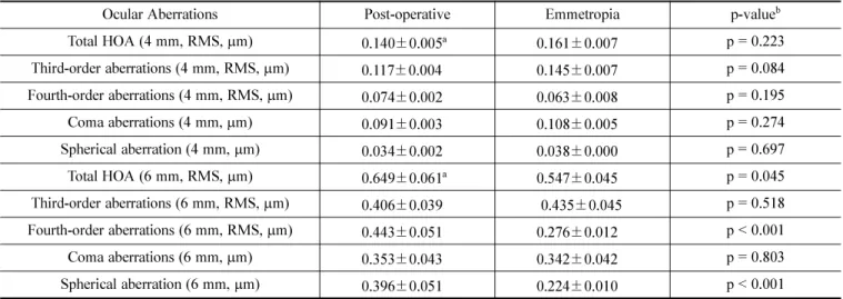
Table 3. Ocular higher-order aberrations for 4 mm and 6 mm natural pupils in the myopic refractive surgery group and emmetropia group
Ocular Aberrations Post-operative Emmetropia p-valueb
Total HOA (4 mm, RMS, µm) 0.140±0.005a 0.161±0.007 p = 0.223
Third-order aberrations (4 mm, RMS, µm) 0.117±0.004 0.145±0.007 p = 0.084 Fourth-order aberrations (4 mm, RMS, µm) 0.074±0.002 0.063±0.008 p = 0.195
Coma aberrations (4 mm, µm) 0.091±0.003 0.108±0.005 p = 0.274
Spherical aberration (4 mm, µm) 0.034±0.002 0.038±0.000 p = 0.697
Total HOA (6 mm, RMS, µm) 0.649±0.061a 0.547±0.045 p = 0.045
Third-order aberrations (6 mm, RMS, µm) 0.406±0.039 0.435±0.045 p = 0.518 Fourth-order aberrations (6 mm, RMS, µm) 0.443±0.051 0.276±0.012 p < 0.001
Coma aberrations (6 mm, µm) 0.353±0.043 0.342±0.042 p = 0.803
Spherical aberration (6 mm, µm) 0.396±0.051 0.224±0.010 p < 0.001
aMean±standard deviation; HOA, higher-order aberrations; p-valueb, by independent t-test.
respectively, and coma and spherical aberrations were 0.353±0.043 µm and 0.396±0.051 µm. We found that the amount of ocular aberrations in the postoperative eyes for a 6 mm pupil was generally greater than those in emme- tropic eyes. There were significant differences in total HOA, fourth-order and spherical aberrations for a 6 mm pupil with ocular aberrations between two groups (p=0.045, p<0.001 and, p<0.001, respectively). However, for a 4 mm pupil size, the mean values of ocular aberrations in the postoperative eyes were lower than those in emmetropic eyes. There was no significant difference in any ocular aberrations for a 4 mm pupil between two groups.
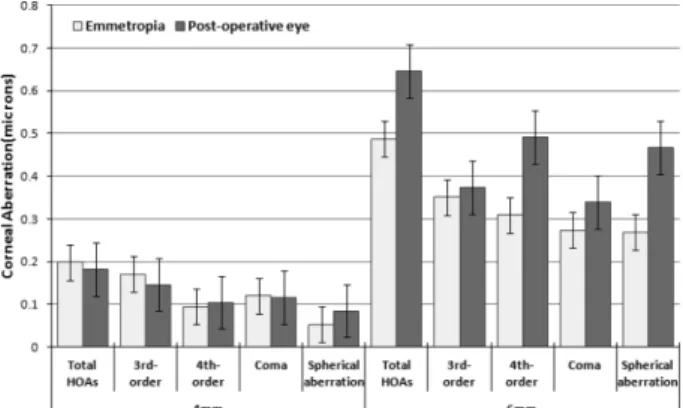
Corneal aberrations of total HOA, third-order aberrations, fourth-order aberrations, coma aberration and spherical aberration for 4 mm and 6 mm pupils are compared in Fig. 2. For a 6 mm pupil size, in emmetropic eyes with corneal aberrations, the RMS values of total HOA, third- order and fourth-order aberrations were 0.486±0.024 µm, 0.351±0.019 µm and 0.309±0.011 µm respectively, and coma and spherical aberrations were 0.273±0.018 µm and 0.269±0.007 µm. In myopic refractive surgery eyes, the RMS values of total HOA, third-order and fourth-order aberrations were 0.646±0.027 µm, 0.373±0.030 µm and 0.492±0.020 µm respectively, and coma and spherical aberrations were 0.339±0.029 µm and 0.467±0.018 µm.
We also found that there were significant differences in total HOA, fourth-order and spherical aberrations for a 6 mm with corneal aberrations between two groups (p<0.001, p<0.001 and, p<0.001, respectively). On the other hand, for a 4 mm pupil, in emmetropic eyes with corneal
aberrations, the RMS values of total HOA, third-order and fourth-order aberrations were 0.198±0.040 µm, 0.170±
0.011 µm and 0.094±0.005 µm respectively, and coma and spherical aberrations were 0.120±0.073 µm and 0.053
±0.012 µm. In myopic refractive surgery eyes, the RMS values of total HOA, third-order and fourth-order aberrations were 0.181±0.062 µm, 0.146±0.052 v and 0.104±0.020 µm respectively, and coma and spherical aberrations were 0.1156±0.042 µm and 0.084±0.002 µm. There was a statistically significant difference in only spherical aberration with corneal aberration for a 4 mm pupil between the emmetropia and postoperative eyes(p=0.001).
Comparison of internal aberrations of total HOA, third- order aberrations, fourth-order aberrations, coma aberration Fig. 1. Comparison of ocular higher-order aberrations (HOAs)
for 4 mm and 6 mm natural pupils in emmetropia and myopic refractive surgery groups. Total HOA, fourth- order and spherical aberrations for a 6 mm pupil show significant difference between two groups.
Fig. 2. Comparison of corneal higher-order aberrations (HOAs) for 4 mm and 6 mm natural pupils in emmetropia and myopic refractive surgery groups. There were significant differences in total HOA, fourth-order and spherical aberrations for a 6 mm pupil and spherical aberration for a 4 mm pupil between two groups.
Fig. 3. Comparison of internal higher-order aberrations (HOAs) for 4 mm and 6 mm natural pupils in emmetropia and myopic refractive surgery groups. There was significant difference in spherical aberration for a 4 mm pupil between two groups.
and spherical aberration in the myopic refractive surgery and emmetropia for 4 mm and 6 mm natural pupils is shown in Fig. 3. For a 6 mm pupil size, in emmetropic eyes with internal aberrations, the RMS values of total HOA, third-order and fourth-order aberrations were 0.396
±0.034 µm, 0.322±0.026 µm and 0.168±0.012 µm respectively, and coma and spherical aberrations were 0.280±0.024 µm and −0.049±0.013 µm. In myopic refractive surgery eyes, the RMS values of total HOA, third-order and fourth-order aberrations were 0.383±0.019 µm, 0.283±0.024 µm and 0.173±0.007 µm respectively, and coma and spherical aberrations were 0.248±0.024 µm and −0.070±0.022 µm. In emmetropic eyes with internal aberrations for a 4 mm pupil size, the RMS values of total HOA, third-order and fourth-order aberrations were 0.141± 0.010 µm, 0.119±0.006 µm and 0.063±0.005 µm respectively, and coma and spherical aberrations were 0.090±0.003 µm and −0.020±0.002 µm. In myopic refractive surgery eyes, the RMS values of total HOA, third-order and fourth-order aberrations were 0.123±0.002 µm, 0.097±0.002 µm and 0.066±0.000 µm respectively, and coma and spherical aberrations were 0.076±0.002 µm and −0.050±0.000 µm.
There was only one significant difference in spherical aberrations for a 4 mm with internal aberrations between two groups (p<0.001).
3. Corneal asphericity
The mean values of corneal asphericity(Q) in emmetropia and myopic refractive surgery groups were 0.483±0.036 and −0.254±0.011, respectively. The distributions of the Q-values in emmertoia and laser refractive surgery are
Fig. 4. Distribution of corneal asphericity (Q-value). Zero Q- value means the cornea is a sphere; negative Q-value indicates a prolate cornea; and positive Q-value indicates an oblate cornea.
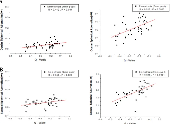
Fig. 5. Effect of corneal asphericity on spherical aberration. (A) The correlation between Q-value and ocular spherical aberration of emmetropia for 4 mm and 6 mm pupil sizes, respectively. (B) The correlation between Q-value and corneal spherical aberration of emmetropia for 4 mm and 6 mm pupil sizes, respectively.
illustrated in Fig. 4. Q-value was significantly different between two groups (p<0.001).
The relationship between Q-value and spherical aberra- tion of emmetropia is shown in Fig. 5. In emmetropia with ocular aberrations, a statistically significant correlation was found between Q-value and spherical aberration in ocular aberrations for 4 mm and 6 mm pupils (r=0.442, p=0.004, and r=0.519, p<0.001, respectively). In emmetropia with corneal aberrations, there was also a significant correla- tion between Q-value and spherical aberration in corneal aberrations for 4 mm and 6 mm pupils (r=0.358, p=0.023, and r=0.646, p< 0.001, respectively). On the other hand, for post-operative eyes, no significant effects were found between Q-value and spherical aberration in any higher- order aberrations.
There was no significant relationship between the shape of the cornea and the subjects’ VA except for one that is significant correlation between Q-value and HCVA (100%) under photopic condition (r=0.519, p<0.001).
DISCUSSION
After conventional LASIK or LASEK, many myopic eyes experience an increased spherical aberration.[14] Previous studies have reported the importance of HOAs, especially coma or spherical aberration, after refractive surgery, which is associated with halos or glare deteriorated visual qual- ity,[15,16] and the role of HOAs in visual performance have been shown to impact on visual outcomes.[17-19] Our find- ings agree with previous study that showed the induced spherical aberration plays a significant role for the change in the wavefront error.[20] Increasing of spherical aberra- tion caused an increase in total HOAs with both ocular and corneal aberrations. In fact, ocular HOAs were evalu- ated rather than corneal HOAs[21] because the ocular HOAs include changes to the posterior cornea and internal path- way of light into the eye even though the refractive effect was performed on the anterior cornea.[22] Our study com- pared ocular, corneal and internal aberrations between emmetropia and myopic refractive surgery eyes. The find- ing showed that ocular, corneal and internal HOAs for a 6 mm pupil were greater in myopic refractive surgery group than those in emmetropia group. The result was that spher- ical aberration had more strongly effect on the amount of total aberrations than other HOA terms.
Internal optics can be understood as crystalline lens. In internal aberrations, our findings showed that only spheri- cal aberration presents unequally in magnitude to that of the cornea but with opposite sign and the magnitude of other HOA terms was relatively less than those of the ocu- lar or corneal aberrations.
In the present study, the results demonstrated that ocular and corneal HOAs for a 6 mm pupil were greater than that for a 4 mm pupil in both emmetropia and myopic refractive surgery groups. This can be explained by the fact that the effect of ocular and corneal aberrations, mainly spherical aberration, on visual quality depends on pupil size,[10,11] on which the magnitude of spherical aber- ration depends.
The corneal surface is prolate when −1<Q<0, oblate when Q>0 and a perfect sphere when Q= 0. In our results, the mean of Q-value in emmetropia indicated a slightly prolate cornea (range −0.08 to −0.45), whereas, the aver- age cornea in laser refractive surgical eyes presented an oblate cornea (range 0.06 to 0.83). In the present study, the result of Q-value in emmetropia was similar to previous study that conducted in adults with early 20s to 30s.[23,24] This study was analyzed the effect of Q-value from spherical aberration with 4 mm and 6 mm pupils. It was found that Q-value is closely related to spherical aberration rather than other aberrations. For the emmetropia with prolate corneal surface, the correlations between Q-value and spherical aberration showed a statistically significant differ- ence in both ocular and corneal spherical aberration for 4 mm and 6 mm pupils. It was also found higher correla- tion between Q-value and spherical with a 6 mm pupil than between Q-value and spherical aberration with a 4 mm pupil in both ocular and corneal spherical aberrations.
The shape change in the corneal surface through excimer refractive surgery, which alters asphericity of the cornea from a prolate to an oblate shape, could be explained by the induction of spherical aberration[25]. However, as con- firmed by the results of this study, in laser refractive sur- gery with change in oblate corneal surface, the correlations did not show a statistically significant difference between Q-value and spherical aberration.
This study did not find a significant relationship between the shape of the cornea and the subjects’ VA except for HCVA (100%) under photopic condition. Emmetropia with prolate cornea and post-operative eyes with oblate cornea
were equally likely to have best-uncorrected HCVA and LCVA at distance. Changes in corneal asphericity had no influence on HCVA and LCVA both under photopic or mesopic conditions. Previous study reported that retaining preoperative corneal Q-value did not guarantee better VA after refractive surgery for treatment.[14] Given the relation- ship between VA and ocular HOAs after laser refractive surgery, this study showed that HCVA and LCVA under photopic and mesopic conditions seem not to be influ- enced directly by ocular HOAs. Therefore, the result that there was no significant difference in VA between emmetropia and laser refractive surgery groups is not surprising, because retinal image quality is the result of combination with various ocular components in optical system, which includes the anterior and posterior corneal surface and crystalline lens.[14] It could be considered the fact that spherical aberration is one of many aberrations in which have an impact on the retinal image quality. Moreover, despite the statistically significant findings of change in spherical aberration, it is still uncertain as to the magni- tude of change in HOAs. Although spherical aberration may not affect VA after laser refractive surgery, it may have an influence on the quality of vision with respect to low contrast conditions or low illumination conditions at night.[26]
Considering the changes in ocular, corneal and internal aberrations with aging, further studies should be required for the longer-term evaluation between changes in ocular aberrations regarding visual quality after refractive surgery.
CONCLUSIONS
We found that VA in mopic refractive surgery group is better than or similar to emmetropia group in spite of changes in corneal asphericity. Nevertheless, in both ocu- lar and anterior corneal wavefront aberrations, the more increasing pupil size in the eye is, the more increasing aberrations in that eye are. Thus, increasing spherical aber- ration after refractive surgery could have an influence on the quality of vision that is related to low contrast sensitiv- ity and low light at night.
REFERENCES
[1] Porter J, Guirao A, Cox IG, Williams DR. Monochro-
matic aberrations of the human eye in a large population.
J Opt Soc Am A Opt Image Sci Vis. 2001;18(8):1793- 1803.
[2] Castejón-Mochón JF, López-Gil N, Benito A, Artal P.
Ocular wave-front aberration statistics in a normal young population. Vision Res. 2002;42(13):1611-1617.
[3] McLellan JS, Marcos S, Burns SA. Age-related changes in monochromatic wave aberrations of the human eye.
Invest Ophthalmol Vis Sci. 2001;42(6):1390-1395.
[4] Liang J, Williams DR. Aberrations and retinal image quality of the normal human eye. J Opt Soc Am A. 1997;
14(11):2873-2883.
[5] Salmon TO, van de Pol C. Normal-eye Zernike coeffi- cients and root-mean-square wavefront errors. J Cataract Refract Surg. 2006;32(12):2064-2074.
[6] Thibos LN, Hong X, Bradley A, Cheng X. Statistical variation of aberration structure and image quality in a normal population of healthy eyes. J Opt Soc Am A Opt Image Sci Vis. 2002;19(12):2329-2348.
[7] Lindskoog Pettersson A, JarkC, Alvin A, Unsbo P, Brau- taset R. Spherical aberration in contact lens wear. Contact Lens & Anterior Eye. 2008;31(4):189-193.
[8] Moreno-Barriuso E, Lloves JM, Marcos S, Navarro R, Llorente L, Barbero S. Ocular aberrations before and after myopic corneal refractive surgery: LASIK-induced changes measured with laser ray tracing. Invest Ophthalmol Vis Sci. 2001;42(6):1396-1403.
[9] Kohnen T, Buhren J. Corneal first-surface aberration anal- ysis of the biomechanical effects of astigmatic keratotomy and a microkeratome cut after penetrating keratoplasty. J Cataract Refract Surg. 2005;31(1):185-189.
[10] Charman WN, Chateau N. The prospects for super-acuity:
limits to visual performance after correction of monochro- matic ocular aberration. Ophthalmic Physiol Opt. 2003;
23(6):479-493.
[11] Nguyen-Khoa JLD, Gicquel JJ, Lebuisson DAA, Dighi- ero P, Maille M. Monochromatic ocular aberrations distri- bution in professional pilots. Invest Ophthalmol Vis Sci.
2005;46(5):E-Abstract 1998.
[12] Gonzalez-Meijome JM, Villa-Collar C, Montes-Mico R, Gomes A. Asphericity of the anterior human cornea with different corneal diameters. J Cataract Refract Surg. 2007;
33(3):465-473.
[13] Yoon JH, Avudainayagam K, Avudainayagam C, Swar- brick HA. Validating a new approach to quantify Poste- rior corneal curvature in vivo. J Korean Oph Opt Soc.
2012;17(2):223-232.
[14] Tuan KM, Chernyak D. Corneal asphericity and visual function after wavefront-guided LASIK. Optom Vis Sci.
2006;83(8):605-610.
[15] Sharma M, Wachler BS, Chan CC. Higher-order aberra- tions and relative risk of symptoms after LASIK. J Refract Surg. 2007;23(3):252-256.
[16] Chalita MR, Chavala S, Xu M, Krueger RR. Wavefront
analysis in post-LASIK eyes and its correlation with visual symptoms, refraction, and topography. Ophthalmology.
2004;111(3):447-453.
[17] Applegate RA, Marsack JD, Ramos R, Sarver EJ. Interac- tion between aberrations to improve or reduce visual per- formance. J Cataract Refract Surg. 2003;29(8):1487-1495.
[18] Chen L, Singer B, Guirao A, Porter J, Williams DR. Image metrics for predicting subjective image quality. Optom Vis Sci. 2005;82(5):358-369.
[19] Cheng X, Thibos LN, Bradley A. Estimating visual qual- ity from wavefront aberration measurements. J Refract Surg. 2003;19(5):S579-584.
[20] Buhren J, Nagy L, Yoon G, MacRae S, Kohnen T, Huxlin KR. The effect of the asphericity of myopic laser ablation profiles on the induction of wavefront aberrations. Invest Ophthalmol Vis Sci. 2010;51(5):2805-2812.
[21] Mcalinden C, Moore JE. Comparison of higher order
aberrations after LASIK and LASEK for myopia. J Refract Surg. 2010;26(1):45-51.
[22] Marcos S. Aberrations and visual performance following standard laser vision correction. J Refract Surg. 2001;
17(5):S596-S601.
[23] Jeon IC, Jeong WJ, Kang JH. Comparison of corneal asphericity with measuring range. J Korean Oph Opt Soc.
2012;17(4):469-476.
[24] Kim HJ, Lee DH. Corneal asphericity of myopia in Korean. J Korean Oph Opt Soc. 2006;11(2):109-114.
[25] Holladay JT, Janes JA. Topographic changes in corneal asphericity and effective optical zone after laser in situ keratomileusis. J Cataract Refract Surg. 2002;28(6):942- 947.
[26] Cox I, Holden BA. Soft contact lens-induced longitudinal spherical aberration and its effect on contrast sensitivity.
Optom Vis Sci. 1990;67(9):679-683.
굴절교정수술을 받은 근시안의 각막 비구면도와 광학적 특성 평가
김정미1, 이아영1, 이군자1,2,
*
1
을지대학교 대학원 안경광학과, 대전 301-832
2
을지대학교 안경광학과, 성남 461-713
투고일(2013년 4월 30일), 수정일(2013년 5월 23일), 게재확정일(2013년 6월 15일)
목적: 엑시머 레이저 근시굴절교정수술을 받은 사람의 각막 비구면도, 시력, 고위수차를 정시안과 비교 평가하였 다. 방법: 단안의 나안 시력이 1.0 이상인 근시 굴절교정수술을 받은 23명(나이: 23.0±2.5세)과 20명(21.0±2.6세)의 정시안을 대상으로 밝은 조명(photopic)과 중등도(mesopic) 조명상태에서 대비도를 가지는 시력표(100% 및 10%)를 이용하여 시력검사를 하였고, wavefront 수차 분석기(KR-1W, Topcon, Japan)를 이용하여 각막의 비구면계수와 4
mm와 6 mm의 동공크기에 따른 눈 전체의 고위수차와 각막의 고위수차를 측정하여 비교하였다. 결과: 굴절교정 수
술안과 정시안에서 대비도에 따른 시력은 유의한 차이를 보이지 않았다. 안구 수차(ocular aberrations)에서 전체 고 위수차, 4차수차, 구면수차는 동공크기 6 mm상태에서 굴절교정 수술안에서 높게 나타났고, 두 그룹 사이에 유의한 차이를 보였다(p=0.045, p<0.001, p<0.001). 각막의 구면 수차(corneal spherical aberrations)는 동공크기 4 mm와 6 mm상태에서 모두 굴절교정 수술 안에서 더 높게 측정되었고 유의한 차이를 보였다(p<0.01, p<0.001). 각막의 전체 고위수차와 4차수차는 동공크기 6 mm 상태에서만 굴절교정 수술안에서 더 크게 나타났고 통계적으로 유의하였다 (p<0.001, p<0.001). 정시안의 비구면계수와 구면수차는 동공크기 4 mm와 6 mm 상태에서 안구 수차(r=0.442, p=0.004, r=0.519, p<0.001) 와 각막수차(r=0.358, p=0.023, r=0.646, p<0.001) 모두에서 유의한 상관성을 나타내었으 나, 굴절교정 수술안에서는 상관성이 없었다. 결론: 각막굴절교정 수술안의 경우 시력은 정상수준을 나타내고 있으 나 동공크기가 커지면 수차가 증가하여 동공이 커지는 야간에는 시력의 질에 영향을 줄 수 있을 것으로 사료된다.
주제어: 고위수차, 구면수차, 각막 비구면계수, 굴절수술