Ethanol Extracts of Rheum undulatum and Inula japonica Protect Against Oxidative Damages on Human Keratinocyte HaCaT cells through the Induction of ARE/NRF2-dependent Phase II Cytoprotective Enzymes
Ok-Kyung Yoo
1, Yong-Geol Lee
2, Ki-Hoan Do
2* and Young-Sam Keum
1*
1College of Pharmacy, Dongguk University, 32 Dongguk-ro, Goyang, Gyeonggi-do 10326, Korea
2Rich Chemical, 120-15 Gojan-dong, Namdong-gu, Incheon 21686, Korea
Received September 28, 2016 /Revised December 25, 2016 /Accepted March 12, 2017
Mammalian cells control cellular homeostasis using a variety of defensive enzymes in order to combat against environmental oxidants and electrophiles. NF-E2-related factor-2 (NRF2) is a transcription fac- tor that, in response to an exposure to oxidative stress, translocates into the nucleus and modulates the inducible expression of various phase II cytoprotective enzymes by binding to the antioxidant re- sponse element (ARE). In the present study, we have acquired 400 ethanol extracts of traditional me- dicinal plants and attempted to find out possible extract(s) that can increase the NRF2/ARE-depend- ent gene expression in human keratinocytes. As a result, we have identified that ethanol extracts of Rheum undulatum and Inula japonica strongly activated the ARE-dependent luciferase activity in HaCaT- ARE-luciferase cells. Exposure of ethanol extracts of Rheum undulatum and Inula japonica in- creased the viability and activated transcription and translation of NRF2-dependent phase II cytopro- tective enzymes in HaCaT cells, such as heme oxygenase-1 (HO-1) and NAD[P]H:quinone oxidor- ecutase-1 (NQO1). In addition, ethanol extracts of Rheum undulatum and Inula japonica suppressed 12-O-tetradecanoylphorbol-13-acetate (TPA)-induced generation of intracellular reactive oxygen species (ROS), thereby inhibiting the formation of 8-hydroxyguanosine (8-OHG) and 4-hydroxynonenal (4-HNE) in HaCaT cells. Together, our results demonstrate that ethanol extracts of Rheum undulatum and Inula japonica exert anti-oxidant effects via the induction of NRF2/ARE-dependent gene ex- pression in human keratinocytes.
Key words : Antioxidant response element (ARE), Inula japonica, NF-E2-related factor-2 (NRF2),
reactive oxygen species (ROS), Rheum undulatum
*Corresponding authors
*Tel : +82-31-961-5215, Fax : +82-31-961-5206 E-mail : [email protected] (Young-Sam Keum) Tel : +82-32-819-5601, Fax : +82-32-819-0560
*E-mail : [email protected] (Ki-Hoan Do)
This is an Open-Access article distributed under the terms of the Creative Commons Attribution Non-Commercial License (http://creativecommons.org/licenses/by-nc/3.0) which permits unrestricted non-commercial use, distribution, and reproduction in any medium, provided the original work is properly cited.
Journal of Life Science 2017 Vol. 27. No. 3. 310~317 DOI : https://doi.org/10.5352/JLS.2017.27.3.310
Introduction
Organisms are constantly exposed to various types of en- vironmental stresses, which contribute to the accumulation of intracellular reactive oxygen species (ROS) and con- sequent oxidative damages on cellular macromolecules [15].
Although an aberrant production of ROS is considered detri- mental, a relevant amount of ROS is also required for carry- ing out a number of critical cellular functions. Hence, a deli- cate balance between the production and elimination of in- tracellular ROS is necessary for the maintenance of proper
redox homeostasis [11]. In order to counteract the excessive oxidative insults, organisms have developed a variety of phase II cytoprotective enzymes during evolution, such as heme oxygenase-1 (HO-1), NAD[P]H:quinone oxidoreductase- 1 (NQO1), superoxide dismutase (SOD), glutathione S-trans- ferase (GST) and γ-glutamylcysteine ligase (γ-GCL) [7].
Previous mechanism-based studies have demonstrated that NF-E2-related factor-2 (NRF2) is responsible for transcrip- tional activation of phase II cytoprotective enzymes [5].
While it is well accepted that NRF2 activation can reduce the oxidative stress-related damages in normal cells, recent studies have demonstrated that gain of function mutations in Nrf2 gene frequently occur in tumor samples, possibly playing a significant role in the survival of tumors [10].
NRF2 is a Cap’N’Collar (CNC) transcription factor that
contains a basic leucine-zipper (bZIP) domain [9]. Under a
basal condition, NRF2 is sequestered in the cytoplasm and
constantly poly-ubiquitinated by an E3 ubiquitin ligase
adaptor, Kelch-like ECH-associated protein-1 (KEAP1). In re-
sponse to various stresses, NRF2 is relieved from KEAP1
and translocates into the nucleus, thereby resulting in the binding of NRF2 to the anti-oxidant-response element (ARE), a nucleotide motif sequence that exist in 5’-upstream region of phase II cytoprotective genes [13]. Traditionally, plants have been the most harnessed natural resource due to an abundance and easy accessibility. Therefore, the use of novel plant ingredients or extracts that can activate the NRF2/ARE-dependent gene expression has been proposed as a feasible strategy to inhibit or delay the oxidative dam- ages in keratinocytes [1] and, accordingly, numerous plant- derived natural compounds that can activate the NRF2/
ARE-dependent gene expression were shown to exhibit ben- eficial effects in vivo [14]. In line with this idea, we have attempted to find out novel ethanol extract(s) of traditional medicinal plants that can increase the ARE-dependent phase II enzymes in human keratinocytes HaCaT cells and identi- fied Rheum Undulatum and Inula japonica significantly re- duced the oxidative damages by activating the NRF2/ARE- dependent phase II gene expression.
Materials and Methods Cell culture, chemicals and reagents
Ethanol extracts of 400 traditional medicinal plants were provided from Dong-A ST (Yongin, Korea). RPMI-1640 me- dia, heat-inactivated fetal bovine serum (FBS), phosphate- buffered saline (PBS) and 100x penicillin/streptomycin (Pen/Strep) were purchased from Welgene (Daegu, Korea).
Human keratinocyte HaCaT cells were cultured in RPMI-1640 media, containing 10% FBS and 1x Pen/Strep at 37℃ in humidified 5% CO
2incubator. Polyclonal antibodies against HO-1 and NQO1 were purchased from Enzo Life Sciences (Farmingdale, NY, USA) and Abcam (Cambridge, MA, USA), respectively. Primary antibodies against actin and NRF2, and horseradish peroxidase (HRP)-conjugated secondary antibodies were purchased from Santa Cruz Biotechnology (Santa Cruz, CA, USA). MTT and primary an- tibodies against 8’-hydroxyguanosine (8-OH-G) and 4-hy- droxynonenal (4-HNE) were purchased from Sigma (St.
Louis, MO, USA). Fluorescein isothiocyanate (FITC)-conju- gated rabbit secondary antibody was purchased from Jackson ImmunoResearch (West Grove, PA, USA). Parafor- maldehyde, BCA protein assay kit, and PVDF membranes were purchased from Millipore (Billerica, MA, USA).
pGreenFire reporter plasmid was purchased from System Biosciences (Mountain View, CA, USA). pMD2.G and
psPAX.2 lentiviral helper plasmids were acquired from Addgene (Cambridge, MA, USA).
Generation of HaCaT-ARE-luciferase cells and measurement of luciferase activity
In order to generate HaCaT-ARE-luciferase reporter cells, we have ligated 3x tandem ARE oligonucleotides (CACC GTGACTCAGGAATTCACCGTGACTCAGGAATTCACCG TGACTCAGGAATT) into pGreenFire reporter plasmid, in which a core ARE sequence was underlined. 293T cells were then transfected with 3 μg pGreenFire-ARE plasmid in com- bination with 3 μg pMD2.G and 3 μg psPAX.2 plasmids, us- ing JetPEI reagent (Polyplus-Transfection, New York, NY, USA). After 72 hr, lentiviral supernatant was collected and filtered, using a 0.45 μm syringe filter. HaCaT cells were transduced with lentiviral supernatant containing 10 μg/ml polybrene for 12 hr at 37℃. Transduced HaCaT cells were selected with 3 μg/ml puromycin for 48 hr. Established HaCaT-ARE-luciferase cells were seeded on 70% confluence in 24-well plates and exposed to individual medicinal plant extracts. After 24 hr, cells were lysed with luciferase lysis buffer [0.1 M potassium phosphate buffer at pH 7.8, 1%
Triton X-100, 1 mM DTT, 2 mM EDTA] and the resulting luciferase activity was measured by GLOMAX Multi-system (Promega, Madison, WI, USA). The data is depicted as a fold ratio of the firefly luciferase activity, compared with the con- trol after normalization with protein concentration.
MTT assay
HaCaT cells (3×10
4cells/well) were plated in 96-well cul- ture plates. After appropriate treatment, cells were exposed to 50 μl MTT stock solution (2 mg/ml) for 4 hr. HaCaT cells were then washed with 1x PBS and lysed with 50 μl dime- thylsulfoxide (DMSO). Measurement using spectropho- tometer was conducted at the wavelength of 540 nm and the percentage of viable cells was plotted in comparison with the control group.
Real-time RT-PCR assay
HaCaT cells were collected and total RNA was extracted
by Hybrid-R RNA extraction kit (GeneAll, Seoul, Korea). 1
μg of total RNA was subject to cDNA synthesis, using
PrimeScript RT-PCR kit (TaKaRa Korea, Seoul, Korea). Real
time PCR was performed with EvaGreen Supermix (Bio-Rad,
Hercules, CA, USA), using a CFX96 instrument (Bio-Rad,
Hercules, CA, USA). Detailed primer sequences are pro-
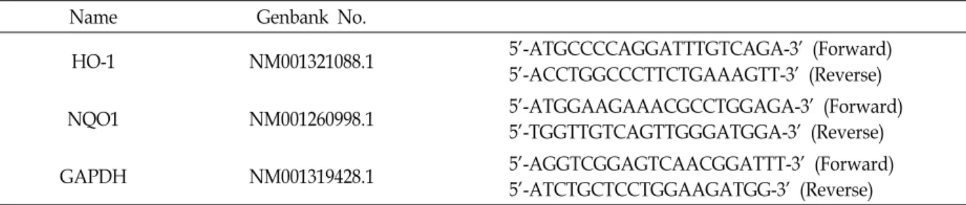
Table 1. List of real-time RT-PCR primers
Name Genbank No.
HO-1 NM001321088.1 5'-ATGCCCCAGGATTTGTCAGA-3' (Forward)
5'-ACCTGGCCCTTCTGAAAGTT-3' (Reverse)
NQO1 NM001260998.1 5'-ATGGAAGAAACGCCTGGAGA-3' (Forward)
5'-TGGTTGTCAGTTGGGATGGA-3' (Reverse)
GAPDH NM001319428.1 5'-AGGTCGGAGTCAACGGATTT-3' (Forward)
5'-ATCTGCTCCTGGAAGATGG-3' (Reverse)
vided in Table 1.
Western blot analysis
After appropriate treatment, HaCaT cells were collected by centrifugation and resuspended with 200 μl RIPA buffer (50 mM Tris-HCl at pH 8.0, 150 mM NaCl, 1% NP-40, 0.5%
sodium deoxycholate, protease inhibitors cocktail) on ice for 1 h. Cell lysates were collected by centrifugation and protein concentration was measured by BCA Protein Assay Kit (Thermo Fisher, Pittsburgh, PA, USA). Equal amounts of cell lysates were resolved by SDS-PAGE and transferred to PVDF membrane. The membrane was incubated in blocking buffer (5% skim milk in 1x PBS-0.1% Tween-20, PBST) for 1 hr and hybridized with the appropriate primary antibodies overnight at 4℃. After washing three times with 1x PBST for 30 min, the membrane was hybridized with appropriate HRP-conjugated secondary antibody for 1 h at room temper- ature and washed three times with 1x PBST solution for 30 min. The membrane was visualized by using an enhanced chemiluminescence (ECL) detection system (GE healthcare, Piscataway, NJ, USA). Actin blot was used as control for an equal loading of samples.
Detection of the Intracellular ROS level and oxida- tive stress markers, 8-hydroxyguanosine (8-OH-G) and 4-hydroxynonenal (4-HNE)
Formation of intracellular ROS was detected using the non-fluorescent probe DCF-DA. DCF-DA, a non-fluorescent substance, passively diffuses into cells and is deacetylated by esterases to form nonfluorescent 2‘,7’-dichlorofluorescein.
In order to measure the changes in the intracellular 8-OH-G and 4-HNE levels, HaCaT cells were grown on a slice glass and incubated with blocking serum (1% BSA) for 30 min.
After washing with 1x PBS three times, cells were hybridized with primary antibodies against 8-hydroxyguanosine (8-OH- G) and 4-hydroxynonenal (4-HNE) overnight at 4°C. The slides were washed with 1x PBS three times and probed with
FITC-conjugated rabbit secondary antibody. The fluorescent images were obtained with a C2 confocal microscope (Nikon Korea, Seoul, Korea).
Results
Identification of ethanol extracts of Rheum un- dulatum and Inula japonica as novel ARE inducers
In order to identify novel traditional medicinal extracts that possess significant stimulatory effects on the ARE-de- pendent gene expression in human keratinocytes, we have exposed individual ethanol extracts of 400 traditional medic- inal plants to HaCaT-ARE-luciferase cells and measured the resulting luciferase activity. Sulforaphane, a chemopre- ventive isothiocyanate, was included as a positive control.
While many of ethanol extracts positively affected the ARE-dependent luciferase activity, we observed that ethanol extract of Rheum undulatum or Inula japonica induced a partic- ularly strong ARE-dependent luciferase activation, whose level was superior to that by sulforaphane (Fig. 1).
Ethanol extracts of Rheum undulatum and Inula japonica increase the NRF2/ARE-dependent phase II cytoprotective enzyme levels in HaCaT cells
We next examined whether ethanol extracts of Rheum un- dulatum and Inula japonica affect the viability of HaCaT cells.
Our MTT assay result shows that both extracts did not in-
hibit, but rather increased the viability of HaCaT cells after
48 hr (Fig. 2A). Based on the observation that ethanol ex-
tracts of Rheum undulatum and Inula japonica increased the
ARE-dependent luciferase activity in HaCaT-ARE-luciferase
cells (Fig. 1), we next examined whether these extracts could
induce the NRF2-dependent phase II cytoprotective enzymes
in HaCaT cells. Our real-time RT-PCR and Western blot re-
sults show that ethanol extracts of Rheum undulatum and
Inula japonica elicited a significant transcriptional activation
(Fig. 2A) and concomitant induction of phase II cytopro-
Fig. 1. Identification of ethanol extracts of Rheum undulatum and Inula japonica as novel inducers of ARE-dependent luciferase ex- pression in HaCaT-ARE-luciferase cells. Individual ethanol extract of traditional medicinal plants (20 μg/ml) were exposed to HaCaT-ARE-luciferase cells and the luciferase activity was measured after 24 hr. Sulforaphane (SFN) was included as a positive control. The statistical analysis was conducted by Student t-test with n=6.
A
0 hr 24 hr 48 hr 0 hr 24 hr 48 hr
B
0 hr 4 hr 8 hr 0 hr 4 hr 8 hr
0 hr 4 hr 8 hr 0 hr 4 hr 8 hr
Time (hr): Time (hr):
Fig. 2. Ethanol extracts of Rheum undulatum and Inula japonica increases transcription and translation of phase II cytoprotective enzymes, heme oxygenase-1 (HO-1) and NAD[P]H:quinone oxidoreductase-1 (NQO1) in human keratinocyte HaCaT cells.
(A) MTT assay demonstrates that ethanol extracts of Rheum undulatum (20 μg/ml) and Inula japonica (20 μg/ml) increases the viability of HaCaT cells after 48 hr. The statistical analysis was conducted by Student t-test with n=5 (B) Exposure of ethanol extracts of Rheum undulatum (20 μg/ml) and Inula japonica (20 μg/ml) to HaCaT cells elicited a transcriptional activation (upper panel), and a subsequent induction of HO-1 and NQO1 proteins (lower panel). The statistical analysis was conducted by Student t-test with n=5. Asterisks indicate a statistical significance with * p<0.05, ** p<0.01, and ***
p<0.05.
A
B
Fig. 3. Ethanol extracts of Rheum undulatum and Inula japonica exert strong anti-oxidative effects on human keratinocyte HaCaT cells. (A) Ethanol extracts of Rheum undulatum (20 μg/ml) and Inula japonica (20 μg/ml) attenuate the production of intracellular reactive oxygen species (ROS). HaCaT cells were exposed to TPA alone or in combination with ethanol extracts of Rheum undulatum and Inula japonica and the intracellular level of ROS was observed after treatment of DCF-DA dye, using fluorescent microscope after 24 hr. (B) Ethanol extracts of Rheum undulatum and Inula japonica suppress the formation of intracellular oxidative stress markers, 8-OH-G and 4-HNE. HaCaT cells were exposed to TPA (10 nM) alone or in combination with ethanol extracts of Rheum undulatum (20 μg/ml) and Inula japonica (20 μg/ml) and the intracellular 8-OH-G and 4-HNE levels were examined by immunofluorescence after 24 hr.
tective enzymes (HO-1 and NQO1) in HaCaT cells (Fig. 2B).
Ethanol extracts of Rheum undulatum and Inula japonica protect HaCaT cells against TPA-induced oxidative DNA damages
Because ethanol extracts of Rheum undulatum and Inula japonica increased the NRF2/ARE-dependent phase II cyto- protective enzymes, we assumed that these extracts would be able to protect HaCaT cells against oxidative stress-medi- ated damages. To examine this hypothesis, we have exposed HaCaT cells to TPA alone or in combination with ethanol extracts of Rheum undulatum and Inula japonica, and meas- ured the levels of intracellular ROS and oxidative damages on cellular macromolecules using immunofluorescence assays.
As a result, we observed that both extracts significantly sup- pressed TPA-induced generation of intracellular ROS (Fig.
3A) and a subsequent formation of 8-OH-G and 4-HNE, as measured by immunofluorescence in HaCaT cells (Fig. 3B).
These results suggest that the induction of NRF2/ARE-de- pendent phase II cytoprotective enzymes by ethanol extracts of Rheum undulatum and Inula japonica was responsible for a decreased oxidative damages in HaCaT cells.
Discussion
As keratinocytes are constantly exposed to oxidative dam- ages by environmental oxidants and electrophiles, it can be envisaged that finding out novel medicinal plants that can boost up the NRF2 activity could be useful in maintaining the integrity of skin. In line with this idea, we have identified that ethanol extracts of Rheum undulatum and Inula japonica exhibit significant anti-oxidant effects through the induction of NRF2/ARE-dependent phase II cytoprotective gene expression. Our preliminary LC/MS/MS study identified that ethanol extracts of Rheum undulatum and Inula japonica contain various types of natural compounds (data not shown): ethanol extract of Rheum undulatum contained 3,4,5- trihydroxystilbene 4-glucoside, rhaponticin, trachrysone-8- glucoside, chrysophanol, and rhein and that of Inula japonica contained nepitrin, axillarin, 1-O-acetylbritannilactone, and inuchinenilide. While it remains to be seen whether any of them were responsible for the stimulation of ARE-dependent phase II gene expression, they could serve, at least in part, as marker compound(s) in the future to ascertain the uni- formity of ethanol extracts of Rheum undulatum and Inula japonica.
Previous studies have demonstrated that Rheum un- dulatum contain various types of biologically beneficial stilbenoids. For example, Dong et al. have illustrated that stilbenoids from Rheum undulatum protected hepatocytes against oxidative stress [4]. Likewise, Lee et al. have demon- strated that Rheum undulatum exhibited anti-obesity and hy- polipidemic effects in vivo via the inhibition of protein ty- rosine phosphatase 1B [8]. Jo et al. have demonstrated that rhapontin and rhapontigenin existing in Rheum undulatum possessed anti-hyperlipidemic effects in rats fed with a high-cholesterol diet [6]. Most notably, Choi et al. have dem- onstrated that desoxyrhapontigenin increased NRF2-mediated HO-1 expression in macrophages [3]: ethanol extract of Rheum undulatum used in the present study contained an analogous compound, rhaponticin. Instead, Inula japonica seem to possess sesquiterpenoids as major metabolites [16]
and previous studies have demonstrated that Inula japonica also exhibit many beneficial effects in vivo and in vitro. For example, Choi et al. have demonstrated that Inula japonica exerted anti-inflammatory responses in RAW 274.7 cells [2].
Shan et al. have demonstrated that Inula japonica exhibited anti-diabetic and hypolipidemic effects in mice [12]. In line with the above reports, we demonstrate in the present study that ethanol extract of Inula japonica exhibits strong NRF/ARE inductive effects on human keratinocytes.
Currently, we are attempting to pinpoint the chemical in- gredient(s) existing in Inula japonica that might be respon- sible for NRF2/ARE activation.
Acknowledgement
This work was supported by Bio-Theme Cluster program of Korea Industrial Complex Corp. (KICOX) (RDNWIC 15005).
References
1. Bryan, H., Olayanju, A., Goldring, C. and Park, B. 2013. The Nrf2 cell defence pathway: Keap1-dependent and -indepen- dent mechanisms of regulation. Biochem. Pharmacol. 85, 705- 717.
2. Choi, J., Park, Y., Li, Y., Jin, M., Lee, J., Lee, Y., Son, J., Chang, H. and Lee, E. 2010. Flowers of Inula japonica attenuate in- flammatory responses. Immune Netw. 10, 145-152.
3. Choi, R., Cheng, M. and Kim, S. 2014. Desoxyrhapontigenin up-regulates Nrf2-mediated heme oxygenase-1 expression in macrophages and inflammatory lung injury. Redox Biol.
2, 504-512.
초록:종대황과 선복화 에탄올 추출물의 인간 피부 세포주인 HaCaT 세포에서 NRF2/ARE에 의존적 인 유전자 발현의 유도를 통한 항산화 효과
유옥경
1․이용걸
2․도기환
2*․금영삼
1* (
동국대학교 약학과,
리치케미칼)
본 연구진은 HaCaT-ARE-luciferase 세포를 이용하여 400 여개의 약용식물 에탄올 추출물 중 NRF2/ARE 유도 효과가 있는 신규 추출물을 검색하였고 이를 통하여 종대황(Rheum undulatum)과 선복화(Inula japonica)의 주정 추 출물이 HaCaT-ARE-luciferase 세포에서 ARE 활성을 강하게 유도하는 것을 관찰하였다. 종대황과 선복화 에탄올 추출물은 HaCaT 세포에서 생존(viability)을 증가시켰고 NRF2/ARE에 의존적인 phase II cytoprotective 효소인 heme oxygenase-1 (HO-1)와 NADPH:quinone oxidoreductase-1 (NQO1)의 전사 및 단백질 발현을 강하게 유도하 였다. 또한 종대황과 선복화 추출물은 HaCaT 세포에서 TPA로 유도한 세포 내 활성 산소 및 이를 통하여 생성되 는 스트레스 마커인 8-hydroxydeoxyguanosine (8-OH-dG)과 4-hydroxynonenal (4HNE)의 발생을 강하게 억제하 였다. 본 연구는 종대황과 선복화의 에탄올 추출물이 인간 피부 세포주인 HaCaT 세포에서 NRF2/ARE에 의존적 인 유전자 발현의 유도를 통하여 강력한 항산화 효과를 발휘한다는 것을 증명한다.
4. Dong, G., Lee, Y., Jeong, J., Zhao, H., Jeon, R., Lee, H. and Ryu, J. 2015. Stilbenoids from rheum undulatum protect hepatocytes against oxidative stress through AMPK Activation. Phytother. Res. 29, 1605-1609.
5. Harder, B., Jiang, T., Wu, T., Tao, S., Rojo de la Vega, M., Tian, W., Chapman, E. and Zhang, D. D. 2015. Molecular mechanisms of Nrf2 regulation and how these influence chemical modulation for disease intervention. Biochem. Soc.
Trans. 43, 680-686.
6. Jo, S., Kim, J. and Lim, Y. 2014. Antihyperlipidemic effects of rhapontin and rhapontigenin from rheum undulatum in rats fed a high-cholesterol diet. Planta Med. 80, 1067-1071.
7. Keum, Y. 2011 Regulation of the Keap1/Nrf2 system by che- mopreventive sulforaphane: implications of posttransla- tional modifications. Ann. N. Y. Acad. Sci. 1229, 184-189.
8. Lee, H., Kim, J., Park, K. and Lim, Y. 2012. Rhapontigenin converted from rhapontin purified from Rheum undulatum enhances the inhibition of melanin synthesis. Biosci.
Biotechnol. Biochem. 76, 2307-2309.
9. Ma, Q. and He, X. 2012. Molecular basis of electrophilic and oxidative defense: promises and perils of Nrf2. Pharmacol.
Rev. 64, 1055-1081.
10. Menegon, S., Columbano, A. and Giordano, S. 2016. The dual roles of NRF2 in cancer. Trends Mol. Med. 22, 578-593.
11. Schieber, M. and Chandel, N. 2014. ROS function in redox signaling and oxidative stress. Curr. Biol. 24, R453-462.
12. Shan, J., Yang, M. and Ren, J. 2006. Anti-diabetic and hypo- lipidemic effects of aqueous-extract from the flower of Inula japonica in alloxan-induced diabetic mice. Biol. Pharm. Bull.
29, 455-459.
13. Sporn, M. and Liby, K. 2012. NRF2 and cancer: the good, the bad and the importance of context. Nat. Rev. Cancer 12, 564-571.
14. Surh, Y. 2003. Cancer chemoprevention with dietary phytochemicals. Nat. Rev. Cancer 3, 768-780.
15. Taguchi, K., Motohashi, H. and Yamamoto, M. 2011.
Molecular mechanisms of the Keap1-Nrf2 pathway in stress response and cancer evolution. Genes Cells 16, 123-140.
16. Yang, C., Wang, C. and Jia, Z. 2003. Sesquiterpenes and oth- er constituents from the aerial parts of Inula japonica. Planta Med. 69, 662-666.

![Fig. 2. Ethanol extracts of Rheum undulatum and Inula japonica increases transcription and translation of phase II cytoprotective enzymes, heme oxygenase-1 (HO-1) and NAD[P]H:quinone oxidoreductase-1 (NQO1) in human keratinocyte](https://thumb-ap.123doks.com/thumbv2/123dokinfo/5005374.548575/5.892.143.756.157.994/undulatum-increases-transcription-translation-cytoprotective-oxygenase-oxidoreductase-keratinocyte.webp)

