박성수ㆍ서진원ㆍ최진영
서울대학교 치과대학 구강악안면외과학교실
반안면왜소증 환자의 안면비대칭 해소를 위한 늑연골 이식 및 악교정 수술의 동시 이용: 증례보고
Correction of Facial Asymmetry Using Costochondral Graft and Orthognathic Surgery in Hemifacial Microsomia Patient: Case Report
Sung-Soo Park, Jin-Won Suh, Jin-Young Choi
Department of Oral and Maxillofacial Surgery, College of Dentistry, Seoul National University, Seoul, Korea
A 31-year-old woman with hemifacial microsomia presented to the Department of Oral and Maxillofacial Surgery, Seoul National University Dental Hospital. The patient was previously treated with distraction osteogenesis device to elongate right maxilla and mandibular ramus. But, the result was not satisfactory, to correct residual facial asymmetry due to hemifacial microsomia we planned costochondral graft for recon- struction of ramus and condyle, Le Fort I osteotomy and sagittal split ramus osteotomy for facial asymme- try. The right mandibular condyle and ramus was reconstructed with right eleventh costochondral graft via submandibular approach. Using costochondral graft and orthognathic surgery the facial asymmetry in hemifacial microsomia patient was corrected. 1-stage treatment consists of costochondral graft and orthog- nathic surgery can achieve function and esthetics at the same time, is timesaving to both patient and sur- geon.
Key words:Hemifacial microsomia, Costochondral graft, Orthognathic surgery Abstract
서 론
반안면왜소증(hemifacial microsomia)은 환자 얼굴의 한 쪽이 다른 쪽보다 작거나 발달이 덜 되었을 때를 의미한 다. 보통 편측성으로 나타나기 때문에 반안면왜소증으로 명 명되지만 안면기형과 두개봉합선(cranial vault)의 기형이 동반되기 때문에 두개안면왜소증으로도 불린다. 반안면왜 소증은 구순구개열 다음으로 흔한 악안면기형으로 알려져 있으며,1) 3500-6000명당 1명 정도의 빈도로 발생하는 것 으로 알려져 있다.2)
다른 악안면기형 환자와 마찬가지로 반안면왜소증 환자의 치료도 다각적 팀 접근에 의한 오랜 치료 기간을 필요로 한 다. 구강악안면외과의사, 교정과의사, 이비인후과의사, 언 어치료사, 안과의사, 소아과의사, 유전학자, 심리학자 등 여 러 전문가들의 협진이 필요하다. 악안면영역에서의 치료 목
적은 듣기, 말하기, 씹기, 삼키기, 숨쉬기 등의 최적의 기능 과 대칭, 균형의 심미를 얻는 것이다.
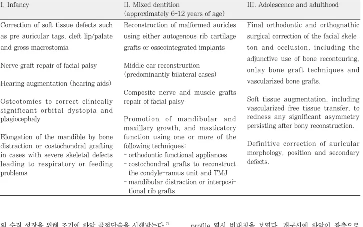
치료 단계에 대해서는 여러 곳에서 발표가 있어왔지만,3) 골격과 연조직 재건에 관한 고려로 제한이 있는 경우가 자 주 있다. Cousley 등4)은 반안면왜소증 환자의 치료와 그 시 기에 관해 간략히 정리해 발표했다(Table 1).
반안면왜소증으로 인해 이형성된 얼굴의 경조직과 연조직 을 재건하는 것은 어려운 일로, 수년에 걸쳐 여러 단계의 치 료를 요한다.1) 따라서 구강악안면영역에 있어 시기에 따라 적절한 치료를 선택하는 것이 항상 관심사였다. 1970년대 중반부터 1990년대 중반까지 사춘기 전의 Pruzansky- Kaban 분류 IIB와 III 환자를 대상으로 결여된 하악 상행 지를 재건하기 위해 늑연골 이식이 많이 사용되었다.5)영향 을 덜 받은 분류 I과 IIA의 어린 아이들은 사춘기 전까지 치 료를 받지 않거나,6) 기능적 교정 장치 치료를 받고 중안면
* 이 논문은 2010년도 정부(교육과학기술부)의 재원으로 한국연구재단의 지원을 받아 수행된 기초연구사업임(2010-0009448).
J Korean Assoc Maxillofac Plast Reconstr Surg 2010;32(4):351-358
의 수직 성장을 위해 조기에 하악 골절단술을 시행받는다.7) 골격 수정 후에 얼굴의 연조직 결핍을 지방이나 진피지방 이식8)을 통해 수정하고, 나중에 유리 혈관화피판을 시행하 기도 한다.7)
이번 증례 보고에서는, 성인 반안면왜소증 환자에서 악교 정 수술과 늑연골 이식을 병행하여 반안면왜소증으로 인한 안면비대칭을 해소한 증례를 보고하고자 한다.
증례보고
31세 여자 환자로 2000년 12월에 서울대학교 치과병원 구강악안면외과에 좌측 턱관절의 통증을 주소로 처음 내원 한 후 우측 하악 과두가 형성 부전된 반안면왜소증으로 진 단받고, 교정 치료 중에 2006년 12월에 재내원하였다. 반 안면왜소증으로 인한 안면비대칭 진단 하에 2007년 3월에 Le Fort I 골절단술, 우측 하악지 골절단술, 구외 골신장장 치 삽입 술식으로, 상하악 동시 신연(bimaxillary distrac- tion)을 시도하였으나 안면비대칭이 잔존하고 우측 하악 상 행지의 골신장이 미흡하였다.
환자는 우측 하악지와 하악 과두가 없는 Pruzansky- Kaban 분류 type IIB 환자로 귀 형성부전이나 skin tag는 없었다. Canting은 상악 양측 제1대구치의 근심협측교두를 기준으로 6 mm 정도였고, 입술과 같은 연조직도 골격과 마 찬가지로 canting을 보였다. 우측 하악 과두 결손으로 인해
profile 역시 비대칭을 보였다. 개구시에 하악이 좌측으로 편위되었고, 하악 우측 근돌기가 과증식되어 있었다(Fig.
1). 처음 치료 계획은 수술을 2단계로 진행하여, 우선 1차 로 Le Fort I 골절단술과 비이환측 하악지시상분할골절단 술을 통해 안면비대칭을 해소하고, 2차로 결여된 하악 과두 의 기능을 재건하기 위해 늑연골 이식을 시행하려고 하였 다. 하지만 환자의 사정 등으로 수술을 동시에 진행하여 상 악의 경우 Le Fort I 골절단술, 하악의 경우 이환측은 늑연 골이식, 근돌기 절제술(coronoidectomy), 골이식, 비이환 측은 하악지시상분할골절단술, 우각부 성형술을 계획하였 다(Fig. 2).
Le Fort I 골절단술을 통하여 상악 좌측 제1대구치의 근 심협측교두를 기준으로 5 mm 올리고, 상악 우측 제1대구 치의 근심협측교두를 기준으로 1 mm 내려 상악골의 cant- ing을 교정하였다(Fig. 3). Intermediate wafer 제작을 위 한 모형 수술은 CAD/CAM (3D Dental Scanning System (Wafer), Orapix, Seoul, Korea)을 이용하였다 (Fig. 4). 수술시에 좌측 zygomatic buttress 부위의 상악 골벽이 얇아 스크류로 고정하기가 여의치 않아 L-자형 금속 판 3개로만 상악골을 고정하였다(Fig. 5).
하악의 경우, 좌측은 하악지시상분할골절단술, 우측은 과 증식된 근돌기 절제술을 시행하고(Fig. 6), final wafer를 통해 교합을 맞추어 고정하였다. 이어서 우측 하악 과두 및 하악지 재건을 위하여 연골과 골연결부분을 포함하는 우측 I. Infancy
Correction of soft tissue defects such as pre-auricular tags, cleft lip/palate and gross macrostomia
Nerve graft repair of facial palsy Hearing augmentation (hearing aids)
Osteotomies to correct clinically significant orbital dystopia and plagiocephaly
Elongation of the mandible by bone distraction or costochondral grafting in cases with severe skeletal defects leading to respiratory or feeding problems
II. Mixed dentition
(approximately 6-12 years of age) Reconstruction of malformed auricles using either autogenous rib cartilage grafts or osseointegrated implants
Middle ear reconstruction (predominantly bilateral cases)
Composite nerve and muscle grafts repair of facial palsy
Promotion of mandibular and maxillary growth, and masticatory function using one or more of the following techniques:
- orthodontic functional appliances - costochondral grafts to reconstruct
the condyle-ramus unit and TMJ - mandibular distraction or interposi-
tional rib grafts
III. Adolescence and adulthood
Final orthodontic and orthognathic surgical correction of the facial skele- ton and occlusion, including the adjunctive use of bone recontouring, onlay bone graft techniques and vascularized bone grafts.
Soft tissue augmentation, including vascularized free tissue transfer, to redness any significant asymmetry persisting after bony reconstruction.
Definitive correction of auricular morphology, position and secondary defects.
Table 1.Principal stages and techniques applicable to the treatment of hemifacial microsomia (Adapted from reference 4)
Fig. 1. A 31-year old woman was a hemifacial microsomia patient with deficient right mandibular ramus and condyle. The patient also had a right hyperplastic coronoid process.
Fig. 2.Comparison of preoperative and postoperative state. Fig. 3.The maxilla was placed desired positioned according to the intermediate wafer. Maxillary occlusal canting was corrected.
Fig. 4. Model surgery for intermediate wafer was made by using CAD/CAM (3D Dental Scanning System (Wafer), Orapix, Seoul, Korea). The maxil- la was impacted 5 mm at left side (reference point:
mesiobuccal cusp tip of left upper first molar), elongated 1 mm left side (reference point:
mesiobuccal cusp tip of right upper first molar). (A : Before the model surgery, B: After the model surgery).
Preop Postop
J Korean Assoc Maxillofac Plast Reconstr Surg 2010;32(4):351-358
11번째 늑연골을 채취하였다. 채취한 오른쪽 11번째 늑연 골의 길이는 약 5 cm 정도였다(Fig. 7 A). 특히 하악 과두 의 역할을 위하여 연골 부분을 과두 형태와 유사하게 다듬 었다. 늑연골 이식의 경우 통상 전이개 접근법과 악하 접근 이나 Risdon 접근법을 통해 시행하나, 이번 증례 보고에서 는 악하 접근법으로만으로 채취한 늑연골을 관절와(gle- noid fossa)로 위치시킬 수 있어 전이개 접근법을 시행하지 않고 늑연골 이식을 시행하였다(Fig. 7 B). 늑연골 이식 후 하악을 개구운동 시켰을 때 좌우측으로 변위없이 잘 가이드 되었다. 추가로 하악 우측 우각부위의 절흔 부위로 인한 좌
우 비대칭을 해소하기 위해 절제된 근돌기를 다듬어 하악 우측 하연에 augmentation하여 대칭을 맞추었다.
상하악 수술 후 좌측 하악각 부위가 비대해 보여 좌측 하 악각 절제술(angle reduction)을 추가 시행하였다. 환자의 안면비대칭이 해소된 것을 확인하고 수술을 종료하였고, 술 후 전후방 방사선사진을 통해서도 이를 확인할 수 있었다 (Fig. 5 A). 수술 다음날 환자에게 final wafer를 물리고 와이어로 악간 고정술을 시행하였다. 5주간 악간 고정을 시 행 후 와이어를 제거하고 개구운동을 연습시켰고, 안정적인 교합을 보였다(Fig. 8).
Fig. 5.Postoperative radiographs (A: Postero-anterior view, B: orthopanogram). Le Fort I osteotomy was used to correct maxil- lary canting. But, left zygomatic buttress area could not be fixed with plates and screws because of thin maxillary anterior wall.
Costochondral graft was used to reconstruct the deficient right mandibular ramus/condyle unit. Sagittal split ramus osteotomy was done on left mandible. To correct asymmetry, resected coronoid process was augmented on right mandibular inferior bor- der and left mandibular angle was reduced.
Fig. 6. Right hyperplastic coronoid process was resected. It was trimmed for bone augmentation on right mandibular inferior border to correct asymmetry and fixed with 3 metal screws.
Fig. 7.Harvested costochodral graft was about 5cm long (A). The right white por- tion of rib is cartilage which is a substitute for mandibular condyle.
Submandibular approach was used for costochondral graft on right mandible (B). It was fixed using onlay bone graft technique with 3 metal screws.
고찰 및 결론
반안면왜소증 환자의 치료 프로토콜을 마련하기 위해서는 경조직, 연조직의 결여 정도에 대한 정확한 분류가 필요하 다. Kaban 등9)은 나이와 골격적 구조에 따른 전반적인 평 가 원칙 및 치료 방법을 요약했다(Table 2). 치료의 개념은 크게 성장기의 어린이에서 하는 경우와 성장이 종료된 반안 면왜소증의 말기에 하는 경우로 나누어 볼 수 있다.
성장 중인 반안면왜소증 환자의 수술적 접근은 반안면왜 소증의 안면비대칭이 진행 중이기 때문에 우선은 지켜 보는 것이다. 그리고 비대칭의 진행 정도는 하악의 변형 정도와 관련이 있다.9)안면 변형의 정도가 성장의 정도보다 심하게 진행되는 경우에 하악의 비대칭에 대한 평가를 사춘기 후에 해야 한다고 주장하는 연구도 있다.10)따라서 성장 중인 안 면비대칭 환자에 대한 수술은 아직도 논란의 여지가 있다.
치료의 접근은 어린이의 하악 골격 type (I, IIA, IIB, III), 중안면의 변형 정도, 연령, 심리사회적인 적응 정도에 영향
을 받는다.
Type I과 IIA 환자에서는 하악을 늘리고 회전시켜서 정중 선을 맞춘다. 하악을 늘리는 것은 보통의 악교정 술식을 이 용하거나 골신장장치를 이용해서 서서히 늘릴 수도 있다.
보통의 골절단술과 골신장 장치를 시행받았으나 상악골 수 술을 시행받지 않은 성장 중인 어린이의 경우는, 이환측에 개교합을 만들어 치아가 맹출함과 동시에 상악이 수직적 성 장을 하도록 함으로써 상악 교합 평면을 맞춘다.11) Type IIB와 III 환자의 경우는 뼈가 충분하고 턱관절이 있을 경우 골신장장치를 이용해 하악을 늘리거나 늑연골 이식을 이용 해 하악지와 턱관절을 재건한다. 개교합을 만들고 상악의 성장과 상악 치아의 맹출을 유도하는 교정 치료는 type I과 IIB의 환자에서와 동일하다. 성장 중인 반안면왜소증 환자 에 있어, 하악을 늘리고 이환측에 개교합을 만들면 성인이 되었을 때 양악 수술이 필요하지 않을 수도 있다.11)
청소년기나 성인 같은 말기 반안면왜소증 환자의 치료는 보통의 악교정 술식을 따라 술전 교정을 하고 양악수술을
Fig. 8. Clinical photography of postoperative 5 weeks. Compared with preoperative clinical photography (Fig. 1), the facial asymmetry was corrected. Occlusion was stable, the patient was instructed to practice mouth opening.
J Korean Assoc Maxillofac Plast Reconstr Surg 2010;32(4):351-358
시행한다. Type I과 IIA 반안면왜소증 환자 치료는 Le Fort I 골절단술을 이용하여 상악골의 위치를 바로 잡고 그 에 맞추어 양측의 하악 골절단술을 시행하는 것이다. 해부 학적 구조와 변형 정도에 따라 골절단과 골이식이 동반될 수도 있다. Type IIB와 type III의 경우, 이환 부위의 하악 지와 과두의 재건이 필요하다.
Gilles가 처음으로 턱관절 재건에 늑연골 이식을 사용하 여 보고하였다.12) 이 외에도 비골, 흉쇄관절, 중족골, 장골 등 다양한 자가골 이식 방법이 사용되어 왔다.13-15) 그러나 늑연골 이식이 구강악안면외과 의사들에게서 가장 많이 사 용된다. 늑연골 이식의 장점으로는 생체적합성, 가동성, 기 능적 적합성, 환자에 대한 최소한의 손상 등이 있다.16)특히 늑연골 이식체의 성장 가능성은 어린 아이들에게 사용하기 에 적합하다.17) 자라는 어린 아이와 성인에서의 하악 과두
재건 목표는 뚜렷이 차이가 난다.17) 즉, 성인에서의 관골하 악복합체의 재건은 상실된 기능과 심미를 복원하기 위해서 이지만, 어린 아이에서는 그뿐만 아니라 계속적인 성장을 할 수 있는 골이식을 하기 위한 것이다.
여러 가지 이유로 완전한 늑연골 접합부(costochondral junction)를 동반한 늑골이 최고의 공여부로 생각되고 있 다.17)뼈로 된 부분은 하악 과두 경부를 대체하면서 하악체 에 이식체를 고정할 수 있게 하고, 연골 부분은 관절와에 위 치한다. 뼈와 연골 경계부는 성장의 중심이 된다.18) 늑연골 이식은 성공적으로 임상에 이용되고 있지만 합병증 등 문제 점도 있다. Figueora 등17)은 여러 술자들이 연구한 늑연골 이식의 장점과 문제점들을 요약해 발표하였다(Table 3).
여러 저자들이 반안면왜소증 환자의 결여된 하악지에 늑 연골 이식을 하는 수술 방법에 대해 발표해 왔다.6,11,19) Table 3.Advantages and problems of costochondral graft (Adapted from reference 17)
Advantages Problems
1. Biologic and anatomic similarities to the condyle 1. Pneumothorax
2. Low morbidity of donor site 2. Placement of graft in nonexistent or rudimentary gle- noid fossae, as well as attachment to rudimentary or severely malformed ramus
3. Numerous donor sites 3. Placement in areas with scarring, poor vascularization, and deficient soft-tissue envelop as in severe forms of hemifacial microsomia
4. Easy to obtain and quick to adapt 4. Difficulty in orientation of rib in graft site 5. Regeneration of donor site 5. Dislodgment of graft laterally or superiorly 6. Retains integrity after transplantation 6. Pain
7. Demonstrated growth potential in juvenile series 7. Infection
8. Uncontrolled and unpredictable growth 9. Fracture of the costochondral joint 10. Facial nerve damage
Table 2.General assessment principles by age and skeletal type (Adapted from reference 9)
Skeletal type Deciduous Mixed Permanent
Type I Skin tag removal Functional appliance Presurgical orthodontics
Mandibular elongation osteotomy/distraction Bimaxillary surgery Creation of posterior open bite
Type IIA As above As above As above
Functional appliance Mandibular distraction
Type IIB As above Construction of ramus, condyle, glenoid fossa Presurgical orthodontics Creation of posterior open bite Bimaxillary surgery
Construction of
temporomandibular joint
Type III As above Level occlusal plane As above
Construction of ramus, condyle, glenoid fossa Creation of posterior open bite
Level occlusal plane
Munro는 반대편 가슴에서의 늑연골 채취를 주장하였다.
그는 하악지의 재건에 여섯 번째 늑골의 만곡이 가장 적절 하다고 하였고, 세 개의 절개를 주었다. 수여부인 하악을 노 출시키기 위한 구강내 절개, 관골궁, 안와측벽, 관절와의 재 건을 위한 반두부관상절개(hemicoronal incision), 턱관절 을 노출시키기 위한 전이개 절개를 준다고 하였다.20) 이번 증례에서는 여자 환자의 오른쪽 유방 하방에 흉터를 가장 적게 남기기 위해 11번째 늑연골을 이용하였다.
Mulliken 등은 턱관절을 노출시키기 위한 전이개 절개와 하악 우각부 노출을 위한 악하 절개를 선호하였다.19)그들은 하악골의 전진, 신장 그리고 회전을 위한 광범위한 연조직 박리를 위해 구강외 접근법이 필요하다고 하였고, 안면 신 경 손상 가능성에 주의해야 한다고 하였다. 구강내나 구강 외로 노출된 하악지에서 이환측의 근돌기 절제술이 필요할 수도 있고, 비이환측의 보상적인 골절단술이나 근돌기 절제 술이 필요할 수도 있다고 하였다. 여러 저자들이 전이개 접 근법과 악하 접근법이나 Risdon 절개를 같이 사용한 것을 보고하였으나,19,21) 이번 증례에서는 전이개 접근없이도 관 절와에 늑연골을 위치시키는 것을 확인할 수 있어서 악하 접근만을 이용하였다.
Obwegeser는 반안면왜소증이 심한 성인 환자의 경우에 만 늑연골 이식을 시행할 것을 주장하였다.6) 그는 어린 나 이에서의 골절단이 안면 골격의 성장을 방해한다고 믿었다.
그리고 수술은 두 단계로 진행되어야 한다고도 주장하였다.
첫 번째 단계에서, 턱관절과 하악지를 늑연골로 재건하고, 만약 괜찮다면 저형성된 측두골, 완와 측벽, 관골궁을 골이 식하여 보강할 수도 있다. 두 번째 단계에서 골이식을 동반 한 Le Fort I 골절단술과 하악지시상분할골절단술, 이부성 형술을 시행한다. Obwegeser는 이 방법이 합병증 위험이 적고, 좀더 예측 가능한 결과를 가져온다고 하였다.
Lauritzen 등은 반안면왜소증을 5가지로 구분하였고, 분 류는 턱관절의 해부학적 구조와 안구로의 침범 정도에 따라 구분했다. 그리고 그 5가지 분류 형태는 각각에 따라 특이 적인 수술 방법이 적용된다고 하였다.22)이번 증례 보고에서 는 Lauritzen과 동료들이 분류한 형태들 중 type II에 해당 한다. 그들의 분류에 따르면, Type II는 하악지가 결여되어 있고 과두가 없으나, 관골궁은 존재하고 재건될 과두를 지 지하기에 충분한 경우다. Type II의 수술은 과두를 재건하 고 하악지의 수직 고경을 높이기 위해 늑연골 이식을 한다.
그리고 Le Fort I 골절단술을 하면서 골이식을 하고, 비이 환측에서 하악지시상분할골단술을 시행하여 보상하고, 이 부성형술도 보통 필요하다.
하악지와 과두를 재건하기 위해 늑연골 이식을 받은 7명 의 성장 중인 환자와 19명의 성장이 끝난 환자를 대상으로 한 장기간의 후향적 연구에 따르면, 선형 과성장으로 인한 부정교합은 없었고, 3명의 환자에서만 관절면 측면에 과성
장을 보였다고 한다.23) 그리고 성장 중이거나 성장이 끝난 환자에서의 늑연골 이식은 하악지와 과두를 재건하는 데 적 합하다고 하였다.
이번 증례에서는 반안면왜소증 환자에 있어서 늑연골 이 식을 악하 접근법만으로 시행하여 구강외 반흔 발생을 최소 화하였고, 늑연골 이식과 동시에 악교정 수술을 시행함으로 써 추가 수술이 필요하지 않아 환자와 술자가 시간을 절약 하고, 악안면영역의 기능과 심미를 동시에 달성할 수 있었 다. 하지만, Onlay bone graft가 흡수에 약하고 교합과 아 래턱 기능 향상에 실패를 가져올 수도 있다는 보고24)도 있기 때문에 장기간의 추적 검사를 통하여 예후를 관찰하는 것이 필요하다.
References
1. Mommaerts MY, Nagy K : Is early osteodistraction a solu- tion for the ascending ramus compartment in hemifacial microsomia? A literature study. J Craniomaxillofac Surg 30 : 201, 2002.
2. Poswillo D : Otomandibular deformity: pathogenesis as a guide to reconstruction. J Maxillofac Surg 2 : 64, 1974.
3. David DJ, Mahatumarat C, Cooter RD : Hemifacial micro- somia: a multisystem classification. Plast Reconstr Surg 80 : 525, 1987.
4. Cousley RR, Calvert ML : Current concepts in the under- standing and management of hemifacial microsomia. Br J Plast Surg 50 : 536, 1997.
5. Padwa BL, Mulliken JB, Maghen A et al : Midfacial growth after costochondral graft construction of the mandibular ramus in hemifacial microsomia. J Oral Maxillofac Surg 56 : 122, 1998.
6. Obwegeser HL : Correction of the skeletal anomalies of oto-mandibular dysostosis. J Maxillofac Surg 2 : 73, 1974.
7. Kaban LB : Pediatric oral and maxillofacial surgery, 1st ed. Philadelphia, WB Saunders, 1990, p.273.
8. Davis WB : Reconstruction of hemiatrophy of face. Case report. Plast Reconstr Surg 42 : 489, 1968.
9. Kaban LB, Padwa BL, Mulliken JB : Surgical correction of mandibular hypoplasia in hemifacial microsomia: the case for treatment in early childhood. J Oral Maxillofac Surg 56 : 628, 1998.
10. Polley JW, Figueroa AA, Liou EJ et al : Longitudinal analysis of mandibular asymmetry in hemifacial microso- mia. Plast Reconstr Surg 99 : 328, 1997.
11. Murray JE, Kaban LB, Mulliken JB : Analysis and treat- ment of hemifacial microsomia. Plast Reconstr Surg 74 : 186, 1984.
12. Gillies HD : Plastic surgery of the face based on selected cases of war injuries of the face including burns with origi- nal illustrations. London, Oxford University Press, 1920, p.177.
13. Converse JM, Coccaro PJ, Becker M et al : On hemifacial microsomia. The first and second branchial arch syndrome.
Plast Reconstr Surg 51 : 268, 1973.
14. Snyder CC, Levine GA, Dingman DL : Trial of a stern- oclavicular whole joint graft as a substitute for the tem- poromandibular joint. Plast Reconstr Surg 48 : 447, 1971.
15. Smith AE, Robinson M : A new surgical procedure in bilateral reconstruction of condyles, utilizing iliac bone
J Korean Assoc Maxillofac Plast Reconstr Surg 2010;32(4):351-358
grafts and creation of new joints by means of non-elec- trolytic metal; a preliminary report. Plast Reconstr Surg 9 : 393, 1952.
16. Bell WH : Modern practice in orthognathic and reconstruc- tive surgery, Philadelphia, WB Saunders, 1992, p.872.
17. Figueroa AA, Gans BJ, Pruzansky S : Long-term follow- up of a mandibular costochondral graft. Oral Surg Oral Med Oral Pathol 58 : 257, 1984.
18. Ware WH, Taylor RC : Growth center transplantation to replace damaged mandibular condyles. J Am Dent Assoc 73 : 128, 1966.
19. Mulliken JB, Ferraro NF, Vento AR : A retrospective analysis of growth of the constructed condyle-ramus in children with hemifacial microsomia. Cleft Palate J 26 : 312, 1989.
20. Grabb WC, Smith JW : Plastic surgery, 3d ed. Boston, Little, Brown, 1979, p.131.
21. Guyuron B, Lasa CI, Jr. : Unpredictable growth pattern of costochondral graft. Plast Reconstr Surg 90 : 880, 1992.
22. Lauritzen C, Munro IR, Ross RB : Classification and treatment of hemifacial microsomia. Scand J Plast Reconstr Surg 19 : 33, 1985.
23. Perrott DH, Umeda H, Kaban LB : Costochondral graft construction/reconstruction of the ramus/condyle unit:
long-term follow-up. Int J Oral Maxillofac Surg 23 : 321, 1994.
24. Longacre JJ, Destefano GA, Holmstrand K : The early versus the late reconstruction of congenital hypoplasias of the facial skeleton and skull. Plast Reconstr Surg Transplant Bull 27 : 489, 1961.
저자 연락처
우편번호 110-749
서울특별시 종로구 연건동 275-1
서울대학교 치과대학 구강악안면외과학교실 최 진 영
원고 접수일 2010년 03월 29일 게재 확정일 2010년 05월 13일
Reprint Requests Jin-Young Choi
Department of Oral and Maxillofacial Surgery, College of Dentistry, Seoul National University
#275-1 Yeongun-dong, Jongno-gu, Seoul, 110-749, Korea Tel: +82-2-2072-2631 Fax: +82-2-766-4948 E-mail: [email protected]
Paper received 29 March 2010 Paper accepted 13 May 2010