Introduction
Intertidal fishes live at the area of land and sea where they are alternately submerged in water and exposed to the air as tide recedes.
Rocky shores include boulder flats, rock pools, or combinations of all of these, along with gravel, sand, and shell substrate. By the tide causing the presence or absence of water, the intertidal fishes come out water during a low tide. Accord- ing to these emergence behaviors, Martin (1995) classified the intertidal fishes into three groups:
the skippers, the tidepool emergers and the re- mainers. In the rocky shores, the remainer fish may become out of water passively, simply by remaining in a site that is usually covered with water, but during a low tide is exposed to air
(Horn and Riegle, 1981; Martin, 1995; Horn et al., 1999). These sites may be protected from desiccation by cover, such as a boulder or sea- weed, and there may be an extremely shallow layer of water on the substrate. Finally, these fishes may be found under boulders or in crevices during low tides, resulting in aquatic hypoxia in tide pools (Horn and Riegle, 1981; Horn et al., 1999).
Therefore, intertidal fishes have an additional air-breathing mechanism to overcome their hy- poxia (Bridges, 1988; Graham, 1997; Horn et al., 1999). However, most marine intertidal fishes, unlike freshwater air-breathing fishes, have no specialized or enclosed air-breathing organ (Gra- ham, 1997). According to Horn et al. (1999), res- piratory gas exchanges in the marine fishes must take place across the same surfaces in air as it does in water: the gills, the skin, and perhaps the linings of the opercular and buccal cavities. A
Structure and Histochemistry of the Skin of a Flat-headed Goby, Luciogobius guttatus
(Gobiidae, Pisces) from Korea
Jong-Young Park*, Yong-Joo Lee1and Ik-Soo Kim Faculty of Biological Sciences and Institute for Biodiversity Research,
Chonbuk National University
1Jeonju National University of Education, Korea
To investigate the skin of the flat-headed goby, Luciogobius guttatus, it was used 8 body regions such as the head, the upper and lower jaw, the chin, the back, lateral region, abdomen, and the operculum. The epidermis consisted of three layers: the outermost layer, middle layer and stratum germinativum (basal layer). The outer- most layer consisted of rather flattened cells arranged in 1 to 4 layers and mucous cells. The middle layer consisted of large epidermal cells occupying the height of the epidermis with 1 to 10 layers, causing a web-shaped structure. Due to the large epi- dermal cells of the middle layer, L. guttaus had a thick epidermis. The large epider- mal cell contains tonofilaments, lucent contents, and desmosome. The basal layer was comprised of a single layer having cuboidal cells. A large number of fine blood capillaries were found just under the basal layer. The dermis consisted mostly of stratum compactum with numerous blood capillaries but had no scale.
Key words : epidermis, mucous cell, swollen cell, intertidal fish, Luciogobius guttatus
*Corresponding author: [email protected]
─
─ 120 ──
flat-headed goby, Luciogobius guttatus, are small and demersal, and they distribute in Northwest Pacific of China, Korean Peninsula, and Japan (Nelson, 1994). Like other intertidal fishes, L.
guttatus inhabits tidepools and river mouths, and stays under stones on the dried bottom for the duration of the low tide. Although about 60 species in 12 families among the intertidal fishes were reported on air emergences and aerial res- pirometry (Graham, 1997; Horn et al., 1999), L.
guttatus had not been studied. Through the his- tology of the skin, therefore, we are going to get information related cutaneous air respiration in L. guttatus.
Materials and Methods
Two males and three females, ranging from 59.8 mm to 71.6 mm in standard length, were collected in June, 2004 by a hand and a small net from Gigo-ri, Changseon-myeon, Hanam-gun, Jeollanam-do, in the southern coast Korea, 34�
20′30′′N and 126�30′10′′E. The specimens were anaesthetized with MS222. For histological examination, the skin fragments, 5×5 mm2, were fixed in 10% neutral buffered formaldehyde and 8 regions of the skin were taken from the following regions of the body: the top of the head, the cheek near the operculum, the upper jaw and lower jaw, the outer operculum close to the pec- toral fin, the dorsal region near the dorsal fin, the lateral region just beneath the dorsal fin, and the ventral region near the anal fin.
We dehydrated these sections through a stan- dard ethanol series to 100%, cleared in xylene and then embedded in wax (Paraplast, Oxford).
We deparaffinized 5µm sections and stained them with Harris hematoxylin, Ehrlich hema- toxylin, and counter-stained with eosin, and Masson trichrome stain (Gurr, 1956) for general histology. Mucins of gland were demonstrated by alcian blue solution (AB) at pH 1.0 and 2.5 (Steed- man, 1950; Lev and Spicer, 1964), and the peri- odic acid-schiff (PAS) method. In addition, the PAS technique was employed in combination with AB and vice-versa for neutral and acid mucins. Acid mucin was shown by metachromat- ic reactions with toluidine blue (Tock and Pearse, 1965). Also, high iron diamine (HID) and with AB (Spicer, 1965) were used for nature of the acid mucins.
For evaluations of the epidermis, we took two
skin fragments by each region per specimen by Video Test-Master (VT image analysis program, USA) on hematoxylin and eosin preparations.
More than 10 sections were used for measure- ment per two skin fragments.
Results
1. Epidermis
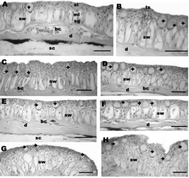
In 8 different body regions, the structures of the epidermis exhibited always a stratified epi- thelium which consisted fundamentally of the superficial layer, the middle layer and the stra- tum germinativum (Fig. 1, A to F). This principle structure is seems in all 8 regions although there are differences in their thickness or stratum es- pecially in the middle cell layer.
In light microscopy, the superficial layer, outer- most layer, of epidermis is composed of rather flattened cells arranged in one to five layers. Bet- ween these cells, small spherical or flask-shaped unicellular mucous cells are present (Fig. 1, A to H). The muocus cells with a large or small spher- ical body open to the exterior by a short narrow neck that opens on the surface by a wide pore.
They have a basal, spherical or oval nucleus with a thin rim of slightly basophilic cytoplasm. The mucous cells with highly vacuolated and baso- philic secretory matter are varied ranging from 14.0 to 22.6µm in height in the 8 regions (Table 1). Of them, the mucous cells in the abdomen were the largest, mean 22.6±5.8µm (10.0 to 37.1 µm) and in the upper jaw the smallest, 14.0±3.9 µm (6.9 to 25.5µm). Whereas, the number of the mucous cell was much, 1 to 10, in the upper jaw, but the rest was mostly similar. Some mucous cells extend to the upper middle layer. The mu- cous cells gave a deep red color reaction with PAS technique, which is diastase resistant,γ-meta- chromasia with toluidine blue, blue with the AB at pH 1.0 and 2.5 (Fig. 2, A to E, Table 3). The mucous cells, giving a red color with aldehyde fucshin and black color with high iron diamine, were likely to be sulfomucins. In mild methyla- tion/AB and acetylation/PAS techniques they were negative, and in methylation /saponifica- tion-AB they were stained blue. Their nuclei were purple or red color with AB-PAS reaction and Masson trichrome stain (Fig. 2, D to E)
The middle layer of epidermis is simpler in structure (Fig. 1, A to H). This layer consists of smaller and voluminous cells. Due to the volumi-
nous cell, the so-called swollen cell, the middle layer exhibits a web-like structure in appearance (Figs. 1 and 2). The swollen cells have an oval nucleus and a homogeneous cytoplasm, and their
boundary is clear (Fig. 1). Occasionally, they appeared to vesicles or vacant acellular struc- tures due to a loss of nucleus. The swollen cell does not any reactions against histochemical
Fig. 1. Transverse sections of the epidermis of Luciogobius guttatus with Ehrlich haematoxylin and eosin (bars indicate 50 µm). A, The skin of the back consisted of epidermis, dermis (d) and subcutis (sc). The epidermis consisted of super- ficial layer (sl), middle layer (ml) and stratum germinativum (sg). bc, blood capillary; d, dermis; sw, swollen cell;
arrow, mucous cell. B, The base of the operculum. d, dermis; ts, taste bud; sw, swollen cell; arrow, mucous cell. C, The base of the abdomen. bc, blood capillary; sw, swollen cell; arrow, mucous cell. D, The head. bc, blood capillary;
d, dermis; sw, swollen cell; arrow, mucous cell. E, The base of the cheek. bc, blood capillary; d, dermis; sc, subcutis;
sw, swollen cell; arrow, mucous cell. F, The lateral region. d, dermis; sw, swollen cell; arrow, mucous cell. G, The upper jaw. See the stratified swollen cell (sw) in the middle layer. a, mucous cell. H, The lower jaw. The stratified swollen cell (sw) occupies the entire height of the epidermis. arrow, mucous cell.
tests.
Towards the outermost layer of the epidermis these swollen cells become stratified. They occu-
pied the entire height of the epidermis (Fig. 1).
The swollen cells are various in height, reaching 15.7µm to 27.0µm and arranged in several layers
Fig. 2. Special staining reactions on the mucous cell of the epidermis (A-E) (bars indicate 50µm) and transmission electron micrographs (F and G) of the skin of Luciogobius guttatus. A, The operculum. PAS rection. sw, swollen cell; arrow, mucous cell. B, The abdomen. HID/AB stain. sw, swollen cell; arrow, mucous cell. C, The operculum. AB (1.0). d;
dermis; arrow, dermis. D, The cheek. Masson trichrome stain. d, dermis; sw, swollen cell; arrow, mucous cell. E, The cheek. AB/PAS stain. D, dermis; sw, swollen cell; arrow, mucous cell. F, The swollen cell of the epidermis in the dorsum. dorsum, ds, desmosome; tf, tonofilaments. Bar indicates 0.5µm .G. The dermis of the dorsum. bl, basal layer; cf, collagen fibers; cp, chromatophores; ep, epidermis; mp, melanophores. Bar indicates 4µm.
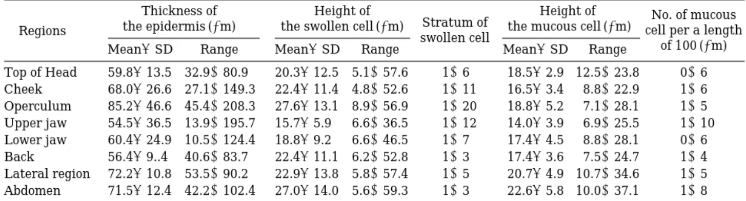
Table 1. The thickness of epidermis, height of the swollen cell and mucous cell in the 8 body regions of the epidermis of Luciogobius guttatus
Thickness of Height of
Stratum of Height of No. of mucous Regions the epidermis (µm) the swollen cell (µm)
swollen cell the mucous cell (µm) cell per a length
Mean±SD Range Mean±SD Range Mean±SD Range of 100 (µm)
Top of Head 59.8±13.5 32.9~80.9 20.3±12.5 5.1~57.6 1~6 18.5±2.9 12.5~23.8 0~6
Cheek 68.0±26.6 27.1~149.3 22.4±11.4 4.8~52.6 1~11 16.5±3.4 8.8~22.9 1~6
Operculum 85.2±46.6 45.4~208.3 27.6±13.1 8.9~56.9 1~20 18.8±5.2 7.1~28.1 1~5 Upper jaw 54.5±36.5 13.9~195.7 15.7±5.9 6.6~36.5 1~12 14.0±3.9 6.9~25.5 1~10 Lower jaw 60.4±24.9 10.5~124.4 18.8±9.2 6.6~46.5 1~7 17.4±4.5 8.8~28.1 0~6
Back 56.4±9..4 40.6~83.7 22.4±11.1 6.2~52.8 1~3 17.4±3.6 7.5~24.7 1~4
Lateral region 72.2±10.8 53.5~90.2 22.9±13.8 5.8~57.4 1~5 20.7±4.9 10.7~34.6 1~5 Abdomen 71.5±12.4 42.2~102.4 27.0±14.0 5.6~59.3 1~3 22.6±5.8 10.0~37.1 1~8
(Table 1). Among the 8 regions, the swollen cells are the largest in the abdomen, mean 27.0±14.0 µm (5.6 to 59.3µm), and the smallest in the upper jaw, mean 15.7±5.9µm (6.6 to 36.5µm), as in the number of mucous cell. The stratum of swollen cell arranged in the middle layer was much in the operculum (1 to 20 layers), the upper jaw (1 to 12 layers) and the chin (1 to 11 layers), and less in the back (1 to 3 layers), the abdomen (1 to 3 layers) and the lateral region (1 to 5 layers) (Table 1). In transmission electron microscopy, the swollen cells s contain few organells, and they have tonofilament, lucent contens, and des- mosome. Pigment cells are present in this layer.
The basal layer, the stratum germinativum was composed of a single layer of either cuboidal cells, or more or less columnar cells (Fig. 1). Blood vessels located adjacent to the sense organs of the epidermis are found in the intraepithelial layer. A number of blood capillaries are present just under the basal membrane.
The epidermis has taste buds, which is typical- ly composed of sensory cells and is a bottle-shaped expansion (Fig. 1, B).
2. Dermis
The dermis lacks scales and there was no clear differentiation between the stratum laxum and the stratum compactum. However, the upper region just under basal membrane was supplied with fine blood capillaries and nerve fiber (Fig. 1, A and E). The stratum compactum consists of bundles of coarse collagenous fibres arranged compactly in several layers (Fig. 1, A and E).
This layer is weakly PAS positive and become a deep green color in Masson trichrome-stained preparations for collagen. This layer contains a few pigments cells, nerve cells and blood capil- laries.
The thickness of the dermis is varied from 10.9 to 30.8µm in the 8 regions (Table 2). In the 8 region, the lateral region is the thickest, 30.8±
10.8µm (15.2 to 56.6µm), whereas the upper jaw and the lower jaw are thin, 10.9±4.0µm (4.8 to 24.1µm) and 12.8±3.8µm (7.1 to 21.7µm), res- pectively.
The dermis has two kinds of chromatophores, melanophore and colorless pigmented cells (Fig.
2, F and G). Melanosomes of melanophores are electron-opaque ellipsoidal structures, whereas colorless pigments, reflecting platelets, appear a stack of empty space. Reflecting platelets are cuboidal, polygonal, or squamous, and have highly
Table 2. The thickness of the epidermis, the dermis, and the subcutis in 8 the body regions of the skin of Luciogobius guttatus
Regions Thickness of the epidermis (µm) Thickness of the dermise (µm) Thickness of the subcutis (µm)
Mean±SD Range Mean±SD Range Mean±SD Range
Top of Head 59.8±13.5 32.9~80.9 26.7±7.1 12.6~43.3 26.2±8.4 14.5~43.9
Cheek 68.0±26.6 27.1~149.3 27.1±8.5 10.7~42.5 26.4±10.1 12.9~52.0
Operculum 85.2±46.6 45.4~208.3 22.8±7.2 14.8~43.8 14.9±10.1 10.0~19.0
Upper jaw 54.5±36.5 13.9~195.7 10.9±4.0 4.8~24.1 8.8±1.9 2.5~11.5
Lower jaw 60.4±24.9 10.5~124.4 12.8±3.8 7.1~21.7 8.9±1.3 2.7~10.9
Back 56.4±9.4 40.6~83.7 27.6±9.4 13.9~57.5 25.5±7.5 18.4~34.2
Lateral region 72.2±10.8 53.5~90.2 30.8±10.8 15.2~56.6 24.3±6.9 8.1~39.1
Abdomen 71.5±12.4 42.2~102.4 25.7±8.2 9.8~42.5 20.5±5.5 10.4~30.4
Table 3. A summary of the histochemical tests performed to show the nature of the mucous cell of epider- mis in Luciogobius guttatus
Techniquesemployed Mucous cell Remarks
Weigerts iron Haemaoxylin ± B
Masson trichrome ++ G
PAS ++++or ++++++ R
PAS after digestion in
malt diastase/PAS ++++ R
AB (1.0) ++or ++ B
AB (2.5) ++++ B
AB/PAS ++++ B, BR, R
PAS/AB ++++ B, BR, R
Toluidine blue ++ γ-meta
Acetylation/PAS -
Acetylation/Saponication /PAS ++
Methylation/AB -
Methylation/Saponification/AB ++ B
Aldehyde fucshin ± R
Aldehyde fucshin/AB (2.5) ++++ BR
HID ± N
HID/AB ++++ BN
B, blue; BN, bluish black; BR, bluish red; G, green; N, black; R, red; Intensity: -, no reaction; ±, weak reaction; ++clear reaction;
++ strongly reaction.
variable dimensions.
3. Subcutis
This layer is situated under dermis (Fig. 1, A and E). The average thickness is approximately 56.9µm, ranging from 29.7 to 79.2µm. Fine col- lagen fiber bundles from the stratum compactum traverse this layer. The main branches of the nerves and blood vessels are found in this layer.
In hematoxylin and eosin preparations, this layer has numerous empty spaces composed of fat cells (Fig. 1, A and E). The thickness of the subcutis ranges from mean 8.8µm to 26.4µm in the 8 regions (Table 2), and the values are somewhat less than those of the dermis. Their thickness show similar values except for the upper jaw, the lower jaw and the operculum, mean 8.8µm, 8.9 µm, and 14.9µm, respectively. The subcutis of the upper jaw, the law jaw and the operculum is not clear differentiation.
Discussion
The skin of cutaneously respiratory fishes, have been documented as the following structures: a thicker epidermis due to several types of glands, intraepithelial blood capillaries, abundant blood capillaries in the superficial dermis, a well-devel- oped vascularization, reduction or absence of scales, and a definite area with acid mucopolysac- charides in the dermis (Jakubowski, 1958; Liem, 1967; Johansen, 1970; Mittal and Munshi, 1971;
Mittal and Banerjee, 1974; Mittal et al., 1980;
Whitear, 1986; Suzuki, 1992; Yokoya and Tamu- ra, 1992; Park and Kim, 1999, 2000; Park, 2002a, b; Zhang et al., 2003; Park et al., 2003a, b, 2006).
Of characters in well-known cutaneously respi- ratory fishes, a thicker epidermis was dependent on several kinds of glands and a specific large epidermal cell. Firstly, epidermis having two kinds of gland cells, both a small mucous cell and a large club cell, or a small mucous cell and a large sacciform cell, were known in Mastacembe- lus, Amphipnous, Misgurnus, Iksookimia, and Liobargus (Mittal and Munshi, 1971; Mittal and Banerjee, 1974; Mittal et al., 1980; Park and Kim, 1999; Park, 2002a; Park et al., 2003b). Because two gland cells occupy the most height of the middle layer in their epidermis, their epidermis was thicker. Secondly, the synbranchoid fish, Monopterus albus, has only mucous gland cells.
Nevertheless, the epidermis has thicker due to a
large mucous cell occupying the entire height of the epidemis (Liem, 1967). Thirdly, although the mucous cell in the epidermis is small or absent, there was specific large and voluminous epider- mal cells swollen by epidermal cell, swollen cell.
Multi-stratified swollen cells occupy the entire height of the epidermis, which it shows web-struc- ture in appearance. The swollen cell in the mid- dle layer of the epidermis was characteristic of amphibious mudskippers, Periophthalmus, Scar- telaos and Boleophthalmus, which undergo cuta- neous respiration using air (Whitear, 1986; Al- Kadhomity and Hughes, 1988; Yokoya and Ta- mura, 1992; Zhang et al., 2000; Park, 2002b; Park et al., 2000, 2006).
The swollen cell found in the epidermis of L.
guttatus was only reported in mudskipper fish, Scartelaos, Boleophthalmus, and Periophthalmus.
The epidermis of L. guttatus have found mucous cell and swollen, as reported in Scartelaos and Boleophthalmus. Otherwise, Periophthalmus has only swollen cell without mucous cell. In particu- lar, the swollen cell was known as a modification of epidermal cell which undergo cutaneous respi- ration using air, in Periophthalmus, Scartelaos and Boleophthalmus (Whitear, 1986; Al-Kadhomi- ty and Hughes, 1988; Yokoya and Tamura, 1992;
Park et al., 2000; Zhang et al., 2000; Park, 2002;
Park et al., 2006). Also, it was known that the swollen cell has desmosome in some Periophthal- mus (Whitear, 1986; Suzuki, 1992; Park, 2002).
Also, the swollen cell of L. guttatus contains tonofilaments, lucent contents, and desmosome, as in the above three genera mudskipper fishes.
The size and abundance of mucous cells play an important role in supporting and maintaining the normal relationships of the cutaneous epithe- lium (Liem, 1967). Also, the appearance of the mucous cell in the epidermis is specifically adapt- ed to a common ecology in which protection form desiccation and lubrication during burrowing (Liem, 1967; Mittal and Munshi, 1971).
No the epidermis of L. guttatus have intraepi- thelial blood capillaries, unlike Periophthalmus and Liobagrus. Nevertheless, breathing is pos- sible through the blood vessels in the dermis because diffusion of oxygen take place readily across the mucous coat of the epithelium (Jaku- bowski, 1958; Liem, 1967; Mittal and Munshi, 1971; Whitear, 1986; Graham, 1997; Horn et al., 1999). Liem (1967) had been experimentally demonstrated in the epidermis of Monopterus albus with only large mucous cells devoid of intra-
epithelial blood capillaries. However, L. guttaus has plenty of blood capillaries just beneath the basal layer of the epidermis. It means that oxy- gen obtained through the mucous cell is suffici- ent to be able to diffuse to the deeper dermis.
L. guttaus has no well-defined lymphatic spaces in the epidermis and definite areas with acid mucopolysaccharides in the dermis, as described in other cutaneously respiratory fishes as Hetero- pneustes, Mastacembelus, Amphipnous, Iksoo- kimia, Misgurnus, Periophthalmus, and Boleo- phthalmus (Mittal and Munshi, 1971; Park and Kim, 2000; Park, 2002b; Park et al., 2003a).
L. guttatus has no scale. Absence and reduction of the scales was mainly found in cutaneously respiratory fishes such as Misgurnus, Perioph- thalmus, Heteropneustes, Mastacembelus, Iksoo- kimia, Amphipnous, Monopterus, and Liobargrus considered as an adaptation for movement or burrowing (Liem, 1967; Mittal and Munshi, 1971;
Whitear, 1986; Park, 2002; Park et al., 2003a, b).
On the basis of the ecological aspect and struc- tural characteristics of the skin, L. guttatus seems to be related to a cutaneous respiratory system for adaptation to its environments.
Acknowledgements
This work was supported by Jeonju Nationa- tional University of Education Research Grant (2005).
References
Al-Kadhomiy, N.K. and G.M. Hughes. 1988. Histological study of different regions of the skin and gills in the mudskipper, Boleophthalmus boddarti with respect to their respiratory function. J. Mar. Biol. Ass. U.K., 68 : 413~422.
Bridges, C.R. 1988. Respiratory adaptations in intertidal fish. Amer. Zool., 28 : 79~96.
Graham, J.B. 1997. Air breathing fishes: Evolution, diver- sity, and adaptation. Academic Press, San Diego, 299pp.
Gurr, G.T. 1956. A practical manual of medical and biologi- cal staining techniques. Interscience, New York.
Horn, M.H., K.L.M. Martin and M.A. Chotkowski. 1999.
Intertidal fishes. Academic Press, San Diego, 399pp.
Horn, M.H. and K.C. Riegle. 1981. Evaporative water loss and intertidal vertical distribution in relation to body size and morphology of stichaeoid fishes California. J.
Exp. Mar. Biol. Ecol., 50 : 273~288.
Jakubowski, M. 1958. The structure and vascularization of the skin of the pond-loach (Misgurnus fossilis L.). Acta
Biol. Cracoviensia, 1 : 113~127.
Johansen, K. 1970. Air breathing in fishes. In: Hoar W.S.
and D.J. Randall (eds), Fish Physiology IV. Academic Press, New York, pp. 361~411.
Lev, R. and S.S. Spicer. 1964. Specific staining of sulphated groups with alcian blue at low pH. J. Histochem. Cyto- chem., 12 : 309.
Liem, K.F. 1967. Functional morphology of the integumen- tary, respiratory, and digestive systems of the synbran- choid fish, Monopterus albus. Copeia, (1967) : 375~388.
Martin, K.L.M. 1995. Time and tide wait for no fish: Inter- tidal fishes out of water. Environ. Biol. Fish., 44 : 165~ 181.
Mittal, A.K. and J.S.D. Munshi. 1971. A comparative study of the structure of the skin of certain air-breathing fresh -water teleosts. J. Zool. Lond., 163 : 515~532.
Mittal, A.K. and T.K. Banerjee. 1974. Structure and kerati- nization of the skin of a fresh-water teleost Notopterus notopterus (Notopteridae, Pisces). J. Zool. Lond., 174 : 314~355.
Mittal, A.K., M.A. Whitear and S.K. Agarwal. 1980. Fine structure and histochemistry of the epidermis of the fish, Monopterus cuchia. J. Zool. Lond., 191 : 107~125.
Nelson, J.S. 1994. Fishes of the World. 3rd ed. John Wiley and Sons, New York, 600pp.
Park, J.Y. 2002a. Morphology and histochemistry of the skin of the spined cobitid fish, Iksookimia koreensis, in relation to respiration. Folia Zool., 51 : 241~247.
Park, J.Y. 2002b. Structure of the skin of an air-breathing mudskipper fish, Periophthalmus magnuspinnatus. J.
Fish Biol., 60 : 1543~1550.
Park, J.Y. and I.S. Kim. 1999. Structure and histochem- istry of skin of mud loach, Misgurnus anguillicaudatus (Pisces, Cobitidae), from Korea. Korean J. Ichthyol., 11 : 109~116.
Park, J.Y. and I.S. Kim. 2000. Structure and cytochemistry of skin in spined loach, Iksookimia longicorpus (Pisces, Cobitidae). Korean J. Ichthyol., 12 : 25~32.
Park, J.Y., I.S. Kim and S.Y. Kim. 2000. Histological study on skin of the amphibious fish, Periophthalmus modes- tus. Korean J. Biol. Sci., 4 : 315~318.
Park, J.Y., Y.I. Lee, I.S. Kim and S.Y. Kim. 2003a. A com- parative study of the regional epidermis of an amphibi- ous mudskipper fish, Boleophthalmus pectinirostris (Gobiidae, Pisces). Folia Zool., 52 : 431~440.
Park, J.Y., I.S. Kim and S.Y. Kim. 2003b. Structure and histochemistry of the skin of a torrent catfish, Liobagrus mediadiposalis. Environ. Biol. Fish., 66 : 3~8.
Park, J.Y., I.S. Kim and Y.J. Lee. 2006. A Study on the vascularization and structure of the epidermis of the air-breathing mudskipper, Periophthalmus magnuspin- natus (Gobiidae, Teleostei), along different parts of the body. J. Applied Ichthyol., 22 : 62~67.
Spicer, S.S. 1965. Histochemistry manual. The university of Kansas Medical Center, Kansas City, 54pp.
Steedman, H.F. 1950. Alcian blue 8G: a new stain for mucin.
Quart. J. Micr. Sci., 1 : 477~479.
Suzuki, N. 1992. Fine structure of the epidermis of the mudskipper, Periophthalmus modestus (Gobiidae). Ja- pan. J. Ichthyol., 8 : 379~396.
Tock, E.P.C. and A.G.E. Pearse. 1965. Preservation of tis- sue mucins by freeze-drying and vapour fixation. J. R.
microsc. Soc., 84 : 519~537.
Whitear, M. 1986. The skin of fishes including cyclostomes:
epidermis. In: Bereiter-Hahn, J., A.G. Matoltsy and K.S. Richards (eds), Biology of the integument Vol. 2 Vertebrates. Springer Verlag, New York, pp. 9~64.
Yokoya, S. and O.S. Tamura. 1992. Fine structure of the
skin of the amphibious fishes, Boleophthalmus pectini- rostris and Periophthalmus cantonensis, with special reference to the location of blood vessels. J. Morphol., 214 : 287~297.
Zhang, J., T. Taniguchi., T. Takita and A.B. Ali. 2000. On the epidermal structure of Boleophthalmus and Scarte- laos mudskippers with reference to their adaptation to terrestrial life. Ichthyol. Res., 47 : 359~366.
Received : May 14, 2007 Accepted : June 4, 2007
한국산 미끈망둑 Luciogobius guttatus(망둑어과) 피부의 구조 및 조직화학적 특성 박 종 영*∙이 용 주1∙김 익 수
전북대학교 생물과학부∙생물다양성연구소,
1전주교육대학교 과학교육학과
한국산 미끈망둑(Luciogobius guttatus)의 피부구조가 머리, 위턱, 아래턱, 뺨, 등, 체측, 배, 아가 미뚜껑 등 모두 8개 부분에서 비교 조사되었다. 이들 모두 표피는 맨바깥층, 중간층, 기저층으로 구성되었다. 맨바깥층은 1~4층의 편평세포와 점액세포로 구성되었으며, 중간층은 1~10층의 대 형 표피세포를 가지며 이러한 표피세포는 마치 거미줄 같은 형태를 보여주고 있다. 이러한 대형 표피세포의 존재는 두꺼운 표피를 갖도록 하며, tonofiilament와 desmosome을 가진다. 한편 기저 층은 한 층의 입방상피로 구성되어 있으며, 많은 모세혈관들이 기저층 바로 아래부분에 존재한 다. 진피에는 비늘이 존재하지 않으며 거의 대부분 모세혈관을 가지는 치밀층으로 구성되어 있 다.