녹차폴리페놀에 노출된 Salmonella typhimurium의 세포반응
최효경․오계헌*
순천향대학교 생명시스템학과
Cellular Responses of Salmonella typhimurium Exposed to Green Tea Polyphenols
Hyo-Kyung Choi and Kye-Heon Oh*
Department of Life Science and Biotechnology, Soonchunhyang University, Asan 336-745, Republic of Korea (Received May 23, 2012 / Accepted June 4, 2012)
The purpose of this study was to examine the cellular response of Salmonella typhimurium exposed to tea polyphenols (TPP) extracted from Korean green tea (Camellia sinensis L.). TPP showed a dose-dependent bactericidal effect on S. typhimurium. Analysis of cell membrane fatty acids of S. typhimurium cultures treated with TPP identified unique changes in saturated and unsaturated fatty acids, while scanning electron microscopic analysis demonstrated the presence of perforations and irregular rod forms with wrinkled surfaces in cells treated with TPP.
Two-dimensional polyacrylamide gel electrophoresis of soluble protein fractions from S. typhimurium cultures showed 16 protein spots increased by TPP. These up-regulated proteins including proteins involved in antioxidants and chaperons, transcript and binding proteins, energy and DNA metabolism were identified by peptide mass fingerprinting using MALDI-TOF. These results provide clues for understanding the mechanism of TPP induced stress and cytotoxicity on S. typhimurium.
Keywords: cellular responses, green tea polyphenols, proteomic analysis, TPP
*For correspondence. E-mail: [email protected]; Tel.: +82-41-530- 1353; Fax: +82-41-530-1350
녹차(Camellias inensis, L)는 전 세계적으로 널리 소비되는 대중적인 천연음료이다. 녹차는 flavanols, flavanoid, phenolic acid 등의 polyphenol류를 함유하는데, 대부분의 녹차의 polyphenol류는 카테킨(catechins)이라고 널리 알려진 flavanol 류이다(Graham, 1992). 녹차에 함유된 카테킨은 혈중 콜레스테 롤 감소, 항균작용, 혈압상승 억제, 혈당억제, 항산화작용, 항암 작용 등의 다양하고 유익한 생리학적 및 약리학적 활성을 가지 는 것으로 알려져 있으며, 이러한 작용들이 녹차의 폴리페놀(tea polyphenols, TPP) 성분에 기인하는 것으로 보고되고 있다. 특 히 TPP의 살균효과는 많은 세균들에서 보고되고 있지만, 어떤 기작에 의해서 세포의 살균이 이루어지는지는 확실하게 밝혀지 지 않고 있다. 사람의 장 및 구강에 서식하는 병원균들에 대한 TPP의 항미생물 효과에 대한 일부 연구가 보고되어있으나(Ahn et al., 1991; Hamilton-Miller, 1995; Yam et al., 1997;
Hirasawa and Takada, 2004), TPP에 노출된 병원성 세균에 대 한 세포반응과 프로테옴 분석과 관련된 연구는 TPP가 가지는 살 균기작을 규명하는데 매우 중요한 의미가 있음에도 불구하고, 현재 활용가능한 연구결과는 미미한 실정이다.
프로테옴 분석은 원핵세포의 유전자 발현에서 변화를 연구하 는데 중요한 실마리를 제공하는 방법으로 알려져 있다(Schmid et al., 1997; Jungblut et al., 1999, 2000). 2-Dimensional gel electrophoresis (2-DE)는 미생물이 어떤 스트레스 유발물질에 노출되었을 때 모든 세균의 수용성 단백질을 보여줄 수 있기 때 문에, 이들 유도 단백질의 선별에 이용될 수 있다.
이러한 배경을 바탕으로 녹차에서 추출된 TPP에 노출됨으로서, Salmonella typhimurium에서 유도된 단백질들의 특성을 조사하 는데 적용하였다. 다양한 농도의 TPP에 노출된 S. typhimurium 에서 생존율과 막 지방산 조성의 변화, 그리고 스트레스 충격단 백질인 DnaK와 GroEL의 유도를 조사하였다. TPP의 노출에 따 른 S. typhimurium의 사멸상태를 주사전자현미경을 통하여 관찰 하였으며, TPP의 작용기작의 규명과 세포반응과 발현된 프로테 옴의 분석을 통한 상관관계를 조사하였다.
재료 및 방법
세균과 배양조건
본 연구에서 사용된 S. typhimurium은 American Type Culture Collection (ATCC)에서 구입한 S. typhimurium 13311을 사용하 였다. 이 균주는 Luria-Bertani (LB) 액체 또는 고체배지에 접종
Tea polyphenols
녹차(Camellia sinensis, L)로부터 tea polyphenols (TPP, 순도 97% 이상)를 추출하였다. 녹차잎을 20% 에탄올에서 물중탕하고, 여과하여 추출액을 수거한 후, 50–60℃에서 감압농축하였다. 농축 후 분사건조기로 분말화하여 녹차추출물을 준비하였다. 준비된 액상의 녹차추출물은 산화를 방지하기 위하여 0.15 mM H3PO4
에서 유지시켰다. 녹차추출물의 TPP에 포함된 카테킨을 HPLC로 분석하여(자료 미제시), 5가지의 주요 화합물을 동정하였으며, 그 성분과 농도는 각각 다음과 같았다: (-)-epigallocatechin gallate (EGCG) (~50%), (-)-epicatechin gallate (ECG) (~24%), (-)-gallocatechin gallate (GCG) (~16%), (-)-epicatechin (EC) (~2%), (-)-epigallocatechin (EGC) (~1%).
TPP에 의한 스트레스 처리와 생존시험
LB broth에서 자란 세포들은 2,000×g로 10분간 원심분리하 여, 얻어진 균체를 세척하고, 30 ml phosphate buffer (pH 7.0)에 서 ~107 cells/ml 되도록 재현탁시켜 추후 실험에 사용될 균체를 준비하였다. 균체는 100 ml Erlenmeyer flask에서 스트레스원 (stressor)으로서 다양한 농도의 TPP에 처리된 후, 37℃에서 배 양되었다. 배양된 시료들은 회수되어 LB 고체평판배지에서 ml 당 colony forming units (CFUs)의 생균수 계수에 의해 결정되 었다(Cho et al., 2002).
Fatty acids methyl ester (FAME) 분석
S. typhimurium 세포에 존재하는 phospholipid ester-linked fatty acids은 MIDI system (Microbial Insights, Inc., USA)를 사 용한 fatty acid methyl esters (FAMEs)의 형태로 분석되었다.
Fatty acid 추출은 사용자 매뉴얼에서 제시한 방법에 따라 수행 되었다(Cho et al., 2002). FAME은 MIDI database에 의해 제공 된 authentic standard의 retention time과 mass spectra을 비교하 여 분석되었다. TPP에 대한 반응으로 변화된 지방산을 조사하기 위하여 세포들은 TSB에서 37℃, 24시간동안 배양하였으며, 수 거된 세포는 potassium phosphate buffer (pH 7.0)에서 2회 세척 하여, 5,000 µg/ml TPP을 포함하는 potassium phosphate buffer 에 재현탁시켰다. 12시간 배양 후, 지방산 조성은 위에서 기술된 방법에 따라 결정하였다.
TPP에 노출된 S. typhimurium 세포의 SEM 분석
LB 고체평판에서 24시간 동안 자란 S. typhimurium의 집락은 0.5 cm3 크기의 작은 한천블록으로 잘랐다. S. typhimurium 세포 를 포함하는 한천블록은 그 후 phosphate buffer에서 37℃, 12시 간 동안 3,000 µg/ml TPP에 노출시켰다. 그후 TPP로 처리된 집 락들은 고정, 탈수, 금으로 코팅 등의 이전에 기술된 방법에 따 라, Hitachi S-2500C 주사전자현미경(Hitachi, Japan)으로 조사 하였다(Ho et al., 2004).
phosphate buffer (pH 7.0)로 3회 세척하였다. 그후 동일 buffer 에 균체를 현탁하여 4℃를 유지하면서, 5 W로 30초간 10회 반 복하면서 파쇄하여 단백질을 추출하였다. 파쇄된 세포는 3,500×g로 30분간 원심분리한 후 단백질을 포함하고 있는 상등 액을 취해 Bradford 방법(1976)을 이용하여 단백질 정량을 실시 하였다.
SDS-PAGE와 Western blot 분석
SDS-PAGE 분석은 Bollag 등(1996)의 방법에 의해 실시되었 다. Separating gel은 12% acrylamide gel을 사용하였으며, stacking gel은 4% acrylamide gel을 사용하였다. 전기영동을 한 후 gel은 staining solution으로 2시간 염색하였고, destaining solution I destaining solution II로 탈색하였다. 스트레스 충격단 백질(DnaK와 GroEL)을 분석하기 위하여 Sambrook 등(2001) 의 방법에 따라 Western blot을 실시하였다.
이차원 젤 전기영동(2-DE)과 in-gel digestion
2-DE는 이전에 기술된 방법에 따라 실시되었다(Ho et al., 2004).
염색된 protein spot은 젤에서 잘라내어, 30 mM potassium ferricyanide와 100 mM sodium thiosulfate를 사용하여 탈염색 하고, 증류수로 세척하였다. 액체를 제거시키고, 그 젤은 50%
acetonitrile과 100% acetonitrile으로 각각 15분간 수축시키고, 200 µl의 100 mM NH4HCO3 (pH 7.8)로 재수화(rehydration)하 였으며, 마지막으로 100% acetonitrile로 다시 수축시켰다. 진공 원심분리기로 건조시킨 후, 젤은 50 mM NH4HCO3 (pH 7.8)를 포함하는 digestion buffer에서 재팽창시켜, 0.2 µg trypsin (Promega, USA)로 37℃에서 16시간동안 처리하였다. 그 peptides 는 elution buffer [50% acetonitrile, 5% trifluoroacetic acid (TFA)]로 용해시키고, 얻어진 peptide extract는 수거하여 진공 원심분리기로 동결건조한 후 -20℃에 저장하였다.
MALDI-TOF/MS에 의한 peptide sequencing
Digested peptides는 0.1% TFA 용액을 사용하여 다시 녹여, zip-tip C18 pipette tips (Millipore, USA)를 사용하여 탈염하였 다. Peptides는 CHCA matrix solution (10 mg/ml CHCA in 0.5% TFA/50% ACN, 1:1)을 사용하여 MALDI plate로 추출하 였다. Mass spectra는 4700 Proteomics Analyzer (Applied Biosystems, USA)를 사용하여 얻었다. 그 후, 단백질은 NCBI database에 대한 MASCOT (http://www.matrixscience.com)를 사용한 MALDI fingerprint data로부터 동정되었다(Pertins et al., 1999).
결과 및 고찰 TPP 노출에 따른 세균의 생존
TPP의 농도증가에 따른 S. typhimurium의 노출은 TPP가 살
Fig. 1. Survival of S. typhimurium after TPP shock. The cells were maintained at TPP concentrations of 0 µg/ml (●), 1,000 µg/ml (■), 2,000 µg/ml (▲), 3,000 µg/ml (◆), 4,000 µg/ml (▼) TPP, respectively. At intervals, the numbers of colony-forming units per ml (CFU/ml) of culture were determined
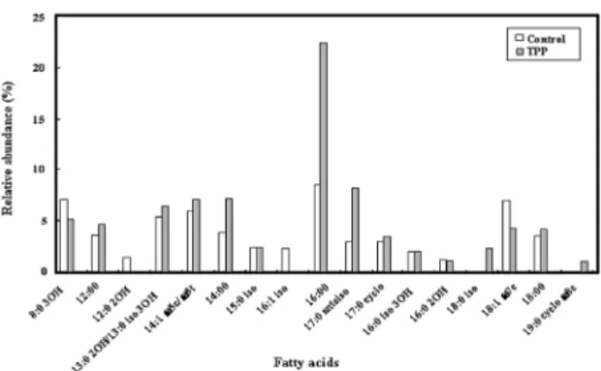
Fig. 2. Fatty acid profiles of S. typhimurium analyzed by GC-FID and GC-MS when grown for 24 h in TSB (control) or after TSB-grown cells were washed and re-suspended for 40 min in phosphate buffer containing 3,000 µg/ml of TPP.
Fig. 3. Scanning electron micrographs of S. typhimurium treated with TPP. (A) untreated cells; (B) cells treated with 4,000 μg/ml TPP for 80 min.
균력을 가지고 있다는 것을 보여주었다. S. typhimurium의 집락 형성단위(colony-forming unit)는 TPP의 노출시간이 경과함에 따라 점차적으로 감소하였으며, 그 감소비율은 사용된 TPP의 농 도에 비례하였다(Fig. 1). ~107 CFU/ml의 접종으로 시작하여, 각 각 3,000 µg/ml의 농도에서 80분간 노출 또는 4,000 µg/ml TPP 에서 60분간 노출 후 S. typhimurium의 집락은 발견되지 않았다.
TPP의 농도에 따른 임상환자들에서 분리된 nalidixic acid에 내 성인 S. typhimurium의 nalidixic acid에 의한 병용사용시 살균상 승과 세포반응에 대한 연구에서 유사한 결과가 보고된 바 있다 (Lim et al., 2008).
TPP 노출에 따른 세포 지방산 조성 변화
TSB에서 24시간동안 자란 S. typhimurium 세포에서, 그리고 3,000 µg/ml TPP를 포함하는 phosphate buffer에서 40분 동안 TSB에서 자란 세포에서 총 세포 지방산의 양을 비교하였다.
Fig. 2에서 보여주는 바와 같이, 주요한 지방산인 12:00, 13:0 2OH/13:0 iso 3OH, 14:1 ω5c/ω5t, 14:00, 16:00, 17:0 17:0 anteiso, 17:0 cyclo, and 18:0는 TPP에 노출된 S. typhimurium 세포에서 증가하였지만, 지방산인 8:0 3OH and 18:1 ω7c는 동 일한 조건하에서 감소하였다. TSB에서 자란 S. typhimurium 세 포에서 검출되지 않았던 지방산 18:1 iso, 19:0 cyclo ω8c는 TPP 에 노출된 세포의 총 세포 지방산들 가운데 각각 2.3%, 1.5% 증 가하였다. 더욱이 TSB에서 자란 S. typhimurium 세포에 존재하 였던 지방산인 12:0 2OH와 16:1 iso는 TPP에 노출된 세포에서 검출되지 않았으며, 부가적으로 분석된 15:0 iso, 16:0 iso 3OH, 16:0 2OH 등의 지방산에서는 TPP 노출에 따른 변화가 관찰되지 않았다. 이와 유사한 실험이 5,000 µg/ml TPP에 12시간 노출된 E. coli 세포에 대하여 실시되었다. TPP에 노출된 S. typhimurium 세포에서 14:1 ω5c/ω5t, 14:00, 16:00, 17:0 cyclo 등의 지방산이 증가하였고, 18:0와 19:0 cyclo ω8c는 감소하였으며, 17:0 anteiso 와 18:1 iso, 20:20 ω6,9c는 차이가 없었다고 보고하였다(Cho et al., 2007). 본 연구에서 수행된 S. typhimurium의 결과를 E. coli
세포와 비교하여 볼 때, TPP 노출에 따른 지방산의 변화는 균주의 종류와 노출시간에 따라 달라지는 것으로 확인되었으며, 이러한 세포막 성분인 여러가지 지방산 변화 증감의 차이는 균주의 종류 에 따른 세포막의 파괴 형태와도 연관이 있을 것으로 사료된다.
TPP 노출에 따른 세포 외부형태 변화
생존율 실험과 관련하여 TPP 노출에 따른 S. typhimurium 세 포의 외부형태의 변화를 주사전자현미경을 사용하여 관찰하였 다. 주사전자현미경 사진에서 보여주는 바와 같이, TPP에 노출 되지 않은 상태에서 복합배지(LB)에서 자란 정상 세포들은 매끈 한 표면의 전형적인 막대형을 보여주었다(Fig. 3A). 그러나 3,000 µg/ml TPP에서 120분 동안 처리된 세포들은 세포 표면에 구멍이 생기거나 움푹 패이고, 표면이 쭈그러지는 것이 관찰되 었다(Fig. 3B). 녹차폴리페놀이 구강에 존재하는 Streptococcus mutans, Streptococcus salivarius, Staphylococcus aureus, Neisseria meningitidis, Lactobacillus plantarum 등의 다양한 세 균에 대한 주사전자 현미경 관찰에서 처리된 모든 세균들이 제 각기 다른 양상으로 세포가 파괴되는 것이 보고된 바 있으며, 본 연구에서 관찰된 S. typhimurium에서 관찰된 것과도 커다란 차 이가 있는 것으로 확인되었다(Cho et al., 2010). 몇가지 연구에 서 고농도의 다양한 화학물질과 천연허브추출물이 막성분의 파괴
20 DnaK + + + ++
GroEL + + + +
40 DnaK + ++ ++ +
GroEL + + + ++
60 DnaK + + + +
GroEL + ++ ++ +
80 DnaK ++ + - -
GroEL ++ + + -
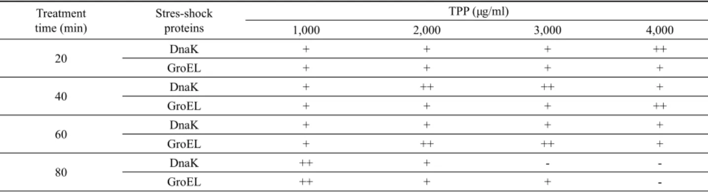
Fig. 4. Induction of stress shock proteins in S. typhimurium treated with 2,000 µg/ml TPP concentrations for different times. The stress shock proteins were analyzed by SDS-PAGE (A), and western blot with anti-DnaK (B), and anti-GroEL (C) monoclonal antibodies, respectively.
Fig. 5. 2-DE analysis of stress-induced proteins of S. typhimurium:
(A) control cells, and (B) cells treated with 2,000 µg/ml TPP for 60 min. Circled spots, up-regulated proteins.
에 의하여 세포에 독성이 있다는 것을 보여준 바 있다(Park et al., 2001; Cho et al., 2002, 2007, 2008; Chang et al., 2004). Ikigai 등(1993)은 TPP가 세균의 세포막을 손상시키고, EGCG의 살균 효과가 막에서 발생한 심각한 손상에 기인한다고 보고하였다.
SDS-PAGE와 Western blot
S. typhimurium 세포를 다양한 농도의 TPP (1,000–4,000 µg/ml)에 노출시킨 후에 경과시간에 따른 세포내에서 발현되는 스트레스 충격단백질의 발현을 조사하였다(Table 1). 충격단백 질 DnaK와 GroEL의 발현정도를 알아보기 위하여 주어진 농도 에서 20분 간격으로 TPP 충격에 의해 유도 발현되는 DnaK와 GroEL을 조사하였다. Fig. 4에서 보여주는 바와 같이, TPP 농도 가 낮을 때에 세균에서 발현되는 충격단백질은 노출시간에 경과 함에 따라 발현량이 증가하였으며, 또한 노출 TPP의 농도가 증 가함에 따라 발현량도 증가하였다. S. typhimurium 세포가 2,000 µg/ml TPP에 노출되었을 때, DnaK와 GroEL은 각각 40분과 60 분에 최대 발현을 보여주었다(Table 1). 고농도의 TPP와 장시간 의 노출에서 S. typhimurium 세포에서 유도되는 스트레스 충격 단백질은 크게 감소하였으며, 이 상태의 세포를 주사전자현미경 으로 관찰하였을 때 형태의 변형과 손상이 이루어져 죽게되는 것이 관찰되었다.
TPP 노출에 따른 프로테옴의 변화
2-DE protein profiles 분석은 S. typhimurium에서 세포 단백
질이 TPP에 노출된 후에 증가 또는 감소여부를 조사하기 위하 여, TPP 미처리 세균세포와 2,000 µg/ml TPP로 60분간 처리된 세포를 사용하여 실시되었다(Fig. 5). 대략 150 protein spots가 isoelectric focusing (pH 4-7)의 2-DE gel과 12% polyacrylamide SDS gel상에서 검출되었다. 30 protein spots 이상이 TPP로 처 리된 세균의 2-DE 상에서 발현되었으며, 이들 protein spots 가 운데 TPP 처리에 의해 유도되어 크기가 증가된 16개를 동정하 기 위하여 선발하였다.
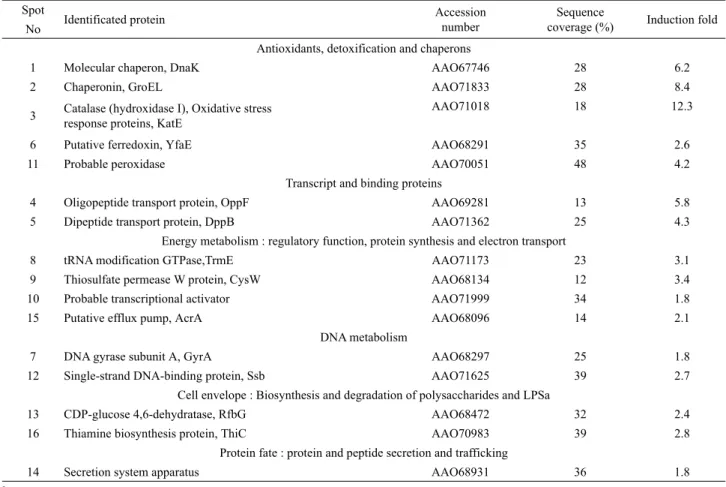
MALDI-TOF를 사용한 peptide mass fingerprinting은 2-DE 에서 변화된 발현을 보여준 단백질을 동정하기 위하여 실시되었 다. MALDI-TOF 분석으로부터 얻어진 결과는 Table 1에 열거 되었다. 16개의 단백질은 성공적으로 동정되었으며, elucidated amino acid sequences를 사용하여 sequence homology에 의해 특성조사를 실시하였다. 2,000 µg/ml TPP에서 60분간 처리된 S.
typhimurium 세포에서 항산화 및 무독성화(KatE, YfaE), chaperone protein HSP (DnaK, GroEL), 전사단백질, 에너지 대 사에 관여(Trm, CysW, AcrA), DNA 대사(GyrA, Ssb) 등에 관 여하는 단백질들이 1.8–12.3배의 범위에서 유도되었다(Table 2). 이들 발견은 세포에서 방어기작과 관련된 TPP-유도 단백질 들이 TPP 노출에서와 같은 좋지 않은 환경조건에 노출되었을 때 세포를 방어하거나 세포의 생존을 돕는데 중요한 역할을 한다는 것을 시사하였다.
녹차추출물은 음료로서 마시게 되면 기분을 상쾌하게 하고, 부드러운 청량감을 제공하는 것으로 알려져 있지만, 일정한 농
Spot Identificated protein Accession number
Sequence
coverage (%) Induction fold No
Antioxidants, detoxification and chaperons
1 Molecular chaperon, DnaK AAO67746 28 6.2
2 Chaperonin, GroEL AAO71833 28 8.4
3 Catalase (hydroxidase I), Oxidative stress response proteins, KatE
AAO71018 18 12.3
6 Putative ferredoxin, YfaE AAO68291 35 2.6
11 Probable peroxidase AAO70051 48 4.2
Transcript and binding proteins
4 Oligopeptide transport protein, OppF AAO69281 13 5.8
5 Dipeptide transport protein, DppB AAO71362 25 4.3
Energy metabolism : regulatory function, protein synthesis and electron transport
8 tRNA modification GTPase,TrmE AAO71173 23 3.1
9 Thiosulfate permease W protein, CysW AAO68134 12 3.4
10 Probable transcriptional activator AAO71999 34 1.8
15 Putative efflux pump, AcrA AAO68096 14 2.1
DNA metabolism
7 DNA gyrase subunit A, GyrA AAO68297 25 1.8
12 Single-strand DNA-binding protein, Ssb AAO71625 39 2.7
Cell envelope : Biosynthesis and degradation of polysaccharides and LPSa
13 CDP-glucose 4,6-dehydratase, RfbG AAO68472 32 2.4
16 Thiamine biosynthesis protein, ThiC AAO70983 39 2.8
Protein fate : protein and peptide secretion and trafficking
14 Secretion system apparatus AAO68931 36 1.8
a LPS, lipopolysaccharide.
Table 2. Relative protein expression profiling in S. typhimurium treated with TPP
도 이상에서는 다양한 종류의 병원성 세균에 대하여 시험관(in vitro) 또는 생체 내(in vivo)에서 강력한 항균효과를 가지는 것이 명백하다(Yang and Landau, 2000). 오늘날 화학항생제의 오남 용으로 인한 문제점을 해결하고자, 천연허브로부터 항균력을 가 지는 성분을 추출하여 신약으로 개발하고자 하는 연구가 많은 관심의 대상이 되고 있다. 녹차추출물의 구성성분인 EGCG가 화학항생제인 imipenem-내성 Klebsiella pneumoniae에 대한 탁 월한 항균효과가 있음이 입증된 바 있다(Cho et al., 2011). 향후 의 연구는 녹차추출물을 구성성분이 가지는 항균활성에 대한 연 구를 통하여 단일 천연 phytochemical의 항균활성을 비교평가 하고, 그 기작을 밝히는 방향으로 추진되어야 할 것이다.
적 요
이 연구의 목적은 국산 녹차(Camellia sinensis L.)에서 추출 한 차폴리페놀(tea polyphenols, TPP)에 노출된 Salmonella typhimurium의 여러 가지 세포반응을 조사하는 것이다. TPP는 S. typhimurium에 대하여 투여량에 비례한 살균효과를 보여주었 다. TPP로 처리된 S. typhimurium 배양에서 세포막을 구성하는 포화 및 불포화 지방산은 조성에서 상당한 변화가 일어난 것으 로 분석되었으며, 주사전자현미경 분석에서 아치사 농도의 TPP
로 처리된 세포는 세포표면에 구멍이 나고, 속이 움푹 패인 불규 칙한 모양으로 관찰되었다. TPP에 노출된 S. typhimurium 배양 의 수용성 단백질 부분에 대한 이차원 폴리아크릴아미드 젤 전 기영동에서 16개의 단백질이 TPP 노출에 의해 증가하는 것이 확인되었다. 항산화 및 chaperons, 전사 및 결합단백질, 에너지 및 DNA 대사 등에 수반되는 단백질을 포함하는 이들 유도된 단 백질은 MALDI-TOF를 사용한 peptide mass fingerprinting에 의해 동정되었다. 이들 결과는 S. typhimurium에 대한 TPP 유도 스트레스와 세포독성의 기작을 이해하는데 중요한 단서를 제공 할 수 있다.
참고문헌
Ahn, Y.J., Kawamura, T., Kim, M., Yamamoto, T., and Mitsuoka, T.
1991. Tea polyphenols: Selective growth inhibitors of Clostridium spp. Agric. Biol. Chem. 55, 1425–1426.
Bollag, D.M., Rozycki, M.D., and Edelstein, S.J. 1996. Protein methods. 2nd ed. New York, Wiley-Liss, USA.
Bradford, M.M. 1976. A rapid and sensitive method for the quantitation of microgram quantities of proetin utilizing the principle of protein-dye binding. Anal. Biochem. 72, 248–254.
Cho, Y.S., Kahng, H.Y., Kim, C.K., Kukor, J.J., and Oh, K.H. 2002.
Physiological cellular responses of the 2,4-D degrading bacterium, Burkholderia cepacia YK-2, to the phenoxyherbicides 2,4-D and 2,4,5-T. Curr. Microbiol. 45, 415–422.
Cho, Y.S., Oh, J.J., and Oh, K.H. 2010. Antimicrobial activity and biofilm formation inhibition of green tea polyphenols on human teeth. Biotechnol. Biopro. Engr. 15, 359–364.
Cho, Y.S., Oh, J.J., and Oh, K.H. 2011. Synergistic anti-bacterial and proteomic effects of epigallocatechin gallate on clinical isolates of imipenem-resistant Klebsiella pneumoniae. Phytomedicine 18, 941–946.
Cho, Y.S., Schiller, N.L., Kahng, H.Y., and Oh, K.H. 2007. Cellular responses and proteomic analysis of Escherichia coli exposed to green tea polyphenols. Curr. Microbiol. 55, 501–506.
Cho, Y.S., Schiller, N.L., and Oh, K.H. 2008. Antibacterial effects of green tea polyphenols on clinical isolates of methicillin-resistant Staphylococcus aureus. Curr. Microbiol. 57, 542–546.
Graham, H.N. 1992. Green tea composition, consumption, and polyphenol chemistry. Prev. Med. 21, 334–350.
Hamilton-Miller, J.M.T. 1995. Antimicrobial properties of tea (Camellia sinensis L.). Antimicrob. Agents Chemother. 39, 2375–
2377.
Hirasawa, M. and Takada, K. 2004. Multiple effects of green tea catechin on the antifungal activity of antimycotics against Candida albicans. J. Antimicrobiol. Chemother. 53, 225–229.
Ho, E.M., Chang, H.W., Kim, S.I., Kahng, H.Y., and Oh, K.H. 2004.
Analysis of TNT (2,4,6-trinitrotoluene)-inducible cellular responses and stress shock proteome in Stenotrophomonas sp.
OK-5. Curr. Microbiol. 49, 346–352.
Ikigai, H., Nakae, T., Hara, Y., and Shimamura, T. 1993. Bactericidal catechins damage the lipid bilayer. Biochim. Biophy. Acta 1147,
Microbiol. 36, 710–725.
Jungblut, P.R., Schaible, U.E., Mollenkopf, H.J., Zimny-Arndt, U., Raupach, B., Mattow, J., Halada, P., Lamer, S., Hagens, K., and Kaufmann, S.H.E. 1999. Comparative proteome analysis of Mycobacterium tuberculosis and Mycobacterium bovis BCG strains: Toward functional genomics of microbial pathogens. Mol.
Microbiol. 33, 1103–1117.
Lim, Y.J., Cho, Y.S., and Oh, K.H. 2008. Antibacterial synergic effect and cellular responses of nalidixic acid-resistant Salmonella typhimurium exposed to tea polyphenols and nalidixic acid. Kor. J. Microbiol. 44, 122–129.
Park, S.H., Oh, K.H., and Kim, C.K. 2001. Adaptative and cross-protective responses of Pseudomonas sp. DJ-12 to several aromatics and other stress shocks. Curr. Microbiol. 43, 176–181.
Pertins, D.N., Pappin, D.J., Creasy, D.M., and Cottrell, J.S. 1999.
Probability-based protein identification by searching sequence databases using mass spectrometry data. Electrophoresis 20, 355–
3567.
Sambrook, J.K., Fritsch, E.F., and Maniatis, T. 2001. Molecular cloning, 3rd ed., Cold Spring Harbor, New York, N.Y., USA.
Schmid, R., Bernhardt, J., Antelmann, H., Volker, A., Mach, H., Volker, U., and Hecker, M. 1997. Identification of vegetative proteins for a two-dimensional protein index of Bacillus subtilis.
Microbiology 143, 991–998.
Yam, T.S., Shah, S., and Hamilton-Miller, J.M.T. 1997. Microbiological activity of whole and fractionated crude extracts of tea (Camellia sinensis), and tea of components. FEMS Microbiol. Lett. 152, 169 –174.
Yang, C.S. and Landau, J.M. 2000. Effects of tea consumption on nutrition and health. J. Nutr. 130, 2409–2412.