• 검색 결과가 없습니다.

NAD(P)H-quinone oxidoreductase-1 silencing modulates cytoprotection related protein expression in cisplatin cytotoxicity

N/A
N/A
Protected

Academic year: 2021

Share "NAD(P)H-quinone oxidoreductase-1 silencing modulates cytoprotection related protein expression in cisplatin cytotoxicity"

Copied!
7
0
0

로드 중.... (전체 텍스트 보기)

전체 글

(1)

Korean J Vet Res(2016) 56(1) : 15~21 http://dx.doi.org/10.14405/kjvr.2016.56.1.15

15

<Original Article>

NAD(P)H-quinone oxidoreductase-1 silencing modulates cytoprotection related protein expression in cisplatin cytotoxicity

Se Ra Park

1,†

, Ju Young Jung

1,†

, Young-Jung Kim

1

, Da Young Jung

1

, Mee Young Lee

2

, Si Yun Ryu

1,

*

1

Department of Veterinary Medicine, Chungnam National University, Daejeon 34134, Korea

2

Basic Herbal Medicine Research Group, Korea Institute of Oriental Medicine, Daejeon 34054, Korea (Received: March 7, 2016; Revised: March 18, 2016; Accepted: March 25, 2016)

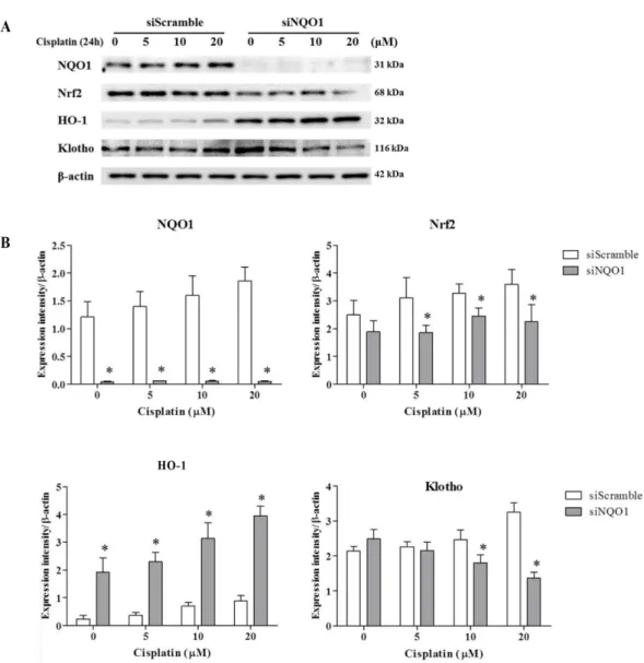
Abstract : NAD(P)H-quinone oxidoreductase-1 ( NQO1) is a down-stream target gene of nuclear factor erythroid 2- related factor 2 (Nrf2), and performs diverse biological functions. Recently, NQO1 is recognized as an effective gene for the cytotoxic inserts with its diverse biological functions, which is focused on antioxidant properties. The aim of present study was to assess the impact of NQO1 knockdown on cytoprotection-related protein expression in cisplatin cytotoxicity by using small interfering (si) RNA targeted on NQO1 gene. Cytotoxicity of cisplatin on ACHN cells was assessed in a dose- and time-dependent manner after siScramble or siNQO1 treatment. After cisplatin treatment, cells were subjected to cell viability assay, western-blot analysis, and immunofluorescence study. The cell viability was decreased in the siNQO1 cells (50%) than the siScramble cells (70%) after 24 h of cisplatin (20 µM) treatment.

Moreover, cytoprotection-related protein expressions were markedly suppressed in the siNQO1 cells after cisplatin treatment. The expression of Nrf2 and Klotho were decreased by 20% and 40%, respectively, of that in siScramble cells. Nrf2 and Klotho activation were also decreased in cisplatin treated siNQO1 cells, confirmed by cytoplasm-to- nuclear translocation. Our findings demonstrate that the increased cisplatin-induced cytotoxicity was accompanied by suppressed Nrf2 activation and Klotho expression in siNQO1 cells.

Keywords : NAD(P)H-quinone oxidoreductase-1, cisplatin, cytotoxicity, nuclear factor erythroid 2-related factor 2

Introduction

Cisplatin is a highly effective anticancer drug, used in the treatment of various types of solid tumors. Despite its clini- cal effectiveness, the application of cisplatin is limited, owing to its severe side effects such as dose-related nephrotoxicity [4]. Cisplatin induces mitochondrial damage and in turn, gen- erates oxidative stress that could aggravate cisplatin cytotox- icity [18]. Several studies have focused on reducing oxidative stress to relieve cisplatin-induced cytotoxicity [2, 11, 15]

NAD(P)H-quinone oxidoreductase-1 ( NQO1) is a flavoen- zyme that has various biological functions. In general, NQO1 is recognized as a detoxification enzyme with its reactive quinone reducing activity and known to play an antioxidant role through endogenous quinone reduction which generates hydroquinones that exerts antioxidant properties [26, 27].

Moreover, NQO1 found to stabilize p53 proteins, which reg- ulates various proteins involved in protection from oxidative stress [24]. The expression of NQO1 is highly inducible and regulated by nuclear factor erythroid 2-related factor 2 ( Nrf2)

[25]. Nioi et al. [22] reported that promoter region of NQO1 gene contains an Nrf2-binding consensus regulatory sequence, antioxidant response element (ARE), which implicates in NQO1 promoter activity. Nrf2/ARE is a major defense mech- anism pathway against oxidative stress in cellular system, which regulates protein expression that involves in the reac- tive oxidants eradication [21].

Previously, several studies have explored protective role of NQO1 by assessing its antioxidant properties [14, 23]. NQO1 was found to be highly expressed in systems that demand high levels of defense against antioxidants [31]. Further- more, knockdown of NQO1 was found to exacerbate cispl- atin-induced cytotoxicity [12]. Aggravated cell damage under NQO1 deleted conditions might result from the absence of NQO1 function, and this absence could affect NQO1-related cytoprotective signals. In the present study, we explored whether NQO1 silencing affects cytoprotection related pro- tein expressions including Nrf2, hemoxigenase1 (HO-1), and Klotho in cisplatin treated adenocarcinoma (ACHN) cells by using small interfering (si) RNA treatment.

*Corresponding author

Tel: +82-42-821-6758, Fax: +82-42-821-7926 E-mail: [email protected]

The first two authors contributed equally to this work.

(2)

16 Se Ra Park, Ju Young Jung, Young-Jung Kim, Da Young Jung, Mee Young Lee, Si Yun Ryu

Materials and Methods

Cell culture and NQO1 silencing

The human kidney ACHN cells were from the American Type Culture Collection (USA). Cells were cultured in Dul- becco’s modified Eagle medium supplemented with 10%

fetal bovine serum, 100 U/mL penicillin, and 100 µg/mL streptomycin (Gibco-BRL, USA) at 37

o

C under 5% CO

2

. For NQO1 silencing, siRNA was used to ACHN cells. Cells were seeded (1 × 10

5

cells/mL) in 6-well plates and incubated with scrambled siControl or siRNA targeting human NQO1 (5'- AAACCAGCCUUUCAGAAUGGCUGGC-3', 20 nM; Invit- rogen, USA) and hyperfectant reagent (Qiagen, USA) for 30 h. After silencing, cells were further treated with serial con- centration of cisplatin (0, 5 10, and 20 µM for 24 h) for west- ern-blot analysis. For a time-dependent assessment in change, cells were harvested at 0, 1, 3, 6, 12, and 24 h after cisplatin (20 µM) treatment.

Cell viability assay

After siScramble or siNQO1 treatment, cells (1 × 10

4

cells/

mL) were seeded in 96-well plates for cell viability assay.

The day after, 3-(4,5-dimethythiazol-2-yl)-2,5-diphenyltetra- zolium bromide (MTT) assay was performed by using cell viability assay kit (EZ-cytox; DaeilLab Service, Korea) accord- ing to the manufacturer’s protocol.

Western blot analysis

Cells were lysed in lysis buffer (20 mM Tris-HCl pH 8 with protease inhibitor cocktail) and electrophoresed in a 10–

15% SDS-polyacrylamide gel. The proteins were transferred onto nitrocellulose membrane in a Semi-Dry Transfer sys- tem (Bio-Rad Laboratories, USA). The membranes were blocked with 5% dry milk with 20% TWEEN-20 (TBS-T).

After blocking, the membranes were incubated with anti–

Nrf2 (1 : 1,000 dilution; Abcam, USA), anti-NQO1 (1 : 1,000 dilution; Santa Cruz Biotechnology, USA), anti–HO-1 (1 : 1,000 dilution; Stressgen Biotechnologies, USA), anti-klotho (1 : 1,000 dilution; Abcam), or β-actin (1 : 3,000; Abcam) antibodies, followed by a horseradish peroxidase-conjugated anti-rabbit or anti-mouse IgG antibody. Horseradish peroxi- dase activity was detected using an enhanced chemilumines- cence detection kit (Amersham ECL Western Blotting Detection Kit; GE Healthcare, UK). Band intensities (n = 3) were quan- tified by an ImageJ program (US National Institutes of Health, USA) and normalized to β-actin.

Immunofluorescence study

ACHN cells (8 × 10

3

/well) were seeded on glass chamber slides and NQO1 silencing was achieved as described above.

After cisplatin treatment (20 µM for 30 min), the cells were fixed with alcohol and washed. After blocking with 3%

bovine serum albumin, the cells were incubated anti–Nrf2 (1 : 100 dilution; Abcam) overnight. After washing and block- ing, the cells were incubated with goat anti-rabbit Alexa

Fluor 594 (Life Technologies, USA) for 60 min and mounted with ProLong Gold Antifade Mountant with DAPI (4',6-dia- midino-2-phenylindole) (Life Technologies). The cells were viewed using an Olympus IX-71 microscope (Olympus, Japan) and images were captured using Olympus DP Controller software. Merged images were achieved using ZEN 2009 software (Carl Zeiss Microscopy, Germany).

Statistical analysis

The data were expressed as the means ± SD. The data were analyzed using one way analysis of variance (ANOVA) or non-parametric Kruskal-Wallis test, as appropriate, followed by a Tukey’s multiple comparison test or Mann-Whitney U test. P values less than 0.05 were considered statistically sig- nificant.

Results

Cisplatin-induced cytotoxicity in NQO1-knocked down ACHN cells

In this study, we investigated whether deletion of NQO1 affects cytoprotection-related protein expression in cisplatin cytotoxicity, using in vitro assay adapted to silencing method.

First, we tested the MTT assay for assessing the cisplatin induced cytotoxicity on ACHN cells (1 × 10

5

cells/mL) in the presence or absence of NQO1. High cisplatin activation has been reported to occur one day treated with cisplatin in this earlier study [30]. Therefore, here we treated the cells with cisplatin for 24 h. Furthermore, we explored changes in cyto- toxicity upon cisplatin treatment for 48 h. Our results revealed that cisplatin treatment reduced the viability of both siScram- ble and siNQO1 cells in a time-dependent manner. More- over, as expected, siNQO1 cells showed significantly lower viability ( p < 0.05) than did the siScramble cells 12, 24, and 48 h after cisplatin treatment (Fig. 1).

Fig. 1. NAD(P)H-quinone oxidoreductase-1 ( NQO1) silencing aggravates cisplatin-induced cytotoxicity in adenocarcinoma (ACHN) cells. After siScramble or siNQO1 treatment, cells (1

× 10

5

cells/mL) were treated with 20 µM of cisplatin for 0, 12,

24, and 48 h and subjected to cell viability assay (MTT). Values

are expressed as means ± SD of triplicate experiments.

*

p < 0.05

indicated significant difference when compared to the siScram-

ble counterpart.

(3)

Dose- and time-dependent changes in cytoprotection- related protein expression cisplatin-treated ACHN cells under NQO1-knocked down

Results of western blot analysis indicated that cisplatin induced the expression of cytoprotective proteins such as Nrf2 and NQO1 in the siScramble cells (1 × 10

5

cells/mL).

Figure 2 shows that NQO1 was knocked-down in these cells.

As expected, the expression of NQO1 was enhanced upon cisplatin treatment in the siScramble cells. On the other hand, cisplatin-induced Nrf2 expression was relatively decreased in the siNQO1 cells (1 × 10

5

cells/mL) than in the siScramble cells. Additionally, cisplatin-induced HO-1 expression was almost four times higher in the siNQO1 cells than in the siS- cramble cells. In contrast, although Klotho expression was initially increased upon cisplatin treatment in the siScramble

cells, it later decreased in a dose-dependent manner ( p < 0.05).

The expressions of NQO1, Nrf2, and Klotho were gradu- ally enhanced upon cisplatin treatment (20 µM) in the siS- cramble cells (Fig. 3). In the siNQO1 cells, similar to the dose- dependent results, Nrf2 expression was relatively lower than that in the siScramble cells, while the expression of Klotho was remarkably lower than that in the NQO1-intact siScram- ble cells at all the time points. In addition , HO-1 expression was generally higher in the siNQO1 cells than in the siS- cramble cells upon cisplatin treatment.

Nuclear translocation of Nrf2 and Klotho cisplatin- treated ACHN cells under NQO1-knocked down

Immunofluorescence experiments were performed to

assess Nrf2 and Klotho expression in cisplatin-treated ACHN

Fig. 2. Dose-dependent expressions of cytoprotection-related proteins in cisplatin treated NQO1 silenced ACHN cells. After siS-

cramble or siNQO1 treatment, cells (1 × 10

5

cells/mL) were treated with 0, 5, 10, and 20 µM of cisplatin for 24 h and subjected to

western-blot analysis (A). Expression intensities were assessed by densitometry and normalized to that of β-actin (B). Values are expressed

as means ± SD of triplicate experiments.

*

p < 0.05 indicated significant difference when compared to the siScramble counterpart.

(4)

18 Se Ra Park, Ju Young Jung, Young-Jung Kim, Da Young Jung, Mee Young Lee, Si Yun Ryu

cells (8 × 10

3

/well). The nuclear regions of ACHN cells were stained with DAPI (blue). In control cells, most of the Nrf2 and Klotho expression (red dots) were detected at the cyto- plasmic regions (Fig. 4). After cisplatin treatment, however, Nrf2 expression was focalized in the nuclear regions, which indicates nuclear translocation of Nrf2. However, cisplatin- treated siNQO1 cells showed relatively reduced nuclear translocation of Nrf2 than did the cisplatin-treated siScram- ble cells with higher cytoplasmic Nrf2 expression. Mean- while, Klotho expression was focalized in both the nuclear and cytoplasmic regions. Cisplatin-treated siNQO1 cells showed relatively reduced expression of Klotho in the nuclear and cytoplasmic regions than did the cisplatin-treated siScramble cells with higher Klotho expression.

Discussion

In the present study, a part from direct antioxidant prop- erty of NQO1, we explored impact of NQO1 silencing on cytoprotection-related signal expression in cisplatin cytotox- icity. At first, we confirmed cytoprotective effect of NQO1 by assessing cisplatin-induced cell viability decrement under NQO1 absence condition. As expected, deletion of NQO1 gene intensified cisplatin-induced cell viability decrement that supports protective role of NQO1 against cytotoxic insults.

After confirming exacerbated cytotoxicity in siNQO1 cells,

we further assessed changes in cytoprotection-related pro-

teins including Nrf2, HO-1, and Klotho after cisplatin treat-

ment. Nrf2 is an up-stream transcription factor of NQO1 that

regulates various antioxidant protein expression. Activation

Fig. 3. Time-dependent expressions of cytoprotection-related proteins in cisplatin treated ACHN cells. After siScramble or siNQO1

treatment, cells (1 × 10

5

cells/mL) were collected at 0, 1, 3, 6, 12, and 24 h after cisplatin (20 µM) treatment. Total cell lysates were

assessed for western blotting (A). Expression intensities were assessed by densitometry and normalized to that of β-actin (B). Values are

expressed as means ± SD of triplicate experiments.

*

p < 0.05 indicated significant difference when compared to the siScramble counterpart.

(5)

of Nrf2 induces the expression of detoxification enzymes as well as enzymes for antioxidants synthesis, which participate in oxidative damage suppression [17]. In addition, several studies reported that the Nrf2 suppression impairs NQO1 expression [1, 10]. Hu et al. [8] demonstrated that basal NQO1 expression was repressed in tissues obtained from Nrf2- knockout mice and stress conditions failed to induce NQO1 expression; this supports involvement of Nrf2 in NQO1

expression [16]. Interestingly, in this study, deletion of NQO1, the down-stream target gene of Nrf2, resulted in reduced cisplatin-induced Nrf2 expression in a dose depen- dent manner. Furthermore, effect of NQO1 silencing on Nrf2 activation was confirmed by immunofluorescence study.

Under unstressed condition, Nrf2 is constitutively expressed in the cell cytoplasm and degraded, while under oxidative stress, Nrf2 is translocated directly to the nucleus where it binds to a DNA promoter and initiates transcription of anti- oxidative genes and their protein synthesis [3]. In this study, NQO1 silencing reduced cisplatin-induced Nrf2 activation in ACHN cells as well as decreased the nuclear translocation of Nrf2.

HO-1 is an enzyme that controls heme catabolism and its induction is known to be associated with protection against oxidative stress [5]. HO-1 and NQO1 are generally recog- nized as similar down-stream target genes of Nrf2. Previ- ously, Yeligar et al. [29] have demonstrated that ethanol- induced oxidative stress enhances the expressions of both HO-1 and NQO1. However, although Nrf2 is involved in activation of both enzymes, the signal transduction pathways for HO-1 induction is different from that of the NQO1. Fur- ther, HO-1 showed different transcription pattern when com- pared to that shown by NQO1 upon HIF-1α silencing. In the present study, although Nrf2 expression was reduced by NQO1 knock-down, HO-1 showed enhanced expression under NQO1 silenced condition. This might be due to the dif- ferent signaling pathways involveed in HO-1 and NQO1 transcription.

We also assessed changes in Klotho expression under NQO1 deleted condition. Klotho is regarded as an anti-aging protein that is expressed in multiple tissues, especially in the kidney [13]. Multiple age-related disorders were reported in Klotho-knockout mice, and these pathologies were reversed by induction of Klotho expression [19]. Furthermore, previ- ous researches have revealed that Klotho has anti-oxidative, anti-apoptosis, and anti-fibrosis activities [9, 20, 28]. Interest- ingly, in the present study, Klotho was negatively regulated by NQO1 silencing in cisplatin treated ACHN cells. After cisplatin treatment, Klotho expression was evidently sup- pressed by NQO1 silencing with a time- and concentration- dependent manner. Recently, pathophysiologic role of Klotho in acute and chronic renal injury has been reported, and Klotho has emerged as a novel target for the renoprotection [6, 7]. Aggravated cisplatin-induced cytotoxicity in siNQO1 ACHN cells, observed in the present study, might be related to reduce Klotho expression, implicated in renoprotection.

In the present study, we found the intensified cytotoxicity and altered cytoprotection-related signal expressions in the ACHN cells under NQO1 deleted condition. NQO1 deleted cells also showed decreases of cisplatin-induced Nrf2 expres- sion and nuclear translocation together with a significantly suppression of Klotho expression. Although further mecha- nistic studies are needed, taken together, these results indi- cate that compromised cytoprotection upon NQO1 silencing Fig. 4. NQO1 silencing reduced nuclear localization of nuclear

factor erythroid 2-related factor 2 (Nrf2) expression and Klotho

expression in cisplatin treated ACHN cells. After siScramble or

siNQO1 treatment, cells (8 × 10

3

/well) were incubated with cis-

platin (20 µM) for 1 h and subjected to immunofluorescence

study. The nuclei were visualized by DAPI (blue), Nrf2 expres-

sion (A) and Klotho expression (B) ware visualized by Alexa

Fluor 594 conjugate (red, arrows) at 400× magnification.

(6)

20 Se Ra Park, Ju Young Jung, Young-Jung Kim, Da Young Jung, Mee Young Lee, Si Yun Ryu

is associated with altered cytoprotection-related signaling pathway.

Acknowledgments

This work was supported by research fund of Chungnam National University.

References

1. Aleksunes LM, Reisman SA, Yeager RL, Goedken MJ, Klaassen CD. Nuclear factor erythroid 2-related factor 2 deletion impairs glucose tolerance and exacerbates hypergly- cemia in type 1 diabetic mice. J Pharmcol Exp Ther 2010, 333, 140-151.

2. Atessahin A, Yilmaz S, Karahan I, Ceribasi AO, Karaoglu A. Effects of lycopene against cisplatin-induced nephrotoxicity and oxidative stress in rats. Toxicology 2005, 212, 116-123.

3. Bryan HK, Olayanju A, Goldring CE, Park BK. The Nrf2 cell defence pathway: Keap1-dependent and -independent mechanisms of regulation. Biochem Pharmacol 2013, 85, 705-717.

4. Cepeda V, Fuertes MA, Castilla J, Alonso C, Quevedo C, Pérez JM. Biochemical mechanisms of cisplatin cytotoxicity.

Anticancer Agents Med Chem 2007, 7, 3-18.

5. Ghattas MH, Chuang LT, Kappas A, Abraham NG.

Protective effect of HO-1 against oxidative stress in human hepatoma cell line (HepG2) is independent of telomerase enzyme activity. Int J Biochem Cell Biol 2002, 34, 1619- 1628.

6. Hu MC, Kuro-o M, Moe OW. Secreted klotho and chronic kidney disease. In: Kuro-o M (ed.). Endocrine FGFs and Klothos. pp. 126-157, Landes Biosciences/Springer Science+

Business Media, Austin/New York, 2012.

7. Hu MC, Shi M, Zhang J, Pastor J, Nakatani T, Lanske B, Razzaque MS, Rosenblatt KP, Baum MG, Kuro-o M, Moe OW. Klotho: a novel phosphaturic substance acting as an autocrine enzyme in the renal proximal tubule. FASEB J 2010, 24, 3438-3450.

8. Hu X, Roberts JR, Apopa PL, Kan YW, Ma Q.

Accelerated ovarian failure induced by 4-vinyl cyclohexene diepoxide in Nrf2 null mice. Mol Cell Biol 2006, 26, 940- 954.

9. Ikushima M, Rakugi H, Ishikawa K, Maekawa Y, Yamamoto K, Ohta J, Chihara Y, Kida I, Ogihara T.

Anti-apoptotic and anti-senescence effects of Klotho on vascular endothelial cells. Biochem Biophys Res Commun 2006, 339, 827-832.

10. Jódar L, Mercken EM, Ariza J, Younts C, González- Reyes JA, Alcaín FJ, Burón I, de Cabo R, Villalba JM.

Genetic deletion of Nrf2 promotes immortalization and decreases life span of murine embryonic fibroblasts. J Gerontol A Biol Sci Med Sci 2011, 66, 247-256.

11. Kim TW, Song IB, Lee HK, Lim JH, Cho ES, Son HY, Park SJ, Kim JW, Yun HI. Platycodin D, a triterpenoid sapoinin from Platycodon grandiflorum, ameliorates cisplatin- induced nephrotoxicity in mice. Food Chem Toxicol 2012, 50, 4254-4259.

12. Kim YJ, Kim TW, Park SR, Kim HT, Jung DY, Ryu SY, Jung JY. Deletion of NAD(P) H:quinone oxidoreductase 1 represses Mre11-Rad50-Nbs1 complex protein expression in cisplatin-induced nephrotoxicity. Toxicol Lett 2016, 243, 22- 30.

13. Kuro-o M, Matsumura Y, Aizawa H, Kawaguchi H, Suga T, Utsugi T, Ohyama Y, Kurabayashi M, Kaname T, Kume E, Iwasaki H, Iida A, Shiraki-Iida T, Nishikawa S, Nagai R, Nabeshima YI. Mutation of the mouse klotho gene leads to a syndrome resembling ageing. Nature 1997, 390, 45-51.

14. Leonard MO, Kieran NE, Howell K, Burne MJ, Varadarajan R, Dhakshinamoorthy S, Porter AG, O'Farrelly C, Rabb H, Taylor CT. Reoxygenation-specific activation of the antioxidant transcription factor Nrf2 mediates cytoprotective gene expression in ischemia-reperfusion injury. FASEB J 2006, 20, 2624-2626.

15. Lin L, Zheng J, Zhu W, Jia N. Nephroprotective effect of gelsemine against cisplatin-induced toxicity is mediated via attenuation of oxidative stress. Cell Biochem Biophys 2015, 71, 535-541.

16. Lin X, Yang H, Zhou L, Guo Z. Nrf2-dependent induction of NQO1 in mouse aortic endothelial cells overexpressing catalase. Free Radic Biol Med 2011, 51, 97-106.

17. Ma Q. Role of Nrf2 in oxidative stress and toxicity. Annu Rev Pharmacol 2013, 53, 401-426.

18. Martins NM, Santos NAG, Curti C, Bianchi MLP, Santos AC. Cisplatin induces mitochondrial oxidative stress with resultant energetic metabolism impairment, membrane rigidifi- cation and apoptosis in rat liver. J Appl Toxicol 2008, 28, 337-344.

19. Masuda H, Chikuda H, Suga T, Kawaguchi H, Kuro-o M. Regulation of multiple ageing-like phenotypes by inducible klotho gene expression in klotho mutant mice. Mech Ageing Dev 2005, 126, 1274-1283.

20. Mitobe M, Yoshida T, Sugiura H, Shirota S, Tsuchiya K, Nihei H. Oxidative stress decreases klotho expression in a mouse kidney cell line. Nephron Exp Nephrol 2005, 101, e67-e74.

21. Nguyen T, Nioi P, Pickett CB. The Nrf2-antioxidant response element signaling pathway and its activation by oxidative stress. J Biol Chem 2009, 284, 13291-13295.

22. Nioi P, McMahon M, Itoh K, Yamamoto M, Hayes JD.

Identification of a novel Nrf2-regulated antioxidant response element (ARE) in the mouse NAD(P)H:quinone oxidoreductase 1 gene: reassessment of the ARE consensus sequence.

Biochem J 2003, 374, 337-348.

23. Park JS, Jung JS, Jeong YH, Hyun JW, Le TKV, Kim DH, Choi EC, Kim HS. Antioxidant mechanism of isoflavone metabolites in hydrogen peroxide-stimulated rat primary astrocytes: critical role of hemeoxygenase-1 and NQO1 expression. J Neurochem 2011, 119, 909-919.

24. Polyak K, Xia Y, Zweier JL, Kinzler KW, Vogelstein B.

A model for p53-induced apoptosis. Nature 1997, 389, 300- 305.

25. Ma Q, Kinneer K, Bi Y, Chan JY, Kan YW. Induction of

murine NAD(P)H:quinone oxidoreductase by 2,3,7,8-tetrachlo-

rodibenzo-p-dioxin requires the CNC (cap ‘n’ collar) basic

leucine zipper transcription factor Nrf2 (nuclear factor

erythroid 2-related factor 2): cross-interaction between AhR

(7)

(aryl hydrocarbon receptor) and Nrf2 signal transduction.

Biochem J 2004, 377, 205-213.

26. Siegel D, Bolton EM, Burr JA, Liebler DC, Ross D. The reduction of α-tocopherolquinone by human NAD(P)H:

quinone oxidoreductase: the role of α-tocopherolhydroquinone as a cellular antioxidant. Mol Pharmacol 1997, 52, 300-305.

27. Siegel D, Gustafson DL, Dehn DL, Han JY, Boonchoong P, Berliner LJ, Ross D. NAD(P)H:quinone oxidoreductase 1: role as a superoxide scavenger. Mol Pharmacol 2004, 65, 1238-1247.

28. Sugiura H, Yoshida T, Shiohira S, Kohei J, Mitobe M, Kurosu H, Kuro-o M, Nitta K, Tsuchiya K. Reduced Klotho expression level in kidney aggravates renal interstitial fibrosis. Am J Physiol-Renal Physiol 2012, 302, F1252-

F1264.

29. Yeligar SM, Machida K, Kalra VK. Ethanol-induced HO-1 and NQO1 are differentially regulated by HIF-1α and Nrf2 to attenuate inflammatory cytokine expression. J Biol Chem 2010, 285, 35359-35373.

30. Yi YW, Bae I. Effects of solvents on in vitro potencies of platinum compounds. DNA Repair (Amst) 2011, 10, 1084- 1085.

31. Zhu H, Jia Z, Mahaney JE, Ross D, Misra HP, Trush

MA, Li Y. The highly expressed and inducible endogenous

NAD(P)H:quinone oxidoreductase 1 in cardiovascular cells

acts as a potential superoxide scavenger. Cardiovasc Toxicol

2007, 7, 202-211.

수치

Fig. 1. NAD(P)H-quinone oxidoreductase-1 ( NQO1) silencing aggravates cisplatin-induced cytotoxicity in adenocarcinoma (ACHN) cells
Figure 2 shows that  NQO1 was knocked-down in these cells.

참조

관련 문서

Control- and ASTRIN- depleted HeLa cells were untreated or treated with 5 Gy. IR and were then fixed at

Sesn2-luciferase transactivation was determined in the lysates of HEK293 cells transfected with an Nrf2 expression construct along with either a Sesn2 (pGL4-phSESN2)

4 &gt; Effects of Taro on COX-2 expression and iNOS expression(hot water) in human thyroid cancer cells. The cells were pretreated for 48hours with either

Although several studies have reported that photofunctionalization improves the wettability and bioactivity of titanium surfaces, they have mostly examined

The expression of Dynamin II protein expressed strongly at advanced bell stage in cytoplasm of odontoblast. Dynamin II protein localized in adjacent the nuclear

To investigate whether oncogenic H-ras might modulate mdr1b expression in NIH3T3 cells, V12-ras-NIH3T3 and pcDNA3-NIH3T3 cells were transiently transfected with

Collectively, exogenous 8-OHdG decreased the expres- sion of NOX1/2/4, TLR2/4, and NF-κB by decreasing Rac1 activity in DOX-treated cells, which eventually led to

iMSC‑EVs decreased itching, which was supported by reduced inflammatory cell infiltration and mast cells in AD mouse skin; reduced IgE receptor expression and