Protective Effect of Atractylodes macrocephala and Taraxacum spp.
Combination Treatment in Balb/c Mice with Dextran Sulfate Sodium-Induced Ulcerative Colitis
Donghun Lee
#, Hocheol Kim
*Kyung Hee University, College of Korean Medicine, Department of Herbal Pharmacology
ABSTRACT
Objectives : This study aimed to investigate the protective effects of an herbal mixture of Atractylodes macrocephala and Taraxacum spp. (ATC) on ulcerative colitis. We have previously screened traditional medicinal herbs to discover the effective candidate by the animal model. A. macrocephala and T. spp were identified as one of the effective herbs in the screening process.
Methods : Experimental colitis was induced in male Balb/c mice by administering drinking water containing dextran sulfate sodium, which mimics the clinical and histological features of ulcerative colitis in human. ATC at doses of 30, 100 or 300 ㎎/㎏ were orally administered to mice twice per day for 10 consecutive days. To evaluate the damage from experimentla ulcerative colitis, body weight, colon length, disease activity index, myeloperoxidase and histological changes were measured and analyzed.
Results : The administration of dextran sulfate sodium with drinking water resulted in markedly reduced colon length, severe body weight loss, increased levels of myeloperoxidase activity and histological damages in mice. ATC treatment significantly ameliorated the colon shortening, histological damage, body weight loss and disease activity index score in a dose-dependent manner. ATC also attenuated the colonic myeloperoxidase activity which reflects the severity and extent of inflammatory damage of colon.
Conclusions : ATC exerts protective effects against inflammatory colonic structural damage induced by epithelial barrier integrity impairment. ATC also inhibits weight loss and related symptoms of UC which can be considered as the functional recovery of colon.
1)
Key words : Atractylodes macrocephala , Taraxacum spp., ulcerative colitis
Ⅰ. Introduction
Ulcerative colitis (UC) is the major form of chronic inflammatory bowel disease, with a high cancer risk and is characterized by fever, fatigue, abdominal pain, diarrhoea, rectal bleeding and body weight loss
1). Although the prevalence and incidence of UC have reached a plateau in Europe and North America, they continue to increase in countries adopting a Western lifestyle
2). The most widely held hypothesis about the pathogenesis
of UC involves excessive immune responses against intestinal bacteria, leading to damage of the epithelial barrier via abnormal pro-inflammatory signals
3). It is commonly accepted that the overexpression of inflammatory markers, such as interleukin-6 (IL-6), tumour necrosis factor-α (TNF-α ) and myeloperoxidase (MPO), plays a crucial role in the pathogenesis of UC
4-5).
Conventional treatments for UC include corticosteroids and aminosalicylates as the mainstay of therapy.
Immunosuppressive agents, such as methotrexate or
*Corresponding author : Hocheol Kim, Kyung Hee University, College of Korean Medicine, Department of Herbal Pharmacology, Seoul, 130-701, Republic of Korea.
·Tel : +82-2-961-0419 ·E-mail : [email protected]
#First author : Donghun Lee, Kyung Hee University, College of Korean Medicine, Department of Herbal Pharmacology, Seoul, 130-701, Republic of Korea.
·Tel : +82-2-961-0419 ·E-mail : [email protected]
·Received:9 Novemver 2016 ·Revised:10 May 2017 ·Accepted:20 May 2017
80
大 韓 本 草 學 會 誌 ― Vol. 32 No. 3, 2017azathioprine, are used for steroid-dependent or –resistant patients
6). Aminosalicylates are well tolerated but cramps, diarrhoea and abdominal pain are occasional side effects and are accompanied by liver or kidney problems.
Corticosteroids have well-known side effects including facial rounding, acne, diabetes and high blood pressure
7); hence, UC patients are refractory to these drugs or are unable to use current drugs for prolonged periods owing to the side effects. Therefore, there is a pressing need for developing effective and safe therapeutic drugs for UC.
ATC is an herbal complex of Atractylodes macrocephala rhizome and Taraxacum spp. herb. A. macrocephala is a medicinal herb with a long history of use for conditions including gastritis, diarrhoea, indigestion, abdominal pain and fatigue
8). A. macrocephala exerts anti-inflammatory, anti-oxidant, anti-tumour, hepatoprotective and gastric protective effects
9-11). The major components of A.
macrocephala are volatile oils, sesquiterpenoids, polysaccharides and amino acids, and they are responsible for its diverse biological activities
12). The plants of the genus Taraxacum , namely dandelions, have long been used as medicinal herbs for various diseases such as dyspepsia, heart burn, spleen and liver complaints and anorexia
13). These herbs are a good source of vitamins, minerals and oligoelements
14). T. spp. and its congeners have been reported to exhibit various activities, including anti-microbial, hepatoprotective, anti- inflammatory and anti-oxidative activities. In particular, A. macrocephala extracts are reported to downregulate IL-6 and IL-17 in a TNBS-induced colitis rat model
15). T. spp. contains inulin, which is reported to be an effective prebiotic for dextran sodium sulfate-induced experimental colitis
16). However, protective effects of this combination treatment against UC have not yet been studied.
In the present study, we assessed the protective effects of ATC on DSS-induced ulcerative colitis in male Balb/c mice. Many symptoms of DSS-experimental model are similar to those observed in human UC, for example diarrhoea, bloody faeces, body weight loss and shortening of the colon
17-18). The mechanism by which DSS causes colitis is a direct toxic effect on the intestinal epithelium that allows bacteria to penetrate the inner mucus layer and cause ulceration in the colon
19-20). The effect of ATC was evaluated by body weight loss, colon length, disease activity index (DAI) score, myeloperoxidase (MPO) activity and histological score.
Ⅱ. Materials and Methods
1. Materials
The dried root of A. macrocephala and dried whole plant of T. spp. were purchased from Daewoo Hanyak Co. (Seoul, Korea). They were identified by Professor Hocheol Kim and voucher specimens (No. 12112306 and No. 12112304) were deposited in Department of Herbal Pharmacology, College of Korean Medicine, Kyung Hee University, Seoul, Korea.
2. Sample preparation
The dried root of A. macrocephala and dried whole plant of T. spp. were extracted separately with distilled water for 3 h at 100℃ in a reflux apparatus. The extracts were filtered and concentrated under reduced pressure and samples were lyophilized to yield powders. The yields of individual extracts were 38.8% and 17.0%, respectively. The powders were then mixed in a ratio of 1:1.
3. HPLC analysis
Quantitative authentication of ATC was performed with a high-performance liquid chromatography (HPLC) system equipped with a Waters 1525 pump, a 2707 autosampler and 2998 PDA detector. Chromatographic separation was achieved at 35℃ on a Waters Sunfire C18 (4.6 ㎜ × 250 ㎜ i.d., 5 ㎛ particle size) column. The mobile phase consisted of 0.5% phosphoric acid (A) and acetonitrile (B) eluted by the following program for separation: 0–5 min, 10%; 5–10 min, 10%–20%; 10–15 min, 20%–20%; 15–20 min, 20%–50%; 20–30 min, 50%–50%;
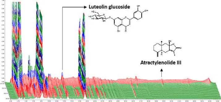
30–35 min, 50%–35%; 35–45 min, 35%–35%; 45–50 min, 35%–10% solvent B. The flow rate was 1 ㎖/min. The injection volume was 10 ㎕ and the eluate was monitored at 220 ㎚ and 348 ㎚ for atractylenolide III and luteolin glucoside, respectively. T. spp. and A. macrocephala extracts were monitored at 348 ㎚ for luteolin glucoside and 220 ㎚ for atractylenolide III. The concentrations of luteolin glucoside and atractylenolide III were 165.6 and 56.1 ㎍/g, respectively (Fig. 1).
4. Animals
Male Balb/c mice (7 weeks old, 20–24 g) were supplied
by Samtako. Mice were housed at 20–22℃ under a 12 h
light–12 h dark cycle and were provided food and water
ad libitum . All procedures were conducted according to
the animal welfare guidelines of the National Institute
of Health (KISTEM-IACUC-2013-001).
Figure 1. Three-dimensional high-performance liquid chromatogram of ATC. X-axis shows retention time; Y-axis shows wavelength and Z-axis shows absorbance units. Analytical conditions were as follows: column, C18;mobilephase, solvent A(0.5%H3PO4) and solvent B(CH3CN);flowrate, 1 ㎖/min.
5. Induction of colitis and experimental design
Colitis was induced by DSS administration. Mice were provided with drinking water containing 5% DSS (MP Biomedicals) ad libitum from day 4 to day 10 for 7 days.
To investigate the effects of ATC, mice were randomly allocated into following groups: normal, DSS treated control and DSS treated with ATC (30, 100 and 300 ㎎/㎏).
Mice were administered distilled water (10 ㎖/㎏) in the DSS group or each dose of ATC in the DSS + ATC group via feeding needle twice per day from days 1 to 10 for 10 days. The animals were sacrificed on day 10 and tissue samples were collected for additional observations.
6. Assessment of body weight and colon length
Body weight was measured daily from day 1 to 10.
Colon was isolated promptly after the last check of body weight. Colon length was measured from anus to caecum using a ruler.
7. Assessment of disease activity index (DAI) score
DAI score was recorded by an investigator blinded to treatment groups, as follows: spontaneous behaviour and posture (0, motionless with hunching [+++]; 1, motion but lethargic with hunching [++]; 2, walking with hunching [+]; 3, running with hunching [-]; 4, running), piloerection (0, piloerection [+++]; 1, piloerection [++];
2, piloerection [+]; 3, piloerection [−]; 4, normal state
[no piloerection]), cleanliness of anal orifice (0, with watery diarrhoea [+++]; anal prolapsed and bleeding [+++]; 1, with loose faeces and bleeding [++]; 2, with slightly loose faeces and bleeding [+]; 3, with faeces and bleeding [-]; 4, healthy state) and edema (0, edema [+++,
>0.35 mm in colon thickness]; 1, edema [++, 0.3–0.35 mm]; 2, edema [+, 0.25–0.30 mm]; 3, edema [±, 0.2–
0.25 mm]; 4, normal [0.1–0.2 mm]). The DAI score was calculated as the average point score for each test.
8. Assessment of myeloperoxidase (MPO) activity
Colonic tissue specimens (50 mg) were thawed and homogenized on ice in 50 mM potassium phosphate buffer pH 6.5 with 0.5% (w/v) hexadecyltrimethylammonium bromide (HETAB) (Sigma) using a homogenizer (50 mg tissue to 1 mL of buffer). The homogenized samples were then frozen and thawed, sonicated three times and centrifuged at 20,000 × g for 30 min at 4℃. Supernatant (10 ㎕) was added to 190 ㎕ phosphate buffer (pH 6.5) containing O-dianisidine (Sigma) (0.167 ㎎/㎖) and 0.0005% (v/v) H
2O
2in 96-well plates. The reaction was terminated after 3 min by addition of 20 ㎍/㎖ catalase and 0.2 M sodium acetate. Absorbance was measured at 470 nm using a microplate reader.
ε: Mallextinction coefficient (M-1cm-1)O-dianisidine=7.5 Vf: finalvolume(L) A: absorbance t: time (min)
82
大 韓 本 草 學 會 誌 ― Vol. 32 No. 3, 20179. Assessment of histological score
Colons were fixed in 10% buffered paraformaldehyde and embedded in paraffin. Histological sections cut from the paraffin blocks were stained with haematoxylin and eosin (H&E). The scoring of histological damage was divided into three parameters in a blind fashion:
severity of inflammation, crypt damage and ulceration.
Severity of inflammation was graded on a scale of 0–3 (0, rare; 1, mild; 2, moderate; 3, severe), obtained from each layer of the colon, including surface epithelium, cryptal glands, stroma, submucosa and transmural layer. The crypt damage was graded on a scale of 0–5 (0, none; 1, loss of the basal one-third; 2, loss of the basal two-thirds; 3, entire crypt loss; 4, change in epithelial surface with erosion; 5, confluent erosion).
Severity of ulceration was graded histologically on a scale of 0–3 (0, none; 1, 1 or 2 foci of ulceration; 2, 3 or 4 foci of ulceration; 3, confluent or extensive ulceration). The overall score was calculated as the sum of all scores.
10. Statistical analysis
All results were expressed as mean values with their standard errors for each group. Data were analysed statistically using Student’s t test. p < 0.05 was considered statistically significant.
Ⅲ. Results
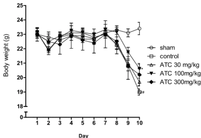
1. Effects of ATC on weight loss in DSS- induced colitis
Body weight changes in the experimental groups are shown in Fig. 2. Compared with the sham group, which showed slow weight gain, the control group showed significant weight reduction beginning on day 8. This reduction in body weight continued for the remaining days. ATC treatment significantly inhibited weight loss compared to the control group at all three doses, with maximal effect at a dose of 100 ㎎/㎏ at day 10 (p < 0.05, 30 ㎎/㎏ group; p < 0.001, 100 ㎎/㎏ group; p < 0.01, 300 ㎎/㎏ group).
2. Effects of ATC on colon shortening in DSS- induced colitis
To determine whether ATC has a beneficial effect on DSS-induced colon shortening, colon length was measured.
As shown in Fig. 3, DSS significantly reduced colon length in the DSS-treated control group (5.9 ± 0.1 ㎝),
compared with that in the normal sham group (7.8 ± 0.1 ㎝). When mice were given ATC orally for 10 days, there was a dose-dependent inhibition of colon shortening due to DSS that reached maximal levels at 300 ㎎/㎏
(6.4 ± 0.1 ㎝, p < 0.01).
1 2 3 4 5 6 7 8 9 10
0 18 19 20 21 22 23 24 25
sham control ATC 30 mg/kg ATC 100mg/kg ATC 300mg/kg
Day
Body weight (g)
*
***
**
###
Figure 2. Effects of ATC on body weight in DSS-induced colitis.
Each group comprised 10–15 mice. Values are means with their standard errors represented by vertical bars. ###, p < 0.001 compared with sham group; *, p < 0.05; **, p < 0.01 and ***, p <
0.001 compared with control group.
sham control 30 100 300
0 5 6 7 8 9 10
ATC (mg/kg)
### * **
Colon length (cm)
Figure 3. Effects of ATC on colon shortening in DSS-induced colitis. ###, p < 0.001 compared with sham group; *, p < 0.05 and **, p < 0.01 compared with control group.
3. Effects of ATC on DAI score in DSS-induced colitis
To determine whether the protective effects of ATC against DSS-induced colitis were associated with any functional recovery, the DAI score, a composite score reflecting clinical signs of the disease including spontaneous behaviour, posture, rectal bleeding, diarrhoea and piloerection, was measured at day 10. Compared with the control group (2.9 ± 0.3), ATC 30, 100 and 300 ㎎/㎏
treatment significantly improved DAI scoring in a dose-
dependent manner by 10.3% (3.2 ± 0.2 score, p < 0.05),
20.7% (3.5 ± 0.3 score, p < 0.001) and 27.6% (3.7 ± 0.3
score, p < 0.001), respectively.
sham control 30 100 300 0
2.0 2.5 3.0 3.5 4.0 4.5
*
*** ***
ATC (mg/kg)
DAI (Disease Activity index)
Figure 4. Effects of ATC on DAI scores in DSS-induced colitis.
*, p < 0.05; **, p < 0.01 and ***, p < 0.001 compared with control group
4. Effects of ATC on DAI score in DSS-induced colitis
To determine whether the protective effects of ATC against DSS-induced colitis were associated with any functional recovery, the DAI score, a composite score reflecting clinical signs of the disease including spontaneous behaviour, posture, rectal bleeding, diarrhoea and piloerection, was measured at day 10. Compared with the control group (2.9 ± 0.3), ATC 30, 100 and 300 ㎎/㎏
treatment significantly improved DAI scoring in a dose- dependent manner by 10.3% (3.2 ± 0.2 score, p < 0.05), 20.7% (3.5 ± 0.3 score, p < 0.001) and 27.6% (3.7 ± 0.3 score, p < 0.001), respectively.
sham control 30 100 300
0 5 10 15
ATC (mg/kg)
*
*
#
MPO (Unit/ mg protein)
Figure 5. Effects of ATC on MPO scores in DSS-induced colitis.
#, p < 0.05 compared with sham group; *, p < 0.05 compared with control group.
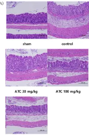
5. Effects of ATC on histological damage to colonic mucosa in DSS-induced colitis
Pathological examinations of colons were performed after H&E staining and representative results are shown in Fig. 6. Administration of DSS resulted in marked histopathological changes in the colon appearing as focal loss of surface epithelium, disruption of the cryptal glands and infiltration of the inflammatory cells.
However, treatment with ATC attenuated DSS-induced histopathological changes in the colon. ATC-treated groups showed protective effects against histological damage of the colonic mucosal layer induced by DSS.
The histological damage observed in the ATC 30 ㎎/㎏
group was similar to that in the control group. Treatment with ATC at 100 or 300 ㎎/㎏ reduced infiltration of the inflammatory cells and showed more intact cryptal glands and surface epithelium than those of the control group (Fig. 6A).
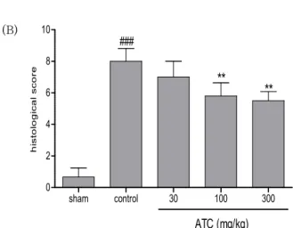
The histological score of the control group was significantly increased compared to the sham group (0.7
± 0.6 vs. 8.0 ± 0.8, p < 0.001). Compared with the control group, the ATC 30, 100 and 300 ㎎/㎏ treatments significantly reduced the histological score in a dose- ependent manner by 12.5% (7.0 ± 1.0, p = 0.2031), 27.5%
(5.8 ± 0.8, p < 0.01) and 31.3% (5.5 ± 0.6, p < 0.01), respectively (Fig. 6B)
(A)
`
84
大 韓 本 草 學 會 誌 ― Vol. 32 No. 3, 2017(B)
sham control 30 100 300
0 2 4 6 8 10
ATC (mg/kg)
###
** **
histological score
Figure 6. Effects of ATC on histological characterization in DSS- induced mouse colitis.
(A) Representative H&E-stained histological sections.
(B) Overall histological scores in these different groups.
###, p < 0.001 compared with sham group; **, p < 0.01 compared with control group.