Abstract (J. Kor. Oral Maxillofac. Surg. 2009;35:494-498)
Introduction
A schwannoma arises from the neural sheath of Schwann cell in cranial, peripheral or autonomic nerves but not in olfac- tory and ophthalmic nerves, which have no Schwann cell. It is a rare benign tumor, well-wrapped in capsule which gradually grows. This schwannoma, also known as a neurilemmoma, is about 6~8% of intracranial tumors and is rarely transformed into malignant tumor. It can arise from all parts of the body, however, 25~45% of intracranial schwannoma arises in the head and neck area such as oral cavity, mastoid, middle ear, larynx and paranasal sinuses
1).
There is no particular age or sex in the onset of this disease and most patients have no symptom. Some patients complain of pain or paresthesia when there is central growth in the exter- nal wall of the nerve sheath compressing adjacent nerves
2). Occasionally, there are symptoms including hearing loss, nasal atresia, dysphagia, hoarseness, headache and tinnitus along with nerves related secondarily, however, it is nonspecific as a
symptom for preoperative diagnosis of a neurologic lesion.
FNA cytology, CT scan and MRI image might be restrictive but helpful to diagnosis of schwannoma, however, final diag- nosis could be made with postoperative histological findings.
Intracranial schwannoma mostly arises from olfactory nerves, followed by trigeminal nerves and it is rare in facial nerves.
Authors experienced a schwannoma within the maxillary sinus originated from infraorbital nerve, the branch of the trigeminal nerve, and that arising in the buccal space originat- ed from buccal branch in the facial nerve. It is determined those two cases of schwannoma in the rare portion is valuable and herein, it is about to report those with literature discus- sions.
Case Report 1
The patient is a 54-years old female with a chief complaint of swelling in the left maxillary gingiva since 2 months ago. In the first visit, she had a history of dental extraction of #26 and
#27 teeth 2 weeks ago and had no pain or discomfort. She had swelling in the left maxillary facial portion and it observed swelling in the left buccal mucosa in the oral cavity. There was no preoperative hypoesthesia in the left face.
It took the with-enhancement facial CT and observed about 6 cm mass, had a relatively good boundary with low density in
Yeong-Cheol Cho290-3 Jeonha-dong Dong-gu, 682-714 Ulsan
Oral and maxillofacial Dept Ulsan university hospital, Korea Tel: 82-52-250-7230
Fax: 82-52-250-7236 E-mail: [email protected]
Schwannoma in the maxillary sinus and buccal space: Case report
Byung-hwan Choi*, Soo-Won Park*, Jang-Ho Son*, Yeong-Cheol Cho*, Iel-Yong Sung*, Ki-Jung Byun*, Young-Min Kim**
*Department of Oral and maxillofacial Surgery, **Department of Pathology, College of Medicine, Ulsan University Hospital, Ulsan University
Schwannomas are tumors which originate from the neuroectodermal Schwann cell of cranial, intraspinal, peripheral and autonomic nerve sheaths, and they are solitary, benign, slow growing and well encapsulated neoplasm. Schwannomas are usually asymptomatic. No strong gender or age pre- dominance exists.
The incidence of extracranial schwannomas in the head and the neck region varies from 25~45%. In addition, schwannomas are rare in the maxil- lary sinus or buccal space.
In this paper, it diagnosed and treated a 54-years old female patient, who had schwannoma in the maxillary sinus derived from infraorbital nerves, the branch of the left trigeminal nerve, and a 19-years old male patient, who had schwannoma arose in the buccal space derived from the buccal branch of the right facial nerve. There was no particular complication except sensory extinction of the nerve in the female patient and paralysis by the nerve in the male patient. It is determined those two cases of schwannoma in the rare portion is valuable and herein, it reports those with literature dis- cussions.
Key words: Schwannoma, infraorbital nerve, facial nerve
[원고접수일 2009. 10. 2 / 1차수정일 2009. 10. 21 / 2차수정일 2009. 10. 30 / 게재확정일 2009. 11. 6]
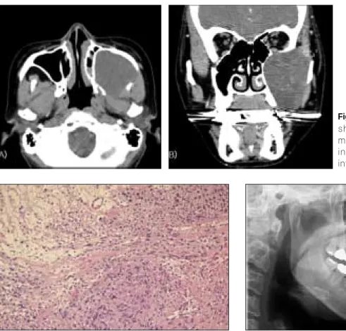
the left masticator space. By that mass, the left maxillary sinus was pushed forward and masseter muscle, pterygoid muscle and lower jaw were pushed backward. In addition, the mass was located up to the infraorbital fissure. There was no calci- fication or ossification within the mass and on the postcontrast enhanced CT scan, that mass showed mild enhancement and there was no destruction of adjacent bones. And then, it was not observed any enlargement of lymph node (Fig. 1).
In outpatient treatment, it performed an incisional biopsy for whitish gray soft tissue in the left maxillary sinus portion and she was diagnosed with chronic inflammation with fibrosis without particularity.
It carried out an operation under general anesthesia. It used an intraoral vestibular incision & Caldwell-Luc approach to minimize postoperative scar after resecting the mass, and used a subciliary approach in order to confirm the status of destruc- tion in the left orbital base. It checked and removed bone chips regarded as the maxillary sinus wall. It could identify the mass strongly connected with infraorbital nerves, the maxillary nerve branches of trigeminal nerve, by approaching the inside
of the maxillary sinus. It was almost impossible to separate the mass and infraorbital nerves so it removed the mass together with the part of nerves. It excised the brownish, focally myx- oid and soft mass (7.5 × 4.3 × 2.5 cm) and the mass had no necrotic lesion. It was diagnosed as a schwannoma by the frozen section.
It was found that the orbital base was slightly expanded toward the eyeball and there was no status of bone destruction in the base. Therefore, it was unnecessary to reconstruct the orbatial base due to this operation.
At histological finding, to some extent, it presented a typical manifestation of schwannoma such as palisaded arrangement of Schwann cell and Antoni A and B area in H&E staining, however, it was confirmed as schwannoma through S-100 pro- tein positive reaction and SMA negative reaction (Fig. 2).
After the operation, there has been no particular symptom except sensory extinction in the left maxillary sinus. After 1 year excision of mass, the alveolar bone of left maxilla was regenerated. The patient received dental implant treatment (Fig. 3).
Fig. 3.Panoramic view of 1st dental implant installation, after 1 year excision of mass.
Fig. 1. Axial(A) and coronal(B) CT of patient showing a well-circumscribed large mass measured approximately 7×4×2.5cm invad- ing left maxillary sinus, floor of orbit and infratemporal fossa.
Fig. 2. Microscopic findings. High-power view shows schwannoma architecture consisting of spindle-type cells in a diffuse Antoni A pattern, with a few areas of Antoni B pattern(H&E stain, ×200).
Case Report 2
A 19-years-old male patient visited the dental clinic for eval- uation of swelling on his right cheek. He said that he recog- nized it about 2~3 years ago. Then, in palpation, it was found a hard indurated, painless, slightly movable mass with nodular form and he reported no pain or discomfort caused by that mass. In his physical examination, he had no facial nerve paralysis or hearing loss.
It performed the FNAC for the mass on his right cheek and there was no evidence of a malignant cell.
It took CT and MRI and observed a well defined oval solid mass with 2 × 2 × 4 cm on the right buccal space on MRI result. The mass showed hyperintense signal than surround muscles in T2 weighted images and isointense signal com- pared with surround muscles in T1 weighted images. The right parotid gland was transposed to the upper side and there was no evidence of cystic change and hematoma. There was a high possibility of lesser salivary gland-derived benign tumor and also of Schwannoma (Fig. 4).
It carried out an operation under general anesthesia. To secure the visual field for this operation, it used a modified blare incision and checked facial nerves using a nerve stimula- tor. Then, it confirmed the mass under the right parotid gland surrounding the buccal branch of facial nerves by blunt dissec- tion. It was inevitable to remove the mass together with the part of the buccal branch for those could not be separated. It resected rubbery nodule, grayish solid mass (6.0 × 2.2 × 2.1cm) wrapped by capsule of bulky and yellowish gray sec- tion. The nerves removed were not reconstructed.
On the result of histological findings, it was firmly diagnosed as a typical schwannoma by H&E staining (Fig. 5).
After the operation, there was a partial paralysis on the right lobule of nose and it conducted postoperative rehabilitation.
Discussion
The schwannoma, firstly introduced by Verocay in 1908, is a benign tumor neurologically originated from Schwann cells wrapped the nervous tissue and slowly grow
3).
In the result of H&E staining, schwannoma is characterized by the pattern of Antoni A and B area and Verocay body. The typical cell shape of schwannoma is fusiform with a long nucleus. Antoni A area is composed of fusiform cells with par- allel rows of a palisade arrangement of the nucleus. Antoni B area has an oviform cell with loose arrangement between mucous substrates and has an intracellular space structure vac- uolized. Occasionally, it causes myxoid change accompanied by vitrification around vessels in the prolonged tumor, called an ancient schwannoma.
In addition, a schwann cell-derived tumor is mostly positive in S-100 immunohistochemical staining and S-100 protein pos- itive reaction is a reliable histological feature in the diagnosis of schwannoma
4).
Due to nonspecific signs and symptoms of schwannoma, it is difficult to diagnose it by laboratory tests. Therefore, it has been introduced FNA cytological technique, MRI and CT image as a diagnostic method for schwannoma. It has been reported that the diagnosis using FNAC characterized by the presence of spindle cells has only 17.6% of accuracy, not so high
5). To increase the accuracy of diagnosis using FNAC, it requires a high quality of specimen and is important to have an experience of a cytolophathologist
5). Therefore, it is recom- mended that FNAC is used for excluding the possibility of
Fig. 5.Photomicrograph of schwannoma showing well- organized Antoni A tissue with adjacent myxoid and less organized Antoni B tissue(H&E stain, ×200).
Fig. 4. T2-weighted magnetic resonance image revealing 2×2×4cm mass involving right side buccal space. Mass is enhanced homogeneously by high signal intensities.
malignancy rather for the exact diagnosis of schwannoma.
Through the FNA conducted in the case 2, it confirmed that there was no malignant cell; however, it had no efficacy on the diagnosis of schwannoma. On CT image, schwannoma was well-wrapped and had a relatively clear fusiform shape. The characteristics of MRI image of schwannoma include the pat- tern of isointense T1 signal and increased and slightly hetero- geneous T2 signal. Besides, after injection of gadolinium, the mass showed better contrast
6). In the case 1, schwannoma was suspected through CT results. The signal pattern of schwanno- ma was determined by MRI image in the case 2 and it could diagnose that as a schwannoma. When the diameter of the nerve derived is big, the ultrasound is a very useful diagnostic tool. It has been reported that it could be used for identifying that the mass is often connected with the nerve related, howev- er, its diagnostic rate by ultrasound is low
4).
Neoplasms originated from cells composed of the nerve sheath are schwannoma and neurofibromatosis. Schwannoma typically arises solely and has a well-wrapped shape. Its trans- formation to malignant cell is very rare and its reoccurrence was low if it is completely removed. Neurofibromatosis is commonly multicentric and only 4% of it is wrapped by cap- sule. Compared to schwannoma, it is more difficult to separate nerves and the mass and has high rate of transformation to malignant cell. It is often related to Von Recklinghausen’s dis- ease, and in this case, it shows about 8% of malignant transfor- mation
7). Schwannoma derived from infraorbital nerves arises within the orbit or maxillary sinus. Intraorbital schwannoma, 1~4% of orbital tumor within the orbit, is generally developed in supraorbital or supratrochlear nerves, and is rare in infraor- bital nerves
8). It is the mass arose from sensory nerves so it does not mostly affect eye movement or visual acuity if it aris- es within the orbital. The main symptoms are exophthalmos and pain. The mass slowly grows and does not invade sur- rounding bone structure; however, it sometimes pushes or cor- rodes the bone wall by pressure.
It is rare to find schwannoma in the paranasal sinus, especial- ly in the maxillary sinus
9). The symptoms of schwannoma in the maxillary sinus are cheek swelling, nasal atresia, down- ward transversion of the palate and pain, rarely accompanying exophthalmosis. For the mass slowly grows, it mostly causes the expansion of the maxillary sinus when the size is bigger, and most bony margin is maintained
7). For the treatment, it should completely resect the mass. If schwwwannoma com- presses the orbital base to elevate the orbital structure and to destroy the orbital base, orbitotomy might be required.
The case 1 presented infraorbital nerve-derived schwannoma in the left maxillary sinus. It completely resect the mass and
there was no complication except paresthesia. The orbital base was elevated to the eyeball by compression; however, there was no destruction of bone. Then, it was unnecessary to recon- struct the orbital base. In addition, there was no limitation for eye movement and no abnormal visual acuity.
Within the buccal space, diverse structures are exists such as salivary gland, blood vessel, lymph glands, connective tissues, muscle and nervous tissue. It has been reported various lesions originated from those structures. Neurofibroma has been often reported as a nervous tissue-derived mass; however, it is very rare to find schwannoma within the buccal space.
It has been known that the facial schwannoma arises along with the route of facial nerves, the clinical symptoms of it within the temporal bone, the majority of facial schwannoma, are facial nerve paralysis and hearing loss, and the main clini- cal symptoms of facial schwannoma outside the temporal bone is a lump of parotid gland
10). It has been also known that facial nerve schwannoma is less than 5% of the cause of facial nerve paralysis and 39% of the schwannoma patient presented facial nerve paralysis.
The patient in the case 2 is a very rare instance of schwanno- ma arose in the buccal space derived from the buccal branches of facial nerves. Before the operation, he had no facial nerve paralysis due to nerve compression by central growth in the external wall of the nerve sheath and it removed a part of the buccal branches during the operation. Then, there was a post- operative right alinasal paralysis. It could consider immediate reconstruction or postoperative rehabilitation for nerve sacri- fice in the incision process of facial schwannoma. In this case, it conducted rehabilitation and followed up the progress, there- fore, it confirmed that the activity of dilator naris muscles, which was decreased by paralysis immediately after the opera- tion, was improved.
There are various operational approaches depending on the location or size of the mass, personal preference of a dentist and related nerves. In our clinic, it used modified blare incision in the case 2 so it secured a good visual field and aesthetic results.
It is still open to discussion in some aspects for the treatment
of facial schwannoma. Several researchers persisted conserva-
tive treatment until there is a precise evidence for facial palsy
if it is little or none or if the mass slowly grows with no symp-
toms. Also, there was a report that, in the case of complete
incision for the aged or the patient unendurable to long opera-
tion, although it needed so long period for follow up, to treat
facial schwannoma in the intratemporal bone by subtotal or
neat total resection is proper to manage it without malignant
transformation
3).
However, the ideal treatment of radioresistant schwannoma is to prevent reoccurrence and to resect it completely while conserving the nerve related in order to minimize nerve dys- function. In addition, total resection with parotidectomy is the most precise treatment when facial schwannoma rarely arises in the parotid gland
5).
In the case of facial schwannoma, it has been reported the easy separation between the mass and nerves related only in several cases. Besides, there are some methods for reconstruc- tion of facial nerves such as greater auricular n. interposition- ing graft, sural nerve interposition graft and hypoglossal-facial nerve anastomosis if facial nerve is resected
10). However, there is the case of postoperative facial nerve dysfunction despite of efforts to conserve or to reconstruct it with the nerve related.
References
1. Samet A, Podoshin L, Fradis M, Simon J, Lazarov N, Boss H:
Unusual sites of schwannoma in the head and neck. J Laryngol Otol 1985;99:523-528.
2. Zachariades N: Schwannoma of the oral cavity. Review of the lit- erature and report of a case. J Oral Med 1984;39:41-43.
3. Kang GC, Soo KC, Lim DT: Extracranial Non-vestibular Head and Neck Schwannoma: A Ten-year Experience. Ann Acad Med 2007;36:233-228.
4. Zhang H, Cai C, Wang S, Liu H, Ye Y, Chen X: Extracracial Head and Neck Schwannomas : A Clinical Analysis of 33 Patients. Laryngoscope 2007;117:278-281.
5. Salemis NS, Karameris A, Gourgiotis S, Stavrinou P, Nazos K, Vlastarakos P, et al.: Large intraparotid facial nerve schwannoma : Case report and Review of the Literature. Int J Oral Maxillofac Surg 2008;37:679-681.
6. Guthikonda B, Theodosopoulos PV, van Loveren H, Tew JM Jr, Pensak ML: Evolution in the assessment and management of trigeminal schwannoma. Laryngoscope 2008; 118:195-203.
7. Shugar JM, Som PM, Biller HF, Som ML, Krespi YP: Peripheral nerve sheath tumors of the Paranasal sinuses. Head Neck Surg 1981;4:72-76.
8. Tezer MS, Ozcan M, Han O, Unal A, Ozlugedik S: Schwannoma originating from the infraorbital nerve: a case report. Auris Nasus Larynx 2006;33:343-345.
9. Sarioğlu S, Ozkal S, Gu¨neri A, Ada E, Sis B, Erdağ TK, et al.:
Cystic schwannoma of the maxillary sinus. Auris Nasus Larynx 2002;29:297-300.
10. Hatziotia JC, Asprides H: Neurilemoma (schwannoma) of the oral cavity. Oral Surg Oral Med Oral Pathol 1967;24:510-526.