영남대학교 의과대학 내과학교실
김혜진, 신경철, 이재웅, 김규진, 홍영훈, 정진홍, 이관호
TNF-α in the Pleural Fluid for the Differential Diagnosis of Tuberculous and Malignant Effusion
Hye Jin Kim, M.D., Kyeong Cheol Shin, M.D., Jae Woong Lee, M.D., Kyu Jin Kim, M.D., Yeong Hoon Hong, M.D., Jin Hong Chung, M.D., Kwan Ho Lee, M.D.
Department of Internal Medicine, College of Medicine, Yeungnam University, Deagu, Korea
Background : Determining the cause of an exudative pleural effusion is sometimes quite difficult, especially between malignant and tuberculous effusions. Twenty percent of effusions remain undiagnosed even after a complete diagnostic evaluation, including pleural biopsy. The activity of tumor necrosis factor-alpha (TNF-α), which is the one of proinflammatory cytokines, is increased in both infectious and malignant effusions. The aim of this study was to investigate the diagnostic efficiency of TNF-α activity in distinguishing tuberculous from malignant effusions.
Methods : 46 patients (13 with malignant pleural effusion, 33 with tuberculous pleural effusion) with exudative pleurisy were included. TNF-α concentrations were measured in the pleural fluid and serum samples using an enzyme- linked immunosorbent assay (ELISA). In addition, TNF-α ratio (pleural fluid TNF-α : serum TNF-α) was calculated.
Results : TNF-α concentration and TNF-α ratio in the pleural fluid were significantly higher in the tuberculous effusions than in the malignant effusions (p<0.05). However, the serum levels of TNF-α in the malignant and tuberculous pleural effusions were similar (p>0.05). The cut off points for the pleural fluid TNF-α level and TNF-α ratio were found to be 136.4 pg/mL and 6.4, respectively. The sensitivity, specificity and area under the curve were 81%, 80% and 0.82 for the pleural fluid TNF-α level (p<0.005) and 76%, 70% and 0.72 for the TNF-α ratio (p<0.05).
Conclusion : We conclude that pleural fluid TNF-α level and TNF-α ratio can distinguish a malignant pleural effusion from a tuberculous effusion, and can be additional markers in a differential diagnosis of tuberculous and malignant pleural effusion. The level of TNF-α in the pleural fluid could be a more efficient marker than the TNF-α ratio.
(Tuberc Respir Dis 2005; 59: 625-630)
Key words : Pleural effusion, Malignancy, Tuberculosis, Tumor factor
이 논문은 2002학년도 영남대학교 학술연구조성비 지원 에 의한 것임
Address for correspondence : Kyeong-Cheol Shin, M.D.
Department of Internal Medicine, College of Medicine, Yeungnam University. 317-7, Daemyeong-dong, Nam-gu, 705-717, Daegu, South Korea
Phone : 82-53-620-3850 Fax : 82-53-654-8386 E-mail : shin@med.yu.ac.kr
Received : Aug. 31. 2005 Accepted : Nov. 7. 2005
서 론
흉수의 진단을 위한 첫 단계는 삼출성 혹은 여출성 여부를 구별하는 것으로, 여출성이라면 더 이상 직접 적인 진단과정은 필요하지 않지만 삼출성흉수라면 부가적인 검사를 하여야 한다1. 삼출성흉수는 흉막의 염증이나 감염, 손상 또는 림프관의 폐색 등의 기전으 로 발생하는데 폐렴, 악성신생물, 결핵, 폐경색이나
류마티스성 관절염 등이 주요 원인 질환이며1, 이뇨제 를 복용하고 있는 심부전 환자도 흉수의 생화학 검사 에서 삼출성흉수로 나타날 수 있다2.
우리나라는 결핵성흉막염이 많으며, 연령이 증가 함에 따라 암성흉수의 빈도가 증가한다. 결핵성흉수 와 악성흉수를 구분하는 작업은 경우에 따라 매우 까 다로운데 적절한 검체를 얻기 위하여 흉막생검, 흉강 경검사 또는 개흉술 등 침습적인 방법을 동원하기도 한다1,3. 그러나 이런 침습적인 진단 방법을 동원하여도 약 20% 정도는 여전히 분명하게 진단되지 않는다4,5.
결핵성흉막염은 흉수 혹은 흉막생검에서 항산성균 을 확인하거나 M. tuberculosis가 배양되는 경우, 흉 막조직에서 괴사성육아종이 확인되면 진단할 수 있 으나 흉수 내 결핵균의 양이 매우 적어 도말염색이나 배양으로 균이 확인되기 어렵고 결핵균배양에 2-6주 의 기간이 필요하며 양성율도 25-37%에 지나지 않는
다6.7. 또한 결핵성흉막염 환자의 10-20%정도는 흉막 조직의 배양이나 조직학적 분석에도 음성이다8. 악성 흉수 역시 흉수 내 악성세포가 없어도 악성흉수의 가 능성을 배제할 수 없다.
결핵성흉막염을 진단하기 위한 방법으로 adenosine deaminase (ADA)가 널리 이용되고 있으나 한계가 있어, 세포성 면역에 관계하는 여러 사이토카인을 이 용하여 결핵성흉막염과 다른 원인에 의한 삼출성흉 수를 구분하려도 시도가 많이 이루어지고 있다.
Tumor necrosis factor-α (TNF-α)는 활성화된 단 핵구나 대식세포에서 분비되는 전염증성 사이토카인 (proinflammatory cytokine)으로서 종양세포나 다른 염증성세포들과 작용하여 숙주의 면역체계에 중요한 역할을 하며9, 다양한 면역세포의 분화와 성장을 조절 하기도 한다10-13.
감염성 혹은 악성흉수는 흉수 내 TNF-α가 증가하
는데9,14,15, Hua 등16은 악성흉수에 비하여 결핵성흉수
에서 proinflammatory cytokine의 농도가 높고 섬유 소용해효소(fibrinolytic enzyme)의 활성이 낮아 흉막 비후를 유도한다고 설명하였다. 그러나 흉수 내 TNF-α 측정이 삼출성흉수의 원인을 진단하는 데에 는 그다지 유용하지 않다는 보고들도 있다9,17.
저자들은 삼출성흉수인 결핵성흉수와 악성흉수 환 자의 혈청과 흉수에서 TNF-α를 측정하여, TNF-α가 이들 질환을 감별하는데 유용한 지표로 사용될 수 있 는지에 대해서 알아보고자 하였다.
대상 및 방법 1. 대 상
1998년 5월부터 2001년 8월까지 영남대학교의료원 호흡기내과에 입원한 삼출성흉수 환자 46명을 대상 으로 하였다. 삼출성흉수의 진단기준은 다음의 세 가 지 경우 중 적어도 한 가지 이상 만족하는 경우로 하 였다. 첫째, 혈청에 대한 흉수의 단백질의 비가 0.5를 초과하거나 둘째, 혈청에 대한 흉수의 lactic acid de
hydrogenase (LDH)의 비가 0.6을 초과하거나 셋째, 흉수의 LDH가 혈청의 정상 상한치 2/3을 초과하는
경우이다18. 악성흉수는 흉수 세포검사나 흉막조직 검 사에서 악성세포가 증명된 경우로 정의하였고, 결핵 성흉막염은 흉막조직에서 전형적인 건락성 괴사가 동반된 육아종성 병변이 관찰된 경우로 정의하였다.
진단에 따라 악성흉수환자 13명, 결핵성흉막염 환자 33명이었다.
2. 방 법
모든 환자는 치료 전 진단적 흉강천자를 하여 흉수 를 채취하였다. 채취한 흉수는 포도당, 총 단백량 그 리고 LDH를 포함한 생화학적 분석과 백혈구백분율 검사를 하였다. 백혈구백분율은 흉수를 EDTA (ethylene diamine tetraacetate-potassium anticoagulant)가 담 긴 튜브에 담은 후 검사하였다.
모든 흉수는 세균배양과 그람염색, 그리고 항산균 염색을 하였고, 종양세포를 확인하기 위하여 세포학 적 분석을 하였다. 흉수채취와 동시에 환자의 혈청을 얻어 흉수뿐만 아니라 혈청의 TNF-α도 측정하였다.
혈청과 흉수는 얻은 즉시 원심분리하여 상층액을 사 용하기 전까지 영하 70℃에서 보관하였으며, 흉막생 검은 승인된 규약에 따라 부분 마취를 한 후 시행하 였다.
3. TNF-α 측정
TNF-α는 Quantine, R&D system (Minneapolis, MN) 에서 생산된 상업적 ELISA kit를 이용하여 공급자들 의 권고에 따라 측정하였다. 흉수와 혈청에서 TNF-α 를 측정하고 혈청에 대한 흉수의 TNF-α 비(이하 TNF-α 비)를 계산하였다.
4. 통계 분석
자료들은 평균±표준편차로 표시하였고 두 그룹간 의 평균치의 통계적인 검정은 Mann-Whitney U test 를 사용하였다. 결핵성흉막염 환자의 혈청 TNF-α와 흉수 TNF-α와의 관련성을 알아보기 위하여 상관분 석을 하여 Spearman 상관계수를 구하였다. 적정 절
Tuberculous PE* Malignant PE* p-value
Serum (pg/mL) 79.16±194.62 98.55±241.81 >0.05
Pleural fluid (pg/mL) 215.84±105.05 84.81±106.90 <0.01
P/S ratio† 15.02±11.17 6.80±7.94 <0.05
*PE : pleural effusion, †P/S ratio : pleural fluid/serum ratio
Table 1. Pleural fluid and serum levels of TNF-α in patients with tuberculous (n=33) and malignant (n=13) effusion
Serum (pg/mL) Pleural fluid (pg/mL) p-value
Tuberculous PE* 79.16±194.62 215.84±105.05 <0.05
Malignant PE* 98.55±241.81 84.81±106.90 >0.05
*PE : pleural effusion
Table 2. Comparison of serum and pleural fluid TNF-α level between tuberculous and malignant effusion
TNF-α in serum
TNF-α in pleural fluid
r=-0.04, p=0.86*
r=0.32, p=0.36†
*Tuberculous effusion, †Malignant effusion
Table 3. Correlation of TNF-α between in the pleural fluid and serum of patients with pleural tuberculosis
사값을 구하기 위해 ROC (Receiver-operating cha
racteristic) 곡선분석을 실시하였다. 언급한 모든 통 계처리는 SPSS for Windows 12.0을 이용하였으며 p
<0.05인 경우를 통계적으로 유의한 것으로 해석하였다.
결 과
혈청의 평균 TNF-α값은 결핵성흉막염 79.16±194.62 pg/mL, 악성흉수 98.55±241.81 pg/mL 이었으나 두 집단사이의 통계적 유의성은 없었다(p>0.05). 흉수 의 TNF-α는 결핵성흉막염 215.84±105.05 pg/mL, 악 성흉수 84.81±106.90 pg/mL로 결핵성흉막염이 유의 하게 높았다(p<0.01). TNF-α 비는 결핵성흉막염 15.02±11.17 로 악성흉수의 6.80±7.94 보다 유의하게 높았다(Table 1).
결핵성흉막염의 흉수 내 TNF-α는 혈청의 TNF-α (215.84±105.05 pg/mL, 79.16±194.62 pg/mL)보다 통 계적으로 유의하게 높았다(p<0.05). 그러나 악성흉
수의 흉수 내 TNF-α와 혈청 TNF-α (84.81±106.90 pg/
mL, 98.55±241.81 pg/mL)는 차이가 없었다(p>0.05) (Table 2). 또한 결핵성흉막염 및 악성흉수환자의 혈 청 TNF-α는 흉수의 TNF-α와 상관관계가 없었다 (r=-0.04, r=0.32, p>0.05)(Table 3).
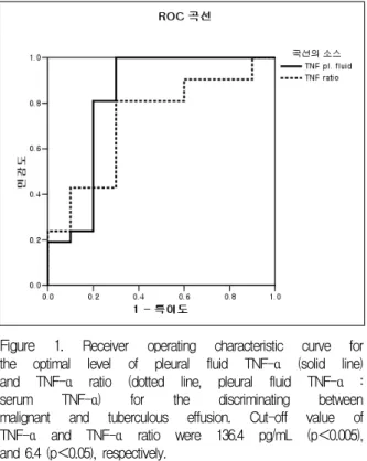
흉수 내 TNF-α의 적정절사값을 136.4 pg/mL로 하였을 때 민감도 81%, 특이도 80%, 곡선밑면적(area under the curve)은 0.82였으며(p<0.005), TNF-α 비 는 적정절사값을 6.4로 하였을 때 민감도 76%, 특이 도 70%, 곡선밑면적 0.72 (p<0.05)였다. 곡선 밑면적 은 흉수 내 TNF-α가 TNF-α 비보다 유의하게 높았 다(Figure 1).
고 찰
저자들의 연구결과 흉수의 TNF-α와 TNF-α 비는 악성흉수와 결핵성흉수를 구별하는데 유용하게 적용 할 수 있을 것으로 생각되며, 흉수의 TNF-α의 적정 절사값을 136.4 pg/mL, TNF-α 비의 적정절사값을 6.4 으로 하였을 때 민감도와 특이도가 비교적 높았다.
혈청 TNF-α는 결핵성흉수와 악성흉수 간 차이가 없었으며, 흉수 내 TNF-α 는 결핵성흉수가 악성흉수 보다 유의하게 높아 이전의 다른 연구결과와 비교적 일치하였다14-16,19,20
. TNF-α 비는 결핵성흉수가 악성 흉수보다 유의하게 높았다.
Figure 1. Receiver operating characteristic curve for the optimal level of pleural fluid TNF-α (solid line) and TNF-α ratio (dotted line, pleural fluid TNF-α : serum TNF-α) for the discriminating between malignant and tuberculous effusion. Cut-off value of TNF-α and TNF-α ratio were 136.4 pg/mL (p<0.005), and 6.4 (p<0.05), respectively.
Hua 등16은 염증이나 감염 때문에 생기는 삼출성흉 수는 플라스미노겐 활성인자 억제자(plasminogen activator inhibitor)가 많이 증가되고, 암성흉수는 플 라스미노겐 활성인자(plasminogen activator)가 증가 됨을 보고하였다. 이들은 결핵성흉수와 암성흉수의 큰 차이점으로 흉막의 섬유소용해(fibrinolysis)를 들 고, 섬유소를 용해하고 흉막을 비후시키데 TNF-α를 포함하는 proinflammatory cytokine이 영향을 미친 다고 설명하였다.
결핵성흉막염은 결핵균 단백질항원에 대한 세포성 면역반응으로 흉막 내 단핵구와 대식세포의 증가하 고 활성화되어 TNF-α를 분비하게 된다16. 또한 결핵 환자의 육아종 내 TNF-α의 농도는 높으나 혈청의 농도는 높지 않은데10, 이는 TNF-α가 대식세포를 활 성시켜 결핵균 감염을 막는 동시에 결핵의 면역반응 인 육아종형성에도 관여하여 발병과 방어기전 모두 중요한 역할을 하는 것으로 생각된다20,21. TNF-α는 부폐렴성흉수나 악성흉수에서도 증가하는데, 악성흉 수의 경우 주로 대식세포자체나 대식세포가 종양세 포와 작용하여 분비되며15,24-27, 중피세포 역시 염증반 응이 진행됨에 따라 TNF-α를 분비하게 된다. 이상
여러 질환에서 TNF-α가 증가한다는 사실은 TNF-α 는 특정질환에서만 생성되는 것이 아니라 염증성질 환에서 일반적으로 증가하며, TNF-α 농도는 염증반 응의 정도를 반영하는 것이라 할 수 있다.
삼출성흉수는 부폐렴성흉수, 결핵성흉수, 악성흉수 등이 대부분을 차지하지만 부폐렴성흉수는 흉수 내 호중구가 대부분이라는 점에서 비교적 구분하기 쉽 다. 그러나 흉수 내 림프구가 우세한 결핵성흉수와 악 성흉수의 구분은 흉수세포검사나 흉막생검 등 침습 적 진단법을 동원하더라도 위음성이 높아 흉수의 약 20%정도는 원인질환이 분명하게 진단되지 않는다4,5. 더욱이 60세 이상 환자의 흉수는 악성의 가능성이 높 기 때문에 더욱 정확한 감별진단이 필요하다.
혈청과 흉수의 TNF-α 관계에 대하여 Odeh 등15은 단순부폐렴성흉수 (uncomplicated parapneumonic effusion)나 울혈성심부전에 의한 삼출성흉수 내 TNF- α는 혈청 TNF-α와 상관관계가 있었으나 암성 흉수 의 경우 이러한 관계는 없었는데, 그 원인을 단순부폐렴 성흉수나 울혈성심부전에 의한 삼출성흉수의 TNF-α는 주로 혈청 TNF-α의 영향을 받기 때문이라고 설명하 였다. 그러나 저자들의 연구는 결핵성흉수 및 암성흉 수의 TNF-α는 모두 혈청 TNF-α보다 높았으나 서로 상관관계가 없어 흉수 내 TNF-α는 혈청 값의 영향 을 받지 않음을 알 수 있다. Odeh 등15의 연구는 결핵 성흉수가 포함되지 않아 저자들의 연구와 직접비교 는 할 수 없으나 결핵성흉수와 암성흉수의 TNF-α는 다른 원인에 의한 삼출성흉수와 달리 흉막의 국소염 증반응에 의하여 생성되며 혈청 내 TNF-α의 영향을 받지 않는다는 것을 의미한다. 또한 TNF-α 비는 암 성 흉수보다 결핵성흉수가 훨씬 높아 흉막 내 국소염 증반응정도는 결핵성흉수가 암성흉수보다 심하며, 이 러한 염증반응의 차이는 결핵성흉수와 악성흉수를 구별하는데 적용할 수 있을 것이다.
TNF-α 비는 결핵성흉수와 비결핵성흉수, 악성흉 수와 비악성흉수의 구분에 대하여 연구되었는데, 그 적정절사값을 0.28과 2.0 으로 보고하였다15,28. 그러나 이들 연구 결과는 악성흉수와 결핵성흉수 이외의 질 환이 포함되어 악성흉수와 결핵성흉수를 구분하는 TNF-α 비의 절사값으로 적절하지 않다. 저자들의 연
구에서 확인된 악성흉수와 결핵성흉수를 구별하는 TNF-α 비의 적정절사값은 6.4로 기존의 연구결과보 다 훨씬 높았다. 이러한 차이는 악성흉수 및 결핵성흉 수를 제외한 삼출성흉수의 흉수 내 TNF-α는 이들 두 질환에 비하여 훨씬 낮기 때문이라 생각된다.
흉수 내 TNF-α와 TNF-α 비에 대한 ROC 곡선분 석을 보면 흉수 내 TNF-α의 곡선 밑면적이 TNF-α 비의 곡선 밑면적보다 넓어 흉수 내 TNF-α가 악성 흉수와 결핵성흉수를 감별하는데 TNF-α 비보다 더 유용할 것으로 생각된다. 이러한 결과는 악성흉수와 비악성흉수를 구분하는데 흉수 내 TNF-α보다 TNF-α 비가 더 유용한 지표로 보고한 이전의 보고와 차이가 있다15. 결핵성흉수와 악성흉수를 구분하기 위하여 흉 수 내 TNF-α의 절사값을 136.4 pg/mL로 하였을 때 민감도 81%, 특이도, 80%, TNF-α 비의 적정절사값 을 6.4로 하였을 때 민감도 76%, 특이도 70%로 확인 되어 두 질환을 구분하는데 유용하게 사용할 수 있을 것으로 여겨진다. 림프구분획이 증가된 흉막 삼출액 의 감별진단을 위하여 현재 널리 이용되고 있는 ade
nosine deaminase (ADA) 와 TNF-α를 동시에 측정 할 경우 민감도는 증가하지만 특이도가 낮아지는 경 향이 있는 등29, 추가적인 진단가치에 대해서 뚜렷하 지 않다는 연구결과가 있다12,30.
그러나 본 연구의 결과를 종합하면 흉수 내 TNF- α와 TNF-α 비는 결핵성흉수와 악성흉수를 구별하는 데 유용하게 사용될 수 있을 것으로 판단되며, 특히 TNF-α 비보다 흉수 내 TNF-α가 더 유용할 것으로 판단된다.
요 약
배 경 :
삼출성흉수의 원인질환 중 흉수 내 림프구가 우세 한 경우는 결핵성흉수와 암성흉수가 가장 흔하다. 그 러나 두 질환의 감별은 흉막생검을 비롯한 침습적 방 법이 필요하며 약 20% 정도는 진단을 명확하게 내릴 수 없다. TNF-α는 종양세포나 다른 염증성세포들과 작용하여 숙주의 면역체계에 중요한 역할을 하며, 감 염성 혹은 악성흉수인 경우 흉수 내 TNF-α는 증가
한다. 저자들은 결핵성흉수와 악성흉수를 구분하는데 세포성 면역에 관계하는 TNF-α의 유용성을 확인하 고자 하였다.
방 법 :
삼출성 흉수로 입원한 환자 46 예(암성흉수 13 예, 결핵성흉수 33 예)를 대상으로 하였다. 혈청과 흉수 의 TNF-α를 ELISA법으로 측정하였으며, 혈청에 대 한 흉수의 TNF-α 농도의 비(TNF-α 비)를 구하였다.
결 과 :
결핵성흉수 및 악성흉수의 혈청 내 TNF-α는 차이 가 없었으나, 흉수 내 TNF-α는 결핵성흉수가 유의하 게 높았으며(p<0.01), TNF-α 비 역시 결핵성흉수가 높았다(p<0.05). 그러나 두 질환의 혈청 및 흉수 내 TNF-α의 상관관계는 없었다. ROC 곡선을 이용하여 이들 질환의 감별할 수 있는 기준치를 구하였는데 흉 수 내 TNF-α의 절사값을 136.4 pg/mL로 하였을 때 민감도 81%, 특이도 80% (p<0.005), TNF-α 비는 절 사값을 6.4로 하였을 때 민감도 76%, 특이도 70% (p
<0.05) 이었다. 곡선밑면적은 흉수 내 TNF-α가 TNF-α 비보다 유의하게 넓었다.
결 론 :
흉수 내 TNF-α측정과 TNF-α 비는 결핵성흉수와 암성흉수를 감별진단 하는데 이용될 수 있으며, 흉수 내 TNF-α가 TNF-α 비보다 더 유용한 지표로 판단 된다.
참 고 문 헌
1. Light RW. Diagnostic principles in pleural disease. Eur Respir J 1997;10:476-81.
2. Chakko SC, Caldwell SH, Sforza PP. Treatment of con
gestive heart failure: its effect on pleural fluid chemistry. Chest 1989;95:798-802.
3. Light RW. Pleural disease. 3rd ed. Baltimore: Williams
& Wilkins; 1995.
4. Storey DD, Dines DE, Coles DT. Pleural effusion: a diagnostic dilemma. JAMA 1976;236:2183-6.
5. Hirsch A, Ruffie P, Nebut M, Bignon J, Chretien J.
Pleural effusion: laboratory test in 300 cases. Thorax 1979;34:106-12.
6. Valdés L, Álvarez D, San José E, Penela P, Valle JM, García-Pazos JM, et al. Tuberculous pleurisy: a study of 254 patients. Arch Intern Med 1998;158:2017-21.
7. Scharer L, McClement JH, Isolation of tubercle bacilli from needle biopsy specimens of parietal pleura. Am Rev Respir Dis 1968;97:466-8.
8. Valdés L, Pose A, San José E, Martínez Vázquez JM.
Tuberculous pleural effusions. Eur J Intern Med 2003;
14:77-88.
9. Xirouchaki N, Tzanakis N, Bouros D, Kyriakou D, Ka
rkavitsas N, Alexandrakis M, et al. Diagnostic value of interleukin-1α, interleukin-6, and tumor necrosis factor in pleural effusions. Chest 2002;121:815-20.
10. Barnes PF, Fong SJ, Brennan PJ, Twomey PE, Ma
zumder A, Modlin RL. Local production of tumor necrosis factor and IFN-γ in tuberculous pleuritis. J Immunol 1990:145;149-54.
11. Barnes PF, Mistry SD, Cooper CL, Pirmez C, Rea TH, Modlin RL. Compartmentalization of a CD4+ T lymphocyte subpopulation in tuberculous pleuritis. J Immunol 1989;142:1114-9.
12. Kim YC, Park KO, Bom HS, Lim SC, Park HK, Na HJ, et al. Combining ADA, protein and IFN-γ best allows discrimination between tuberculous and mali
gnant pleural effusion. Korean J Intern Med 1997;12:
225-31.
13. Pettersson T, Soderblom T, Nyberg P, Riska H, Linko L, Klockars M. Pleural fluid soluble interleukin 2 receptor in rheumatoid arthritis and systemic lupus erythematosus. J Rheumatol 1994;21:1820-4.
14. Hamed EA, el-Noweichi AM, Mohamed AZ, Mahmoud A. Vasoactive mediators (VEGF and TNF-α) in patients with malignant and tuberculous pleural effusions.
Respirology 2004;9:81-6.
15. Odeh M, Sabo E, Srugo I, Oliven A. Tumour necrosis factor alpha in the diagnostic assessment of pleural effusion. QJM 2000;93:819-24.
16. Hua CC, Chang LC, Chen YC, Chang SC. Proinflam
matory cytokines and fibrinolytic enzymes in tuber
culous and malignant pleural effusions. Chest 1999;
116:1292-6.
17. Soderblom T, Nyberg P, Teppo AM, Klockars M, Riska H, Pettersson T. Pleural fluid interferon-γ and tumour necrosis factor-α in tuberculous and rheumatoid ple
urisy. Eur Respir J 1996;9:1652-5.
18. Light RW, Macgregor MI, Luchsinger PC, Ball WC Jr.
Pleural effusions: the diagnostic separation of transu
dates and exudates. Ann Intern Med 1972;77:507-13.
19. Xirouchaki N, Bouros D, Alexandrakis M, Tzanakis
G, Eliopoulos N, Siafakas M. The role of cytokines in pleural effusion differentiation. Eur Respir J 1994;7 (Suppl 18):471s.
20. Orphanidou D, Gaga M, Rasidakis A, Dimakou K, Toumbis M, Latsi P, et al. Tumour necrosis factor, interleukin-1 and adenosine deaminase in tuberculous pleural effusion. Respir Med 1996;90:95-8.
21. Olobo JO, Geletu M, Demissie A, Eguale T, Hiwot K, Aderaye G, et al. Circulating TNF-α, TGF-β, and IL-10 in tuberculosis patients and healthy contacts.
Scand J Immunol 2001;53:85-91.
22. Ishii Y, Uchiyama Y, Hasegawa S, Kinoshita T, Mitsui K, Kojima H, et al. Detection of tumour necrosis factor/cachectin in pleural effusion of patients with lung cancer. Clin Exp Immunol 1990;80:350-3.
23. Baughman RP, Lower EE. An inhibitor of tumor necrosis factor found in pleural effusions. J Lab Clin Med 1991;118:326-31.
24. Gjomarkaj M, Pace E, Melis M, Satafora M, Toews GB. Mononuclear cells in exudative malignant pleural effusions: characterization of pleural phagocytic cells.
Chest 1994;106:1042-9.
25. Lin CC, Liu CC, Lin CY. Changes in cell population and tumor necrosis factor, interleukin-6, and inter
leukin-8 in malignant pleural effusions after treat
ment with intrapleural tetracycline. Am Rev Respir Dis 1993;147:1503-6.
26. Doers J, Baughman RP, Buchsbaum J, Lower EE.
Tumor necrosis factor production by pleural macro
phages. Am Rev Respir Dis 1989;139:A360.
27. Baughman RP, Doers J, Lower EE, Buchsbaum J.
Isolation of functionally active pleural macrophages. J Clin Lab Immunol 1989;30:147-51.
28. Tahhan M, Ugurman F, Gozu A, Akkalyoncu B, Sa
murkasoglu B. Tumor necrosis factor-α in comparison to adenosine deaminase in tuberculous pleuritis. Res
piration 2003;70:270-4
29. NA HJ, Park SC, Kang KW, Park HK, Kim YC, Choi IS, et al. Diagnostic significance of TNF-α in tuber
culous and non-tuberculous pleural effusion. Tuberc Respir Dis 1997;44:611-20.
30. Shim TS, Yang SE, Chi HS, Kim MJ, Chung H, Jegal YJ, et al. TNF-α, TGF-β, and fibrinolytic parameters in tuberculous and malignant pleural effusion. Tuber Respir Dis 2000;49:149-61.