Tuberc Respir Dis 2013;74:215-221
CopyrightⒸ2013. The Korean Academy of Tuberculosis and Respiratory Diseases. All rights reserved.
Identification and Distribution of Nontuberculous Mycobacteria from 2005 to 2011 in Cheonan, Korea
Jae Kyung Kim, Ph.D.1, Insoo Rheem, M.D.2
1Department of Laboratory Medicine, Dankook University Hospital, Dankook University College of Medicine, 2Department of Laboratory Medicine, Dankook University College of Medicine, Cheonan, Korea
Background: Nontuberculous mycobacteria (NTM) are considered opportunistic pathogens, and several species of NTM are associated with human diseases that typically involve the pulmonary, skin/soft tissue, or lymphatic systems; such infection may also cause disseminated diseases. Recent studies have reported increasing rates of NTM-induced disease worldwide.
Methods: Respiratory samples are being analyzed for acid-fast bacilli (AFB) culture and NTM identification at Dankook University Hospital in Cheonan, Korea, from September 2005 to September 2011. Identification is performed by using polymerase chain reaction-restriction fragment length polymorphism analysis targeting a novel region of the rpoB gene.
Results: A total of 25,133 specimens were received for AFB culture, of which 1,014 (4.0%) were NTM-positive.
A total of 267 samples from 186 patients were tested for NTM identifications, and 232 samples from 157 patients were positive for NTM species. Among the patients who tested positive for NTM, 65.6% were men and the average age was 63.3 years. Mycobacterium avium complex, the most commonly detected NTM pathogen, was found in 65.9% of the 232 samples. The annual average percentage of NTM isolates from AFB culture-positive specimens was 31.3%: the highest rate was seen in 2011 (44.3%), followed by 2009 (37.4%) and 2010 (37.2%). An upward trend in NTM incidence was found during the study period.
Conclusion: The prevalence of pulmonary NTM isolates continues to increase in Cheonan, suggesting that pulmonary NTM disease is becoming increasingly common.
Key Words: Nontuberculous Mycobacteria; Mycobacterium avium Complex; Age Distribution
Address for correspondence: Insoo Rheem, M.D.
Department of Laboratory Medicine, Dankook University College of Medicine, 119 Dandae-ro, Dongnam-gu, Cheonan 330-997, Korea
Phone: 82-41-550-6668, Fax: 82-41-550-7055 E-mail: [email protected]
Received: Feb. 13, 2013 Revised: Mar. 14, 2013 Accepted: May. 6, 2013
CCIt is identical to the Creative Commons Attribution Non-Commercial License (http://creativecommons.org/licenses/by-nc/3.0/).
Introduction
Mycobacteria species other than those of the Mycobacterium tuberculosis complex (MTBC) are called nontuberculous mycobacteria (NTM) or “atypical” my- cobacteria. NTM are environmental organisms found in soil and water throughout the world
1,2. NTM are gen-
erally hardy, ubiquitous environmental bacteria that vary in geographic distribution and pulmonary patho- genicity. NTM can also cause disseminated disease
3. Disseminated disease due to NTM is primarily asso- ciated with acquired immunodeficiency syndrome and other forms of severe immunosuppression
4. However, the incidence of NTM disease in patients without human immunodeficiency virus infection is increasing
5. Different NTM species have different antibiotic sus- ceptibility patterns, and their resistance to anti-tuber- culosis drugs is of particular importance. For these rea- sons, the accurate and early differential diagnosis of MTBC and NTM is required for optimal outcomes.
Currently, the identification of clinical isolates of my-
cobacteria at the species level is primarily based on the
characteristics of the cultured bacteria and the bio-
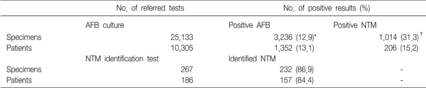
No. of referred tests No. of positive results (%)
AFB culture Positive AFB Positive NTM
Specimens 25,133 3,236 (12.9)* 1,014 (31.3)†
Patients 10,305 1,352 (13.1) 206 (15.2)
NTM identification test Identified NTM
Specimens 267 232 (86.9) -
Patients 186 157 (84.4) -
*Positive rate of AFB culture: positive culture results/referred tests in specimens or patients. †Rate of NTM identification to AFB culture-positive results: positive NTM results/AFB culture-positive results in specimens or patients.
AFB: acid-fast bacilli; NTM: nontuberculous mycobacteria.
Table 1. Basic statistics of AFB culture and NTM identifications chemical test results. These conventional tests can take several weeks to perform and cannot always precisely identify the species
6. Furthermore, these testing proce- dures are complex and laborious, and they are usually impeded by the slow growth of mycobacteria in culture.
There are numerous species of NTM, and recently de- veloped molecular methods have enabled the recog- nition of many of these species
7. In this study, NTM were identified using a polymerase chain reaction (PCR)-restriction fragment length polymorphism (RFLP) method, based on the rpoB gene, at the Korean Institute of Tuberculosis
8,9.
Mycobacterial identification at the species level is both of academic interest and of importance for gaining a better understanding of the organisms' epidemiology and pathogenesis. Geographic variability in the environ- mental exposure and prevalence of NTM disease is of global significance
10.
Given these clinical challenges, further knowledge of the epidemiology of NTM in Korea is needed. The pur- pose of this study was to identify NTM and investigate their distribution in clinical specimens isolated from a tertiary teaching hospital in Cheonan, Korea.
Materials and Methods
This study included the isolation of NTM species from all clinical specimens referred to Dankook University Hospital Laboratory in Cheonan, Korea, from September 2005 to September 2011 (Table 1). Patients for whom
NTM identification tests were requested provided sam- ples of sputum and bronchial washing fluid. For this study, we retrospectively analyzed the results of these samples. Data were analyzed and categorized according to gender, age, and year of analysis.
1. Culture
1) Sample pre-processing: For respiratory samples, N-acetyl-L-cysteine (NALC)-NaOH solution (5% NaOH+
0.5% NALC) was added to the sample to liquefy and decontaminate the mucous sputum. The solution was centrifuged at 3,000 ×g for 18 minutes at 4
oC, and the supernatant was discarded. Phosphate buffered saline 1 mL was added after the sediment had been vortexed.
2) Mycobacterial culture: The processed samples (0.2 mL) were used to inoculate 3% Ogawa medium (Eiken, Tokyo, Japan) and were cultured for 8 weeks in an in- cubator at 35–37
oC under 5–10% CO
2. An interim report was provided at 4 weeks, if necessary.
Samples (0.5 mL each) were used to inoculate MGIT medium (Becton Dickinson, Sparks, MD, USA) after mixing with PANTA/supplement according to the manu- facturer's protocol. The tubes were then incubated for 6 weeks in the BACTEC MGIT 960 system (Becton Dickinson). If fluorescence was detected in the tube, the test was considered positive.
2. NTM identification
NTM identification was performed at the Korean
Institute of Tuberculosis, where PCR-RFLP methods are
Figure 1. Age group distribution of the positive spe- cimens. Forty-four patients (28.0%) were aged 60–69 years, 37 patients (23.6%) were aged 70–79 years, and 34 patients (21.7%) were aged 50–59 years. M., Mycobacterium.
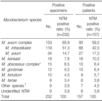
Mycobacterium species
Positive specimens
Positive patients
No.
NTM positive ratio (%) (n=232)
No.
NTM positive ratio (%) (n=157) M. avium complex 153 65.9 93 59.2 M. intracellulare 119 51.3 66 42.0 M. avium 34 14.7 27 17.2 M. kansasii 18 7.8 16 10.2 M. abscessus complex* 15 6.5 10 6.4 M. gordonae 12 5.2 10 6.4 M. fortuitum 10 4.3 9 5.7 M. terrae 8 3.4 6 3.8 Other species† 9 3.9 7 4.5 Unidentified NTM 9 3.9 6 3.8
Total 232 100 157 100
*M. abscessus complex: M. abscessus and M. massiliense.
†Other species: M. szulgai, M. chelonae, M. ulcerans, M. non- chromogenicum, M. peregrinum, and M. simiae.
Table 2. Distribution of species after nontuberculous my- cobacteria (NTM) identification according to specimens and patients
used to target a novel region of the rpoB gene.
3. Statistical analysis
The statistical analyses were performed using SAS version 9.2 (SAS Institute, Cary, NC, USA). The Cochran-Armitage test for trend was used to evaluate the tendency of isolating NTM from respiratory acid-fast bacilli (AFB)-culture specimens over the years studied.
Results
1. Characteristics of NTM-positive patients
A total of 25,133 specimens were received for AFB culture, of which 3,236 (12.9%) were AFB positive and 1,014 (4.0%) were NTM positive. A total of 267 samples from 186 patients were collected for NTM identification, and 232 samples from 157 patients were positive for NTM species (Table 1).
Gender analysis showed that 66.7% of all patients were men, and that 65.6% of the NTM-positive patients were men. The average patient age was 63.3 years (range, 33.3–91.1 years). More than 50% of patients were aged 60–79 years (Figure 1).
2. Distribution of NTM species
Mycobacterium avium complex (MAC), the most com- mon NTM, was detected in 153 samples (65.9%): M. in- tracellulare was detected in 119 and M. avium in 34 samples. M. kansasii was the next most prevalent spe- cies (18 isolates, 7.8%), followed by M. abscessus com- plex ( M. abscessus and M. massiliense ; 15 isolates, 6.5%) and M. gordonae (12 isolates, 5.2%) (Table 2).
The annual distributions of NTM species are detailed in Table 3.
The overall proportion of NTM isolates from among the respiratory AFB-positive specimens was 31.3%
(1,014/3,236); the highest rate was seen in 2011 (192/
433, 44.3%), followed by 2009 (135/361, 37.4%) and 2010 (246/662, 37.2%). There was an upward trend in NTM proportion in respiratory specimens during the study period (Cochran-Armitage test for trend, p<
0.001) (Figure 2).
Mycobacterium species 2005 2006 2007 2008 2009 2010 2011 Total
M. avium complex 3 24 17 17 26 40 26 153
M. intracellulare 2 16 15 13 20 32 21 119
M. avium 1 8 2 4 6 8 5 34
M. kansasii 0 1 2 3 1 6 5 18
M. abscessus complex* 0 0 1 1 3 3 5 13
M. gordonae 0 1 2 3 0 2 4 12
M. fortuitum 0 1 0 3 0 6 0 10
M. terrae 0 1 0 0 0 5 2 8
M. chelonae 0 0 1 1 0 0 0 2
Other species† 0 1 0 0 0 2 4 7
Unidentified NTM 0 1 0 2 1 2 3 9
Total, n (%) 3 (1.3) 30 (12.9) 23 (9.9) 30 (12.9) 31 (13.4) 66 (28.4) 49 (21.1) 232 (100.0)
*M. abscessus complex: M. abscessus and M. massiliense. †Other species: M. szulgai, M. chelonae, M. ulcerans, M. non- chromogenicum, M. peregrinum, and M. simiae.