© Copyright
Keimyung University School of Medicine 2017
Tracheobronchopathia osteochondroplastica (TO) is a rare dysplastic disease of the trachea characterized by cartilaginous or bony nodules in the tracheobronchial lumen. Rigid video-stylet is an intubating device that provides favorable conditions even in the difficult cases. In this report, we describe a successful airway management using the rigid video-stylet in a 62-year-old man with unanticipated difficult intubation later diagnosed for TO. He was planned for elective percutaneous nephrolithotomy under general anesthesia. He was healthy without any airway symptoms. With the rigid video-stylet, we not only performed successful tracheal intubation but also examined endotracheal lumen simultaneously. Using the rigid video-stylet, we noticed multiple whitish projecting nodules in the trachea, which were the typical findings for TO.
Keywords: Airway management, Difficult intubation, Rigid video -stylet, Tracheobronchopathia osteochondroplastica
Introduction
Difficult endotracheal intubation is a clinical challenge for anesthesiologists and is associated with increased morbidity and mortality. Therefore, thorough pre-operative airway assessment and preparation of proper airway management tools are important [1].
Tracheobronchopathia osteochondroplastica (TO) is a rarely observed dysplasia which causes tracheal narrowing leading to difficult intubation. It is signified by multiple calcified cartilaginous masses in the tracheal lumen. Although patients with TO might have symptoms such as dyspnea, stridor, dry cough and hemoptysis, most of the TO
Received: March 27, 2017 Revised: May 10, 2017 Accepted: May 31, 2017
Corresponding Author: Kyoungho Ryu, M.D., Department of Anesthesiology and Pain Medicine, Kangbuk Samsung Hospital,
29 Saemunan-ro, Jongno-gu, Seoul 03181, Korea Tel: +82-2-2001-1376
E-mail: [email protected]
• The authors report no conflict of interest in this work.
Department of Anesthesiology and Pain Medicine, Kangbuk Samsung Hospital, Sungkyunkwan University School of Medicine, Seoul, Korea
Eun Ah Cho, M.D., Keulame Song, M.D., Tae Young Lim, M.D., Kyoung Ho Ryu, M.D., Hyun Soo Kim, M.D.
Successful Tracheal Intubation Using Rigid Video-Stylet in a Patient
with Tracheobronchopathia Osteochondroplastica
cases are asymptomatic [2]. Therefore, TO might result in unexpected difficult tracheal intubation in general anesthesia settings. Here in this report, we represent a TO case which was not detected by preoperative evaluations leading to unanticipated difficult intubation and in which successful endotracheal intubation and tracheal examination were done by rigid video-stylet (OptiScopeⓇ PM 201, Clarus Medical, Minneapolis, MN, USA).
Case
A 62-year-old male (height 161 cm, weight 64.5 kg, BMI 25.2 kg/m2), ASA physical status 1, was scheduled for elective percutaneous nephrolithotomy for renal stone under general anesthesia. The patient didn’t have any medical history or previous surgical history. He was a non-smoker. There were no abnormal respiratory signs and the patient didn’t complain of respiratory symptoms. Preoperative chest X-ray showed non-specific findings. The pulmonary function test revealed normal values.
He had a Mallampati Class Ⅱ airway and showed the normal range of motion of cervical spine in the pre-anesthetic visit. His lung sound was clear at both whole lung fields.
After entering the operating room, application of ASA standard monitoring and preoxygenation with 100% O2 were done. General anesthesia was induced with midazolam 2 mg, propofol 120 mg IV and remifentanil 0.05 mcg/kg/min IV without premedication. After checking successful bag-mask ventilation, rocuronium 50 mg was injected intravenously and face mask bagging was done with 100% O2 and desflurane for 3 minutes until the sufficient muscle relaxation was achieved.
There was no specific event during the mask ventilation. The first intubation trial was performed with internal diameter (ID) 8.0-mm endotracheal
tube (ETT) under direct laryngoscopy with Macintosh #3 blade. Cormack-Lehane score was checked grade 1. The initial attempt failed because it was unable to advance the ID 8.0-mm ETT through the vocal cord even with the clear view of the supraglottic structure. Serial attempts of intubation with smaller ID ETT were tried stepwise with ID 7.0-mm and 6.5-mm tube, however, resistance below the vocal cord kept blocking the advance of the endotracheal tube. Suspecting subglottic airway abnormality, we planned intubation with ID 6.0-mm ETT while inspecting the subglottic tracheal lumen using the rigid video- stylet (Fig. 1). On an endotracheal exam with the rigid video-stylet, multiple cobblestone-like appearance masses were seen in the tracheal lumen (Fig. 2). Successful intubation was done using ID 6.0-mm ETT under rigid video-stylet guidance.
After tracheal intubation, ventilation was effective with peak airway pressure of 17 cm H2O and tidal volume of 450 ml. Dexamethasone 5 mg was injected intravenously to prevent airway edema associated with multiple trials of laryngoscopic manipulation. During the intubation period, the vital signs were stable and SPO2 was maintained 100%. The otorhinolaryngologist was contacted and the bronchoscopic exam was done. Suspecting the TO, the otorhinolaryngologist suggested for the neck computed tomography (CT) for confirmation.
Although the general medical condition of the patient seemed tolerable for the surgery, we had a discussion with the urologist and decided to cancel and reschedule the operation after the evaluation of the airway because the operation time was expected to last over 3 hours in the prone position.
The patient was reversed with pyridostigmine 15 mg and glycopyrrolate 0.4 mg IV. After confirming spontaneous breathing and alert mentality, the patient was extubated and sent to the post anesthesia care unit. He was recovered without any
complications.
The next day, neck CT imaging was done and it showed irregular calcified plaques along the anterolateral wall of the larynx and trachea (Fig. 3).
The narrowest trans-tracheal diameter measured 7.48 mm. Ill-defined infiltration in right upper lobe along the bronchovascular bundle was also seen,
which was a possible finding for bronchial pathology. Finally, the patient was diagnosed for TO. Because the patient refused to proceed the percutaneous nephrolithotomy, he was discharged without further treatment.
Discussion
TO is a rare benign disease involving trachea. It rarely descends down the bronchial trees. It is characterized by cartilaginous or osseous submucosal nodules projecting into the tracheal lumen mostly in anterolateral tracheal walls [2,3].
The incidence of the TO, though not well established, is approximately between 1 in 125 and 1 in 5,000 [4]. Patients are usually sixth or seventh decade of life and there is no discrepancy in incidence by sex [5].
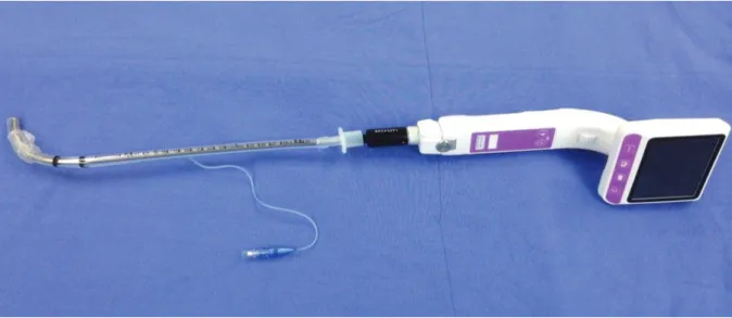
Although most of the TO patients are asymptomatic, there might be symptoms such as a dry cough, voice change, dryness of the throat, moderate dyspnea and recurrent pulmonary infections [6]. Chest radiographic exams are usually normal. However, Fig. 1. Rigid video-stylet (OptiScope® PM 201) preloaded with an internal diameter 7.5-mm endotracheal tube.
Fig. 2. Optiscopic view of the trachea, showing multiple cobblestone-like appearance masses (arrows) protruding into the tracheal lumen, and also diffuse hemorrhage in the subglottic lesion due to multiple intubation attempts.
in some advanced cases, tracheal irregularity and wall thickness or lobar collapse might be evident [7,8]. Likewise, the patient in our case was without any definite symptoms and preoperative tests were in normal range. Therefore, it was difficult to expect uneventful failure of endotracheal intubation.
The definite etiology of the disease is not well known. Various hypotheses including congenital, genetic, chronic inflammatory, metabolic and amyloidosis have been suggested [3]. It might be associated with ecchondroses and enchondroses from tracheal rings or metaplasia of the elastic tissues [9,10].
Bronchoscopy and CT are widely used for diagnosing the TO [3,5,7]. CT of the neck reveals numerous nodules with calcifications involving anterolateral endotracheal walls [7]. Under bronchoscopy, the diagnosis might be confirmed by its typical appearance described as cobblestones [9]. Multiple white projecting masses over the normal mucosa are spread along the anterior and lateral tracheal lumen. Laryngeal and main bronchial involvements are rarely seen [5].
T h e r e a r e a f e w c a s e s r e p r e s e n t i n g
unanticipated difficult intubations in anesthesia settings [3,5,7-10]. Most of the cases were incidentally found on intubation due to difficult intubation, and there were no severe adverse events. Patients were safely intubated with smaller endotracheal tubes than average size after multiple trials of intubation [7-10]. However, there is one extreme case with the large size of TO, which was intubated with ID 4.0-mm tube. In the case report, bougination and carbon dioxide laser removal of the mass were done under rigid bronchoscopy [11]. A laryngeal mask airway (LMA) could be considered as an alternative device to secure the airway when the attempts to intubate the patient with TO ended in failure. There is a case report that LMA was used successfully in a patient with TO undergoing laparoscopic abdominal surgery, under intermittent positive pressure ventilation [12].
In this case, we used the rigid video-stylet for intubation. It not only provided successful intubation by visualization of the airway but also allowed us to inspect the subglottic area through the vocal cord to find out the lesion. The rigid Fig. 3. Postoperative chest computed tomography with axial scan (A) and coronal scan (B). The images revealed multiple calcified nodules (arrows) from the anterolateral into the tracheal lumen, confirming the diagnosis of tracheobronchopathia osteochondroplastica.
A B
video-stylet is one of the devices facilitating better condition for intubation by visualizing vocal cord through the colored view monitor providing faster intubation and higher success rate for intubation [13]. Intubation maneuver with the rigid video- stylet is conventionally done in the following steps [14]. First, anterior jaw lift is done while putting the non-dominant thumb into the patient’s mouth, hooking the mandible anteriorly. Then, with the dominant hand, the rigid video-stylet is introduced in the midline of the mouth, placing its tip behind the uvula guided by the monitor (Fig. 4). Finally, after the tip passes the vocal cord, the endotracheal tube is advanced through the stylet.
Use of a rigid video-stylet can be effectively done in difficult scenarios such as limited mouth opening or neck extension with minimal trauma [14]. There is a case report presenting an awake intubation with video-stylet for expected difficult intubation due to a short neck, immobility of the head and limited mouth opening [15]. With its rigidity of the device, it is reported that the rigid
video-stylet guided intubation is easily skilled and might be an alternative tool for the flexible bronchoscope [14]. As we can evaluate airway structures through its monitor, the rigid video-stylet is also used as a diagnostic tool for the airway abnormality, as we described in our case [16].
In conclusion, it is important to recognize that rare cases with unanticipated tracheal narrowing such as TO might cause difficult intubation.
Therefore, if conventional airway manipulation fails, the rigid video-stylet might be a suitable option to enable tracheal examination and intubation simultaneously.
References
1. Norskov AK, Wetterslev J, Rosenstock CV, Afshari A, Astrup G, Jakobsen JC, et al. Effects of using the simplified airway risk index vs usual airway assessment on unanticipated difficult tracheal intubation - a cluster randomized trial with 64,273 participants. Br J Anaesth 2016;116:680-9.
2. Gurunathan U. Tracheobronchopathia osteochon- droplastica: a rare cause of difficult intubation. Br J Anaesth 2010;104:787-8.
3. Warner MA, Chestnut DH, Thompson G, Bottcher M, Tobert D, Nofftz M. Tracheobronchopathia osteochon- droplastica and difficult intubation: case report and perioperative recommendations for anesthesiologists. J Clin Anesth 2013;25:659-61.
4. Prakash UB. What is tracheo (broncho) pathia osteo (chondro) plastica? J Bronchology Interv Pulmonol 2001;8:75-7.
5. Coetmeur D, Bovyn G, Leroux P, Niel-Duriez M.
Tracheobronchopathia osteochondroplastica presenting at the time of a difficult intubation. Respir Med 1997;91:496-8.
6. Wagner RB, Barson PK. Tracheobronchopathica osteochondroplastica diagnosed as a result of difficult Fig. 4. Illustration of endotracheal intubation
using a rigid video-stylet with lateral (A) and frontal (B) views.
A B
intubation. Anesthesiology 1979;51:269-70.
7. Tadjeddein A, Khorgami Z, Akhlaghi H. Tracheo- bronchopathia osteoplastica: cause of difficult tracheal intubation. Ann Thorac Surg 2006;81:1480-2.
8. Seo JH, Do SH. Tracheobronchopathia osteochon- droplastica detected during difficult endotracheal intubation : a case report. Anesth Pain Med 2007;2:102-5.
9. Thomas D, Stonell C, Hasan K. Tracheobronchopathia osteoplastica: incidental finding at tracheal intubation.
Br J Anaesth 2001;87:515-7.
10. Sharma A, Shende DK, Vyas V, Byani G, Devi SK, Devara S. Difficult intubation in a patient with carcinoma colon due to tracheobronchopathia osteochondroplastica: an incidental finding or otherwise. Egypt J Anaesth 2015;31:199-202.
11. Birzgalis AR, Farrington WT, O'Keefe L, Shaw J.
Localized tracheopathia osteoplastica of the subglottis.
J Laryngol Otol 1993;107:352-3.
12. Ishii H, Fujihara H, Ataka T, Baba H, Yamakura T, Tobita T, et al. Successful use of laryngeal mask airway for a patient with tracheal stenosis with
tracheo-bronchopathia osteochondroplastica. Anesth Analg 2002:95:781-2.
13. Yang M, Kim JA, Ahn HJ, Choi JW, Kim DK, Cho EA. Double-lumen tube tracheal intubation using a rigid video-stylet: a randomized controlled comparison with the Macintosh laryngoscope. Br J Anaesth 2013;111:990-5.
14. Lee AR, Yang S, Shin YH, Kim JA, Chung IS, Cho HS, et al. A comparison of the BURP and conventional and modified jaw thrust manoeuvres for orotracheal intubation using the Clarus Video System.
Anaesthesia 2013;68:931-7.
15. Gaszynski T, Gaszynska E. The Clarus Video System stylet for awake intubation in a very difficult urgent intubation. Anaesthesiol Intensive Ther 2013;45:153- 4.
16. Costa F, Mattei A, Massimiliano C, Cataldo R, Agro FE. The Clarus Video System as a useful diagnostic tool. Anaesthesia 2011;66:135-6.