Copyright © 2016 The Korean Association of Internal Medicine
This is an Open Access article distributed under the terms of the Creative Commons Attribution Non-Commercial License (http://creativecommons.org/licenses/
by-nc/3.0/) which permits unrestricted noncommercial use, distribution, and reproduction in any medium, provided the original work is properly cited.
pISSN 1226-3303 eISSN 2005-6648 http://www.kjim.org
IMAGE OF INTEREST
Korean J Intern Med 2016;31:807-808 http://dx.doi.org/10.3904/kjim.2015.064
A 48-year-old man visited our hospital due to exertional chest pain. His resting electrocardiogram exhibited a T wave inversion in the inferior leads. A trans- thoracic echocardiogram showed a pre- served left ventricular systolic function without any regional wall motion ab- normalities. Multi-detector computed tomography was performed to evaluate
the existence of any coronary artery dis- ease and it revealed a significant steno- sis in the right coronary artery; however, there was a minimal atherosclerotic le- sion in the left coronary artery (Fig. 1A).
The right coronary angiogram showed a significant stenosis in the mid portion of the right coronary artery and percu- taneous coronary intervention for that
Angiographically minimal but functionally
significant coronary lesion confirmed by optical coherence tomography
Hyuck-Jun Yoon, Yun-Kyeong Cho, Chang-Wook Nam, Kwon-Bae Kim, and Seung-Ho Hur
Department of Internal Medicine, Keimyung University Dongsan Medical Center, Daegu, Korea
Received : March 13, 2015 Revised : March 23, 2015 Accepted : April 22, 2015 Correspondence to Seung-Ho Hur, M.D.
Tel: +82-53-250-7998 Fax: +82-53-250-7034 E-mail: [email protected]
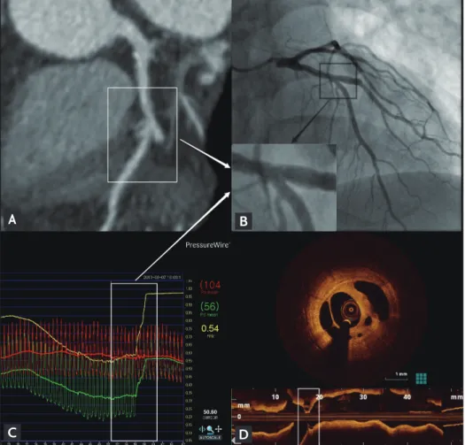
Figure 1. (A) Multi-detector com- puted tomography shows a minimal coronary stenosis in the left anteri- or descending artery (LAD). (B) The coronary angiogram also shows no significant coronary lesions in the LAD. (C) The fractional flow reserve (FFR) measurement using a pressure wire showed a significant function- al defect (FFR, 0.54) in the LAD and steep step up of the FFR value at the bifurcation of the LAD and first diagonal branch. (D) Optical coher- ence tomography revealed a focal (2 mm length) lotus root-like lesion consisting of multiple cavities di- vided by septa within mild fibrous plaque. In spite of the variability in the size of the channel, microchan- nels were seen to communicate with each other.
A
C
B
D
808 www.kjim.org
The Korean Journal of Internal Medicine Vol. 31, No. 4, July 2016
http://dx.doi.org/10.3904/kjim.2015.064
lesion was performed successfully with a drug-eluting stent. The left coronary angiogram showed a focal mini- mal lesion in the mid left anterior descending artery, just proximal to the diagonal bifurcation (Fig. 1B). Contrary to our expectations, the fractional flow reserve (FFR) mea- sured by a pressure wire at that lesion was significantly decreased at 0.54 (Fig. 1C). Intravascular optical coherence tomography (OCT) was performed to evaluate the cause of the discordance between the angiography and FFR. The OCT revealed a focal (2 mm length) lotus root-like lesion consisting of multiple cavities divided by septa within fi- brous plaque (Fig. 1D). In spite of the variability in the size of the channels, microchannels were seen to com- municate with each other. That was a suspicious finding of a recanalized intracoronary thrombus. That lesion was also successfully revascularized and the patient’s symp-
toms no longer occurred after discharge.
Decision making for revascularization based on angi- ography alone without prior vessel specific stress testing can be difficult in patients with multi-vessel coronary disease including intermediate or ambiguous coronary lesions. Therefore, functional and physiological consid- erations of such coronary lesions should be warranted for the selection of the target vessel. This representative case well demonstrated a noteworthy risk of an underestima- tion using coronary angiography, which could be com- pensated by invasive functional or intravascular imaging procedures.
Conflict of interest
No potential conflict of interest relevant to this article was reported.