ORIGINAL ARTICLE
Jae Hyun Park, Si Hyung Lee, Joon Mo Park, Chan Seo Park, Department of Internal Medicine, Yeungnam University College of Medicine, Daegu 705-717, South Korea
Kyung Sik Park, Eun Soo Kim, Kwang Bum Cho, Department of Internal Medicine, Keimyung University College of Medicine, Daegu 705-717, South Korea
Author contributions: Park JH, Park JM and Park CS con- tributed equally to this work; Park JH collected and analyzed the data, and drafted the manuscript; Park JM and Park CS provided analytical oversight; Lee SH designed and supervised the study;
Park KS, Kim ES and Cho KB revised the manuscript for important intellectual content; all authors have read and approved the final version to be published.
Institutional review board statement: The study was reviewed and approved by the Yeungnam University hospital Institutional Review Board.
Data sharing statement: Technical appendix, statistical code, and dataset available from the corresponding author at e-mail:
[email protected].
Open-Access: This article is an open-access article which was selected by an in-house editor and fully peer-reviewed by external reviewers. It is distributed in accordance with the Creative Commons Attribution Non Commercial (CC BY-NC 4.0) license, which permits others to distribute, remix, adapt, build upon this work non-commercially, and license their derivative works on different terms, provided the original work is properly cited and the use is non-commercial. See: http://creativecommons.org/
licenses/by-nc/4.0/
Correspondence to: Si Hyung Lee, MD, Department of Internal Medicine, Yeungnam University College of Medicine, 170 Hyeonchungro, Nam-gu, Daegu 705-717,
South Korea. [email protected] Telephone: +82-53-6203830 Fax: +82-53-6548386 Received: April 1, 2015
Peer-review started: April 2, 2015 First decision: June 4, 2015
Revised: July 27, 2015 Accepted: August 29, 2015 Article in press: August 29, 2015 Published online: October 21, 2015
Abstract
AIM: To find risk factors of lymph node metastasis (LNM) in early gastric cancer (EGC) and to find proper endoscopic therapy indication in EGC.
METHODS: We retrospectively reviewed the 2270 patients who underwent curative operation for EGC from January 2001 to December 2008. EGC was defined as malignant lesions that do not invade beyond the submucosal layer of the stomach wall irrespective of presence of lymph node metastasis.
RESULTS: Among 2270 enrolled patients, LNM was observed in 217 (9%) patients. LNM in intramucosal (M) cancer and submucosal (SM) cancer was de- tected in 38 (2.8%, 38/1340) patients and 179 (19%, 179/930) patients, respectively. In univariate analysis, the risk factors for LNM in EGC were size of tumor, Lauren classification, ulcer, lymphatic invasion, vascular invasion, and depth of invasion. However, in multivariate analysis, size of tumor, lymphatic invasion, vascular invasion, and depth of invasion were risk factors for LNM in EGC. Size of tumor, lymphatic invasion, vascular invasion, and depth of invasion were risk factors for LNM in cases of intramucosal cancer and submucosal cancer. In particular, there was no lymph node metastasis in cases of well differentiated early gastric cancer below 1 cm in size without ulcer regardless of lymphovascular invasion.
CONCLUSION: Tumor size, perilymphatic-vascular invasion, and depth of invasion were risk factors for LNM in EGC. There was no LNM in EGC below 1 cm Retrospective Study
Prediction of the indication criteria for endoscopic resection of early gastric cancer
Jae Hyun Park, Si Hyung Lee, Joon Mo Park, Chan Seo Park, Kyung Sik Park, Eun Soo Kim, Kwang Bum Cho
DOI: 10.3748/wjg.v21.i39.11160
© 2015 Baishideng Publishing Group Inc. All rights reserved.regardless risk factors.
Key words: Early gastric cancer; Lymph node metastasis;
Endoscopic resection
© The Author(s) 2015. Published by Baishideng Publishing Group Inc. All rights reserved.
Core tip: Although the depth of tumor infiltration, tumor size as a maximum tumor diameter, and perilym- phovascular invasion are independent risk factors for lymph node metastasis (LNM) in early gastric cancer (EGC), there was no LNM in intramucosal cancer which was not signet ring cell type and was below 1 cm without ulceration regardless of lymphatic invasion. This means that endoscopic submucosal dissection can be the treatment of choice in patients with intramucosal cancer below 1 cm without ulceration. There was LN metastasis in EGC of extended criteria in this study.
But, the possibility of LNM in intramucosal cancer of extended indication was below 1%.
Park JH, Lee SH, Park JM, Park CS, Park KS, Kim ES, Cho KB. Prediction of the indication criteria for endoscopic resection of early gastric cancer. World J Gastroenterol 2015; 21(39):
11160-11167 Available from: URL: http://www.wjgnet.
com/1007-9327/full/v21/i39/11160.htm DOI: http://dx.doi.
org/10.3748/wjg.v21.i39.11160
INTRODUCTION
Early detection in gastric cancer is increasing with screening endoscopy. Consequently early gastric cancer (EGC) which was resectable with endoscopic resection has increased. In gastric cancer, the most significant factor in endoscopic resection is the absence of lymph node metastasis (LNM) because it determines the treatment. For that reason, prediction of lymph node metastasis in EGC is very important.
The predictors of the absence of LNM in EGC were tumor size of 2 cm or smaller, histologically differentiated type, intramucosal cancer, and no lymphovascular (LV) invasion
[1]. According to the risk factors of LNM, endoscopic submucosal dissection (ESD) is a standard treatment for differentiated- type adenocarcinoma without ulceration, of which the depth of invasion is up to muscularis mucosa and the diameter is below 2 cm (Japanese gastric cancer treatment guidelines 2010
[1]). However, some recent studies have reported extended indications for endoscopic resection
[2-6]in differentiated EGC without lymphatic or vascular involvement, including: (1) mucosal cancers with no ulcerative findings, regardless of tumor size; (2) mucosal cancers with ulcerative findings ≤ 30 mm; and (3) minute (≤ 500 μm from the muscularis mucosae) submucosal invasive cancers
≤ 30 mm.
However, evidence from these studies is limited in South Korea. Thus, the purpose of this study was to determine the risk factors of lymph node metastasis in EGC removed by gastrectomy and to determine the safety of extended criteria for endoscopic treatment of EGC in South Korea.
MATERIALS AND METHODS
A total of 2270 patients who had undergone gas- trectomy with lymph node dissection for EGC at Yeungnam University hospital and Keimyung University hospital and we retrospectively reviewed the patient who has been taken radiologic imaging study and upper gastrofibroscope, and confirmed pathological reports after operation.
The patient profiles were investigated, including sex, age, tumor location, size, ulceration, histological type, lymphovascular invasion, and depth of in- vasion. Well and moderately differentiated tubular adenocarcinoma and papillary adenocarcinoma were classified as differentiated lesions. Poorly differentiated adenocarcinoma, signet ring cell carcinoma, and mucinous carcinoma were categorized as undifferen- tiated types. Lesions with ulcer or ulcer scar within cancer were regarded as ulcerated lesions. The depth of submucosal invasion was checked from the muscularis mucosa to the point of deepest penetration.
The depth of submucosal invasion was subclassified according to two groups: SM1 (≤ 500 μm penetration into submucosa) and SM2 (> 500 μm). The tumor size was measured by the results of the pathological report after surgical resection.
Statistical analysis was performed using the SPSS program. The relationship between lymph node metas- tasis and various factors was assessed using the simple χ
2test and multiple logistic regression analysis.
RESULTS
Baseline characteristics of enrolled patients and EGC Male to female ratio was 1488:782 and mean age was 59.3 ± 11.7. The mean length of major axis was 25.9 ± 16.5 mm. The most common location of early gastric cancer was middle anterior wall of stomach.
Risk factors of LNM
Among 1340 patients with M cancers, 2.8% (39/1340) were diagnosed as LMN; 19.2% (179/930) in SM cancer, 14.5% (55/379) in SM1 lesion, and 22.3%
(123/551) in SM2 lesion. The relationships between
various clinical or histological factors and the risk of
LNM are summarized in Table 1, Table 2 and Table
3. Tumor size, lymphatic or venule invasion, deeper
vertical invasion, and ulceration were the risk factors
of lymp node metastasis. Similar to the finding for
cancer involving intramucosa, significant correlation
was observed between tumor larger than 3 cm and
Table 3 Risk factors for lymph node metastasis in submucosal gastric cancer ( n = 930) by univariate analysis n (%)
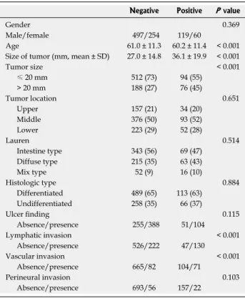
Table 2 Risk factors for lymph node metastasis in intramucosal gastric cancer ( n = 1340) by univariate analysis n (%) Table 1 Risk factors for lymph node metastasis in early gastric cancer ( n = 2270) by multivariate analysis
lymphovascular invasion with an increased risk of LNM. Also, cancer with involvement deep into the submucosa showed greater association with LNM;
and significant correlation was observed between tumor larger than 2 cm and lymphovascular invasion and LNM in submucosal cancer. This meant that the possibility of LNM in cancer with involvement deep into the submucosa was greater than in smaller sized tumor than in intramucosal cancer.
Suggestion for expanded indication of endoscopic resection for EGC
According to extended criteria, 3 (0.8%, 3/378) differentiated intramucosal lesions without lym- phovascular invasion and ulceration regardless of tumor size showed association with LNM. Two (0.9%, 2/230) differentiated intramucosal ulcerative lesions below 3 cm without lymphovascular invasion showed association with LNM. Three (2.7%, 3/113)
differentiated submucosal (≤ 500 μm from the muscularis mucosae) lesions were below 3 cm without lymphovascular invasion (Table 4). Although there were few patients with LNM in cases reflected by extended criteria, the possibility of LNM in EGC remained.
In our study, none (0/102) of the differentiated intramucosal lesions below 1 cm without lympho- vascular invasion and ulceration showed association with LNM. In particular, there was no lymph node metastasis (0/127) in cases of well differentiated early gastric cancer below 1 cm in size without ulcer regardless of lymphovascular invasion. The undifferentiated intramucosal cancer below 1 cm in size with ulcer did not show association with metastasis.
One (1.2%, 1/81) of the undifferentiated early gastric cancers below 1 cm without ulcer regardless of lym- phovascular invasion showed association with LNM.
The cell type of the patient in one case was signet ring.
Thus, there was no LNM in patients with early gastric cancer below 1 cm in the without ulcer group without signet ring type cancer in cell differentiation (Table 4).
Relative contraindication of endoscopic resection for EGC Of 406 patients with undifferentiated M cancer, 2.7%
(11/406) were found to have LNM. Seven patients with perilymphatic-vascular invasion were confirmed LNM and four of them had no ulcerated lesion. In 11 patients with LNM, one case was below 10 mm in size (Table 4).
OR 95%CI P value Tumor size ≤ 30 mm vs > 30 mm 2.1 1.5-3.0 < 0.001 Depth of invasion
M vs SM1 1.6 1.0-2.4 < 0.001
M vs SM2 4.7 3.0-7.2 0.040
Lymphatic invasion 4.1 2.8-6.0 < 0.001
Vascular invasion 4.7 3.1-7.1 < 0.001
Negative Positive P value
Gender 0.217
Male/female 843/458 29/10
Age 58.5 ± 11.8 50.6 ± 14.5 < 0.001
Size of tumor (mm, mean ± SD) 23.5 ± 15.7 37.9 ± 28.6 < 0.001
Tumor size < 0.001
≤ 30 mm 961 (81) 18 (54)
> 30 mm 224 (19) 15 (46)
Tumor location 0.052
Upper 192 (15) 14 (36)
Middle 736 (59) 19 (48)
Lower 324 (26) 6 (16)
Lauren < 0.001
Intestine type 634 (61) 11 (38)
Diffuse type 371 (36) 16 (55)
Mix type 33 (3) 2 (7)
Histologic type 0.715
Differentiated 799 (62) 20 (53)
Undifferentiated 498 (38) 18 (47)
Ulcer finding 0.757
Absence/presence 619/550 14/14
Lymphatic invasion < 0.001
Absence/presence 1202/93 23/15
Vascular invasion < 0.001
Absence/presence 1272/22 22/17
Perineural invasion < 0.001
Absence/presence 1279/13 36/3
Negative Positive P value
Gender 0.369
Male/female 497/254 119/60
Age 61.0 ± 11.3 60.2 ± 11.4 < 0.001
Size of tumor (mm, mean ± SD) 27.0 ± 14.8 36.1 ± 19.9 < 0.001
Tumor size < 0.001
≤ 20 mm 512 (73) 94 (55)
> 20 mm 188 (27) 76 (45)
Tumor location 0.651
Upper 157 (21) 34 (20)
Middle 376 (50) 93 (52)
Lower 223 (29) 52 (28)
Lauren 0.514
Intestine type 343 (56) 69 (47)
Diffuse type 215 (35) 63 (43)
Mix type 52 (9) 16 (10)
Histologic type 0.884
Differentiated 489 (65) 113 (63)
Undifferentiated 258 (35) 66 (37)
Ulcer finding 0.115
Absence/presence 255/388 51/104
Lymphatic invasion < 0.001
Absence/presence 526/222 47/130
Vascular invasion < 0.001
Absence/presence 665/82 104/71
Perineural invasion 0.103
Absence/presence 693/56 157/22
Table 4 Lymph node metastasis according to the presence of ulcer, differentiation, lymphovascular invasion, and the depth of invasion in early gastric cancer patients
In 324 patients with undifferentiated submucosal cancer, 20.3% (66/324) were found to have LNM. Of the subgroup of 16 patients with undifferentiated SM1 lesion, 16.3% (16/98) were diagnosed as LNM. Yet, of 82 undifferentiated SM1 lesions without LNM, 31 cases had ulcerated lesion, no one had perivascular
invasion, 32 cases showed perilymph invasion. And, of 16 undifferentiated SM1 lesions with LNM, no one had perivascular invasion, and perilymph invasion was detected in three cases. Seven patients had lymphatic and vascular invasion.
Based on these results, the treatment of choice of
Ulcer Differentiation VI LI Size
≤ 10 mm > 10 mm > 20 mm > 30 mm M
Ulcer negative Differentiated
No No 0/102 2/112 0/81 1/57
Yes 0/5 0/2 0/1 0/5
Yes No 0 0 0 1/2
Yes 0 1/2 0/0 1/2
Undifferentiated No No 1/66 2/41 0/41 1/45
Yes 0/3 0/3 0/1 0/2
Yes No 0 0 0 0
Yes 0 0 0/1 3/3
Ulcer positive Differentiated No No 1/59 1/104 0/67 1/47
Yes 0/5 1/12 0/1 1/5
Yes No 0 0 0 0
Yes 0/2 0/4 0/0 1/1
Undifferentiated No No 0/40 0/50 1/42 0/46
Yes 0/4 0/2 0/6 1/4
Yes No 0 0 0 0
Yes 0/1 1/2 0/1 1/1
SM1
Ulcer negative Differentiated No No 0/10 1/19 0/12 1/13
Yes 0/3 0/3 1/2 0/1
Yes No 0 0 1/1 2/2
Yes 0 1/3 0/0 1/3
Undifferentiated No No 0/7 0/6 1/8 0/6
Yes 0 0/2 0/0 0/1
Yes No 0 0 0 0
Yes 0 0/1 0/1 2/3
Ulcer positive Differentiated No No 0/14 2/24 0/19 1/13
Yes 0/1 0/0 1/7 1/5
Yes No 0 0/1 0/0 0
Yes 0 0/1 2/3 3/3
Undifferentiated No No 0/6 2/12 2/7 1/6
Yes 0/2 1/7 0/3 1/7
Yes No 0 0 0 1/1
Yes 1/2 3/5 1/4 1/1
SM2
Ulcer negative Differentiated No No 0/4 2/23 1/25 0/18
Yes 0 1/12 2/5 11/27
Yes No 0/1 0/0 0/0 0
Yes 0/1 1/3 0/1 6/8
Undifferentiated No No 0/1 1/14 2/9 1/4
Yes 0/2 1/1 0/2 3/7
Yes No 0/1 0/0 0/0 0
Yes 0/1 0/0 5/6 1/3
Ulcer positive
Differentiated No No 3/16 2/33 1/31 3/31Yes 0/3 5/15 12/27 13/31
Yes No 0 0 1/3 0/3
Yes 0/1 1/2 2/5 4/5
Undifferentiated No No 0/4 2/14 1/19 4/18
Yes 0/2 1/8 1/6 4/11
Yes No 0 0 0/1 1/1
Yes 1/1 3/4 5/8 3/5
undifferentiated SM1 lesion is not endoscopic resection but surgical resection (Table 4).
Percentage of cases in which endoscopic resection was indicated among EGCa patients who underwent surgery
In Yeungnam and Keimyung university hospitals, 1340 patients with M cancer underwent surgery.
Among them, 799 (59.6%) patients were confirmed as differentiated M cancer without LNM. Among 930 patients with SM invasion, 118 (12.6%) differentiated SM1 cancer patients without perilymphatic-vascular invasion were free from LNM; 40.3% (917/2270) of patients with EGC were overtreated (Figure 1).
DISCUSSION
The definition of EGC was suggested by the Japanese Gastroenterologic Endoscopic Society in 1962. In this definition, EGC is defined as gastric cancer which invades within submucosa regardless of lymph node metastasis
[7].
The existence of LNM was association with bad prognosis
[8]. The 5-year survival rate is > 90% in EGC, and the absence of lymph node metastasis is the most significant prognostic factor
[9,10]. The 5-year survival rate was reported to be 87.3% in patients with regional lymph nodes metastasis and 94.2%
in those without
[11]. Maruyama et al
[12]reported that the number of metastatic lymph nodes in patients with EGC was associated with the survival rate. EGC patients with LNM had a lower survival rate than patients without lymph node metastasis
[13].
Because of an increased accuracy of diagnosis of EGC, which in turn leads to a better prognosis, increased interest has been focused on betterment of the quality of life and minimalization of invasive procedures. Furthermore, nowadays, various minimally invasive treatment modalities have been developed and endoscopic resection has enabled rapid restoration
of patient’s health with lower risk of procedure, however, this endoscopic treatment also has risk of disease recurrence and distant metastasis.
Therefore, we suggested investigation of the relationship between various risk factors and LNM, and we think that our research will be helpful in development of more delicate criteria for endoscopic resection of EGC.
The overall incidence of a LNM in EGC ranges from 10% to 15%
[9,10,14,15]. The incidence of lymph node invasion was reported upto 4.8% in mucosal cancers and 23.6% in SM cancers
[9,10,16]. These results are similar to ours generally, and the incidence of LNM was similar to those in mucosal carcinoma reported by Yamao et al
[17]and Tsujitani et al
[18], and lower risk in submucosal carcinoma reported by other researchers
[19]. So far, standard treatment of SM cancer is gas- trectomy with lymphadenectomy
[20]. But, endoscopic resection such as EMR or ESD is widely used standard treatment modality for mucosal cancer
[9,21]. In studies analyzing the outcome of endoscopic resection in undifferentiated EGC, complete resection rate of undifferentiated EGC was relatively lower than that of differentiated EGC
[22].
There were various attempts to clarify risk factors predicting LNM and relationship between these risk factors and prognosis. Maehara et al
[23]found that large tumor, lymphatics involvement, and submucosal invasion were risk factors for LNM in EGC patients.
Yamao et al
[17]also reported that lymphatic invasion, histologic type, and large tumor size were independent risk factors for LNM in intramucosal EGC. In our study, univariate and multivariate analysis showed association of LNM in EGC with large tumor size, submucosal invasion, and perilymphatic-vascular invasion.
Some researchers have advocated that submucosal invasion is the most predictive risk factor for LNM.
Otherwise, of risk factors for metastasis, the most important is perilymphatic invasion
[24]. As for lymphatic vascular invasion, this may be the most important direct route to the regional lymph nodes
[6]. Relation of prognostic factors, including gender, tumor size, depth of invasion, endoscopic findings, and lym- phatic invasion to LNM in SM cancer has been demon- strated
[20,25,26]. Besides pathologic type, Lauren classi- fication and perineural involvement also showed significant and independent association with LNM.
However, Keita Nakahara et al
[6]reported that the histological type was not a risk factor for LNM. In our results, histological type and presence of differentiation were unrelated to LNM. And, in the study by Nakahara et al
[6], no significant difference in invasion depth was observed between SM1 and M cancers among EGC.
Yet, in our studies, a statistically significant difference was observed between SM1 and M, and SM1 and SM2.
With respect to size and ulceration, the possibility of SM invasion is higher and ulceration develops more readily in larger lesions. In contrast, there was no difference between ulcerated lesion and LNM in
Not indicated Indicated 100%
90%
80%
70%
60%
50%
40%
30%
20%
10%
0% M SM Total