∙ Received: October 28, 2008. Accepted: November 25, 2008.
∙ Corresponding author: Young Deok Seo
Department of Nuclear Medicine, National Cancer Center 809 Madu 1-dong, Ilsandong-gu, Goyang-si, Gyeonggi-do, 410-769, Korea
Tel: +82-31-920-0171, Fax: +82-31-920-0179 E-mail: [email protected]
Original Article Lymphoscintigraphy에 이용되는 방사성의약품의
사용제한으로 인한 대체 방사성의약품의 성능평가
국립암센터 핵의학과, 연구소 분자영상치료연구과1
서영덕⋅정재훈⋅문종운⋅윤상혁⋅김영석⋅강세훈1⋅원우재⋅김석기
Performance Evaluation of Substitution
Radiopharmaceutical according to Restriction of the Radiocolloids in Lymphoscintigraphy
Young Deok Seo, Jae Hun Jeong, Jong Woon Moon, Sang Hyeok Yun, Yeong Seok Kim, Se Hun Kang1, Woo Jae Won, Seok-Ki Kim
Departments of Nuclear Medicine and Research Institute1, National Cancer Center, Korea
Purpose: Several radioactive agents were used for lymphoscintigraphy in breast cancer. But recently, due to the change of local radiopharmaceutical licensing policy and discontinuation of supplies from manufacturer, we could not use many radiocolloids such as 99mTc-Antimony Trisulfide Colloid, 99mTc-Tin Colloid, 99mTc-Human Serum Albumin. So it is necessary to use the substitution radiopharmaceutical. Therefore, this study aims to evaluate the performance of substitution radiopharmaceutical (99mTc-Phytate) including the existing radio- colloids and to set up of the appropriate protocol in lymphoscintigraphy. Materials and Methods: For each radiocolloids (99mTc-Antimony Trisulfide Colloid (ASC), 99mTc-Tin Colloid (TC), 99mTc-Human Serum Albumin (HSA), 99mTc-Phytate) were performed the particle size by Zeta Sizer (Nano-ZS) and we compared the radiolabeling time, procedure and acquisition time for each of the radiocolloids (total 200 patients). For the last time, we made an analysis of image for each of the radiocolloids with our previous report (SJ Jang et al, Korean Nucl Med Mol imaging Vol.41, No.6, Dec 2007). Results: The particle size of each radiocolloids showed A.S.C (50 nm), T.C (310 nm), H.S.A (10.8 nm), 99mTc-Phytate (499 nm). The labeling and acquisition time for each of the radiocolloids showed no substantial difference. But there is difference of the labeling time for the A.S.C.
because the ASC procedure need to boiling process. There were no significant differences among those radiocolloids (p>0.005) in the identification rate (IR), false negative rate (FNR), and negative predictive value (NPV). Conclusions: 99mTc-labeled radiocolloids showed equivalent results in lymphoscintigraphy. Therefore, in this exam of each radiocolloids could be applied appropriate protocol in lymphoscintigraphy. (Korean J Nucl Med Technol 2009;13(1):25-29)
Key Words : Sentinel lymphy node, Lymphoscintigraphy, Breast cancer, Antimony sulfide colloid, Tin colloid, Human serum albumin, Phytate
서 론 우리나라에서 유방암은 2005년 현재 전체 암 발생의 15%
(27,049명)이고 여성의 악성 종양 중에서 가장 흔한 암이 되 었다. 본원의 국가 암 정보센터(National Cancer Information Center)에서 조사한 자료에 의하면 1999~2005년에 여자의 연 평균 암 발생자수는 65,044건이고, 유방암(15%), 갑상샘암 (14.6%), 위암(13.7%), 대장암(11.3%)의 순이었다. 매년 평균 25% 암 발생 증가가 관찰되었고, 특히 여자에서는 유방암과 갑상샘암이 매년 빠르게 증가하고 있는 추세이다.1) 이러한
Fig. 1. Zetasizer (Nano ZS).
유방암의 높은 발병률로 인하여 조기검진의 중요성이 증가하 였고, 이에 따라 최근에 이르러서 영상기술의 발달과 정기 검진의 확대로 유방암의 조기발견이 증가하여 액와림프절 전이가 없는 경우가 유방암환자의 과반수 이상이 되고 있다.2,3) 지금까지는 유방암 수술시 액와부림프절 곽청술이 표준 치료 법이었으나, 전이된 림프절을 모두 제거한 후에는 림프부 종이나 견관절 운동장애, 신경손상 등 다양한 합병증을 동 반한다.4,5) 또한 근래에는 환자의 삶의 질에 대한 관심이 높아 지면서 액와림프절 곽청술을 줄이고자 하는 노력으로 감시림 프절 생검술이 보급되었다.6,7) 여기서 전이가 먼저 일어나는 림프절, 즉 감시림프절(sentinel lymph node)이 존재한다는 사실이 알려지고 이를 찾는 방법은 다양하게 시도 되었다.8) 유방암의 감시림프절의 정확도가 향상되면서 액와림프절의 절제 필요여부를 알 수 있게 되었고, 최소 침습적인 수술치료 도 가능해졌다.9)
감시림프절을 찾는 방법은 다양하게 시도되었으나, 현재는 방사성동위원소를 이용한 방법이 표준 술식으로 이용되고 있다. 하지만 그동안 감시림프절을 찾기 위해 사용되었던
99mTc-Antimony Trisulfide Colloid (ASC), 99mTc-Tin Colloid (TC), 99mTc-Human Serum Albumin (HSA)의 국내사용이 제한 되면서 다른 방사성의약품으로의 대체 가능성에 대한 연구가 필요하였다. 본 연구에서는 기존에 국내에서 널리 사용되 었던 99mTc-Antimony Trisulfide Colloid (ASC), 99mTc-Tin Colloid (TC), 99mTc-Human Serum Albumin (HSA)와 현재 사용하고 있는 99mTc-Phytate의 성능을 평가해 보고 그에 적절한 protocol을 확립하고자 하였다.
실험재료 및 방법
1. 연구 대상
① 각 방사성의약품들의 입자크기를 측정하기 위해서 2001년부터 지금까지 사용하고 있는 99mTc-Antimony Tri- sulfide Colloid (ASC), 99mTc-Tin Colloid (TC), 99mTc-Human Serum Albumin (HSA), 99mTc-Phytate의 실제 입자크기를 측정하였다.
② 각 방사성 교질의 표지과정과 시간, 영상획득시간을 비 교하기 위해서 총 유방암환자 200명을 대상으로 50명씩 각 방사성교질별로 측정하였다.
③ 감시림프절 매핑성적은 임상적으로 액와림프절 전이와 원격전이가 없는 유방암환자 총 482명에 대하여 조사하였다.
이 중 202명은 2001년 10월부터 2003년 3월까지 99mTc-ASC를
이용하였으며, 120명은 2004년 4월부터 9월까지 99mTc-TC를 이용하였고, 75명은 2006년 6월부터 12월까지 99mTc-HSA를 이용하였고, 나머지 85명은 2007년 10월부터 2008년 5월까지
99mTc-Phytate로 감시림프절 매핑을 실시하였다.
2. 방법
① 현재 본원에서 사용하고 있는 각 방사성교질들을 표지 한 후 준비된 cuvette(용기)에 12 μL 정도의 볼륨으로 넣고 ZetaSizer (Nano ZS)로 실제 입자크기를 측정하였다.
② 각 방사성의약품의 표지시간과 영상획득시간을 비교 하기 위해서 총 200명의 유방암환자를 대상으로 각각 50명씩 방사성교질별로 측정하였고, 그에 따른 표지과정을 비교해 보면 ASC는 99mTc을 표지한 ASC (Korea Atomic Energy Re- search Institute, Yuseong, Daejeon, Korea) 6 mCi를 용량 0.6 mL로 만들어 HCl용액을 0.4 cc 넣고 수욕상에서 30분간 끓인 후, Buffer용액을 0.4 cc 넣는다. 그리고 400~500 μCi를 0.2 cc의 볼륨으로 뽑아서 주사한다. TC는 99mTc 20 mCi를 2 cc볼륨으로 TC (Amerscan HepatateII Agent, Amersham plc, Little Chalfont, Buckinghamshire, UK)에 표지한 후 220 nm 크기의 필터(Millipore filter, Millipore Corp, Bedford, MA, USA)로 여과한 후 400~500 μCi를 0.2 cc의 볼륨으로 뽑아서 주사한다.
HSA와 Phytate는 99mTc 20 mCi를 2 cc 볼륨으로 HSA (Techne Albumin Kit, Daiichi radioisotope laboratory, Tokyo Japan)와 Phytate (Techne Phytate Kit, Daiichi radioisotope laboratory, Tokyo, Japan)에 표지한 후 500~700 μCi를 0.15 cc 의 볼륨으로 뽑아서 주사한다. 그리고 모두 주사 후에는 1~3 분간 마사지하였고, 그 후 10분, 20분 영상을 얻은 뒤 수술 실로 이동하였다. 림포신티그라피는 고해상도 평행조준기가 장착된 단일헤드 감마카메라(ARGUS, ADAC laboratories,
Table 1. Comparisons in particle size of each radiocolloids
Particle size(nm) 99mTc-ASC 99mTc-TC 99mTc-HSA 99mTc-Phytate
Standard value 3~30 100~1000 10~100 400~500
Measurement value 50 310 10.8 499
ASC; antimony trisulfide colloid, TC; tin colloid, HSA; human serum albumin.
Fig. 2. Results of particle size by ZetaSizer (Nano ZS).
Milpitas, CA, USA), 또는 이중 헤드 감마카메라(VERTEX, ADAC laboratories, Milpitas, CA, USA), (MCD, ADAC laboratories, Milpitas, CA, USA)를 이용하였다.
따라서 표지시간은 각 방사성교질별로 표지과정에 따라 50회씩 실시하여 평균과 표준편차를 구하였고, 영상획득시간 은 주사 후 감시림프절을 모니터로 확인하는 시간까지로 정 하여 각 방사성교질별로 50회씩 측정하여 평균과 표준편차를 구하였다.
③ 감시림프절 매핑을 통해 감시림프절을 성공적으로 절 제한 경우를 전체 수술 건수로 나눈 비율을 발견율(Identifi- cationrate, IR)로 정의하였다. 감시림프절 매핑에 성공한 환 자의 경우 감시림프절에서는 전이소견이 없었으나 감시림프 절 이외의 절제된 액와림프절에서 전이가 확인된 경우를 위 음성이라 하였고 이 위음성의 건수를 전체 액와림프절 전이 환자수로 나누어 위음성률(False negative rate, FNR)을 구하 였다. 감시림프절에서 음성 병리소견이 나온 전체 환자에서 액와림프절에서도 음성 소견이 나온 비율을 음성예측도(Ne- gative predictive value, NPV)로 정의하였다. 각 군별 자료에
서 위의 3가지 값을 구하였고, 검사 결과의 차이 여부를 비교 하기 위해 Fisher의 직접확률법을 시행하였다. 통계학적으로 p값이 0.05 이하인 것은 유의한 차이가 있는 것으로 보았다.
결 과
1. 각 방사성교질별 입자크기
각 방사성교질별 입자크기를 ZetaSizer (Nano ZS)로 측정 한 결과 99mTc-ASC (50 nm), 99mTc-TC (310 nm), 99mTc-HSA (10.8 nm), 99mTc-Phytate (499 nm)로 측정되었다(Table 1).
그림 2는 ZetaSizer (Nano ZS)로 측정된 입자크기를 분포에 따라 보여주고 있고, 또한 이는 3번 측정된 값으로 그 중에 가장 많이 분포하고 있는 값을 보여주고 있다.
입자의 크기가 500 nm 이상일 경우에는 주입된 방사성의 약품이 림프계로 이동되지 않고 주사된 부위에 머무르는 양 이 많으며, 크기가 5 nm 이하일 경우에는 림프계보다는 모세 혈관을 통하여 흡수되므로 감시림프절에 집적되지 않고 배액
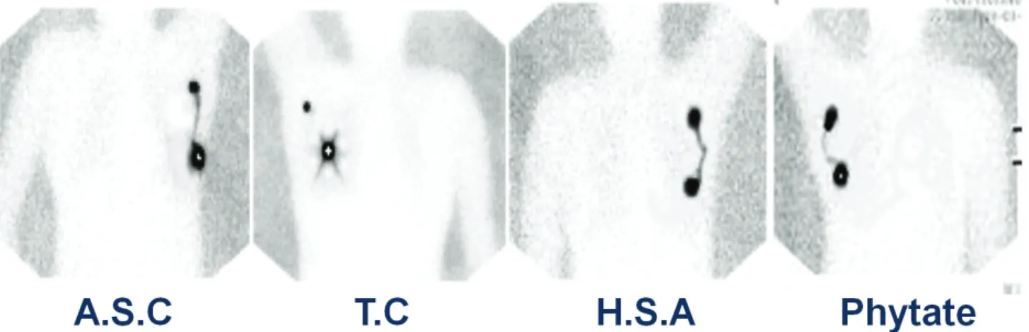
Fig. 3. Limphoscitigraphies for each radiocolloids. ASC; antimony trisulfide colloid, TC; tin colloid, HSA; human serum albumin.
Table 2. Comparisons in labelling time of each radiocolloids
Average Value (n=50) 99mTc-ASC 99mTc-TC 99mTc-HSA 99mTc-Phytate p
Labeling time (min) 40 10 5 5 p<0.05
ASC; antimony trisulfide colloid, TC; tin colloid, HSA; human serum albumin.
Table 3. Comparisons in acquisition time of each radiocolloids
Average Value (n=50) 99mTc-ASC 99mTc-TC 99mTc-HSA 99mTc-Phytate p
Acquisition time (min) 4.5 7.5 2.5 3.5 p>0.05
ASC; antimony trisulfide colloid, TC; tin colloid, HSA; human serum albumin.
Table 4. Results of SLN mapping in each radiocolloids
Unit (%) 99mTc-ASC (n=202) 99mTc-TC (n=120) 99mTc-HSA (n=75) 99mTc-Phytate (n=85)
IR (No) 99.0 (200/202) 96.7 (116/120) 94.7 (71/75) 96.4 (82/85)
FNR (No) 21.5 (14/67) 20.5 (8/38) 17.6 (3/17) 26.3 (5/19)
NPV (No) 90.5 (133/147) 90.7 (78/86) 94.7 (54/57) 94.1 (64/68)
ASC; antimony trisulfide colloid, TC; tin colloid, HSA; human serum albumin, IR; identification rate, FNR; false negative rate, NPV;
negative predictive value.
되는 양이 많다. 이러한 점을 고려해 볼 때 네 가지 교질 모두 기준 값에 포함되어 있어 림포신티그라피 검사를 하는데 유 의한 차이는 없는 것으로 보인다.
2. 표지시간과 영상획득시간
각 방사성교질별 표지과정에 따른 표지시간은 99mTc-ASC (40분), 99mTc-TC (10분), 99mTc-HSA (5분), 99mTc-Phytate (5분) 이다(Table 2). 여기서 ASC는 표지할 때 30분 동안 끓여야 하기 때문에 다른 교질들에 비해서 시간이 조금 더 소요됐고, TC는 입자크기가 크기 때문에 필터링 하는 시간이 포함되어 있어서 시간이 조금 더 걸렸다. 그리고 각 방사성교질별 영상 획득시간 즉, 모니터로 감시림프절을 확인할 수 있는 시간은
99mTc- ASC (4.5분), 99mTc-TC (7.5분), 99mTc-HSA (2.5분),
99mTc-Phytate (3.5분)이다(Table 3). 여기서 주사방법은 모두 피내주사(intradermal injection)로 실시하였고, 결과적으로 볼 때 기존에 사용하였던 ASC, TC, HSA과 마찬가지로 Phytate 도 감시림프절을 확인할 수 있는 시간에는 유의한 차이가 없었다.
3. 각 방사성교질에 따른 매핑성적과 영상분석
감시림프절의 발견율(IR)과 감시림프절 위음성률(FNR) 그리고 음성예측도(NPV)를 평가하면 ASC: IR 99.0%, FNR 21.5%, NPV 90.5%, TC: IR 96.7%, FNR 20.5%, NPV 90.7%, HSA: IR 94.7%, FNR 17.6%,, NPV 94.7%, Phytate:
IR 96.4%, FNR 26.3%, NPV 94.1%로 사용한 교질의 종류에 따 라 통계적으로 유의한 차이를 보이지 않았다(p>0.05) (Table 4).
림포신티그라피 영상에 있어서는 네 가지 교질의 입자크 기가 모두 다르기 때문에 림프관이 영상화되는 것은 각 교질 마다 차이가 있었다. 하지만 이러한 차이로 인해 감시림프절 매핑이 통계적으로 차이를 보이지는 않았다. 림프관이 영상화 되는 것은 감시림프절을 관찰하는데 중요한 요소는 아니다.
하지만 림프관에 교질이 계속 정체되어 있는 경우는 림프절 매핑에 방해가 될 것이다. ASC, HSA, Phytate는 림프관이 잘 영상화되지만 빨리 배액되어 림프절 매핑에는 방해가 되지 않는 것으로 생각된다(Fig 3).
결 론
현재까지 본원에서 림포신티그라피 검사에 사용되었던 방사성의약품은 99mTc-Antimony Trisulfide Colloid (ASC),
99mTc-Tin Colloid (TC), 99mTc-Human Serum Albumin (HSA) 이다. 하지만 ASC, TC, HSA는 제조와 수입이 모두 중단된 상태이기 때문에 최근에 본원에서는 99mTc-Phytate를 사용하고 있다. 림포신티그라피 검사에 이용되는 방사성의약품은 그 밖에도 99mTc-dextran,10)99mTc-hydroxyl ethyl starch,11) 99mTc- colloidal albumin12) 등 여러 가지가 있지만 아직까지 어느 의 약품을 사용하는 것이 림포신티그라피에 가장 적절한지 또는 임상적으로 감시림프절 진단에 영향을 미치는지는 정립된 바가 없다. 다만 이상적인 방사성의약품은 림프관을 통해 빠 르게 배액되고 감시림프절에 정체되어 영상화 할 수 있도록 입자크기가 10~200 nm 정도 되는 것이다.13) 그렇기 때문에 본 연구에서는 전반적으로 지금까지 사용해왔던 방사성교질 들(ASC, TC, HSA)과 현재 사용하고 있는 Phytate가 감시 림프절을 찾아내는데 적절한 입자크기를 가지고 있는지 측 정하였고, 표지시간 및 영상획득시간 비교, 매핑성적 비교 등 여러 가지 성능평가를 실시하였고, 그 결과 어느 방사성교질 을 사용하더라도 감시림프절 진단능에는 유의한 차이가 없는 것으로 확인하였다. 또한 앞으로 림포신티그라피 검사에 다른 방사성의약품이 사용된다 하더라도 그에 따른 적절한 성능평가가 병행되어야 할 것이다.
REFERNCES
1. 2003-2005년 국가 암발생률 및 1993-2005년 암 발생자의 5년 생존 율 발표 자료. 한국 중앙암 등록본부(http://www.ncc.re.kr/index. jsp).
2. Kim BT. Sentinel lymph node imaging in breast cancer. Korean J Nuc Med 1999;33:243-6.
3. Velanovich V, Szymanski W. Quality of life of breast cancer patients with lymphedema. Am J Surg 1999;177:184-187.
4. Weaver DL. Sentinel lymph nodes and breast carcinoma: which micrometastases are clinically significant? Am J Surg Pathol 2003;27:
842-845.
5. Silverstein MJ, Gierson ED, Waisman JR, Senofsky GM, Colburn WJ, Gamagami P. Axillary lymph node dissection for T1a breast carcinoma. Is it indicated? Cancer 1994;73:664-667.
6. Peintinger F, Reitsamer R, Stranzl H, Ralph G. Comparison of quality of life and arm complaints after axillary lymph node dissection vs sentinel lymph node biopsy in breast cancer patients. Br J Cancer 2003;89:648-652.
7. Lee HD, Choi JW, Kim DY, Park BW, Lee IK, Song HJ, et al. Clinical experience for sentinel lymphadenectomy alone in early breast cancer. J Korean Breast Cancer Soc 2003;6:263-270.
8. Giuliano AE, Dale PS, Turner RR, Morton DL, Evans SW, Krasne DL. Improved axillary staging of breast cancer with sentinel lymphade- nectomy. Ann Surg 1995;222:394-399.
9. Boolbol SK, Fey JV, Borgen PI, Heerdt AS, Montgomery LL, Michael Paglia, et al. Intradermal isotope injection: a highly accurate method of lymphatic mapping in breast carcinoma. Ann Surg Oncol 2001;8:20-24.
10. Bennett LR, Lago G. Cutaneous lymphoscintigraphy in malignant melanoma. Semin Nucl Med 1983;13:61-69.
11. Sadek S, Owunwanne A, Abdel‐Dayem HM, Yacoub T. Preparation and evaluation of Tc-99m hydroxyethyl starch as a potential radio- pharmaceutical for lymphoscintigraphy: comparison with Tc-99m human serum albumin, Tc-99m dextran, and Tc-99m sulfur micro- colloid. Lymphology 1989;22:157-166.
12. Pijper R, Meijer S, Hoekstra OS, Collet GJ, Comans EFI, Boom RPA, et al. Impact of lymphoscintigraphy on sentinel node identification with technetium-99m-colloidal albumin in breast cancer. J Nucl Med 1997;38:366-368.
13. Linehan DC, Hill AD, Tran KN, Yeung H, Yeh SD, Borgen PI, et al.
Sentinel lymph node biopsy in breast cancer: unfiltered radioisotope is superior to filtered. J Am Coll Surg 1999;188:377-381.
14. YS Song, et al. Quantitative Assessment of the Radiation Exposure during Pathologic Process in the Sentinel Lymph Node Biopsy using Radioactive Colloid. Nucl Med Mol Imaging Vol. 41, No. 4, Aug 2007.
15. SG Jang, et al. Comparison of the Results for Sentinel Lymph Node Mapping in the Breast Cancer Patients using 99mTc-Antimony Trisulfide Colloid, 99mTc-Tin Colloid, and 99mTc-Human Serum Albumin. Nucl Med Mol Imaging Vol. 41, No. 6, Dec 2007.