• 검색 결과가 없습니다.

Combined Photodynamic Therapy and Intravitreal Bevacizumab Injection for the Treatment of Adult Coats' Disease: A Case Report

N/A
N/A
Protected

Academic year: 2022

Share "Combined Photodynamic Therapy and Intravitreal Bevacizumab Injection for the Treatment of Adult Coats' Disease: A Case Report"

Copied!
3
0
0

로드 중.... (전체 텍스트 보기)

전체 글

(1)

374

Korean J Ophthalmol 2010;24(6):374-376 DOI: 10.3341/kjo.2010.24.6.374 pISSN: 1011-8942 eISSN: 2092-9382

Case Report

Combined Photodynamic Therapy and Intravitreal Bevacizumab Injection for the Treatment of Adult Coats'

Disease: A Case Report

Jongshin Kim

1

, Kyu Hyung Park

1,2

, Se Joon Woo

1,2

1

Department of Ophthalmology, Seoul National University College of Medicine, Seoul, Korea

2

Department of Ophthalmology, Seoul National University Bundang Hospital, Seongnam, Korea

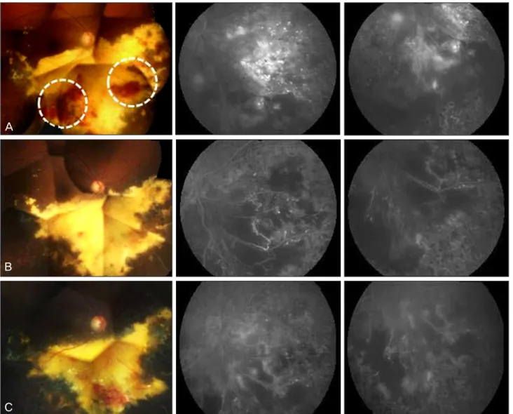
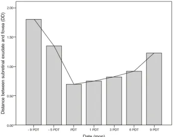
A 68-year-old woman presented with a visual field defect in her right eye. The fundus of her right eye showed multi- ple telangiectatic vessels, retinal hemorrhages, and subretinal exudates in the inferior peripheral retina. Nine months later, the subretinal exudates extended to the fovea despite treatment with laser photocoagulation.

Cryotherapy was not possible at the time because of the posterior location of the retinal telangiectatic vessels. She was treated with a combination of photodynamic therapy (PDT) and intravitreal bevacizumab injection: three in- jections were given at 2-month intervals. After this combined therapy, her right fundus revealed a significant re- gression of abnormal retinal vessels and subretinal exudates. A fluorescein angiography showed no leakage from the abnormal retinal vessels. At 9 months after the combined therapy, she was able to maintain a stable visual acuity and visual field. This is the first case report that demonstrates the efficacy of the combined treatment of PDT and in- travitreal bevacizumab injection in Coats's disease. This combined therapy is a kind of treatment modality for adult Coats’ disease in cases which cryotherapy cannot be employed and are refractory to laser photocoagulation.

Key Words: Bevacizumab, Coats' disease, Photodynamic therapy

ⓒ2010 The Korean Ophthalmological Society

This is an Open Access article distributed under the terms of the Creative Commons Attribution Non-Commercial License (http://creativecommons.org/licenses /by-nc/3.0/) which permits unrestricted non-commercial use, distribution, and reproduction in any medium, provided the original work is properly cited.

Received: October 25, 2009 Accepted: March 26, 2010

Reprint requests to Se Joon Woo. Department of Ophthalmology, Seoul National University Bundang Hospital, #166 Gumi-ro, Bundang-gu, Seongnam 463-707, Korea. Tel: 82-31-787-7377, Fax: 82-31-787-4057, E-mail: sejoon1@daum.net

Coats' disease is an idiopathic condition characterized by telangiectatic and aneurysmal retinal vessels and chronic accumulation of subretinal exudates [1]. It is predominantly unilateral occurring mostly in young males, and can cause se- vere visual loss resulting from exudative retinal detachment [2]. Less commonly, it presents in adulthood and progresses at a slower rate, but with similar features [3].

Current treatment options for patients with Coat's disease are ablation of abnormal retinal vessels, either by laser pho- tocoagulation or by cryotherapy, and surgery for retinal detachment. Recently, the dysregulation of vascular endothe- lial growth factor (VEGF)-mediated angiogenesis was sug- gested to be a component in Coats' disease and a few pedia- tric patients with the disease have been reported to respond well to anti-VEGF therapy [4,5]. However, the involvement of the macula as a result of subretinal exudates or exudative

retinal detachment can produce poor visual acuity despite proper treatment [2]. In this article, we report the first case of an adult patient with macula-threatening Coats' disease, treated with a combined treatment of photodynamic therapy (PDT) and intravitreal bevacizumab injection.

Case Report

A 68-year-old woman presented with symptoms of a con- stricted visual field in her right eye. Although the best-cor- rected visual acuity (BCVA) was 20/25 in both eyes, a fundus examination revealed multiple telangiectatic and aneurysmal retinal vessels, retinal hemorrhages, and subretinal exudates at the inferior mid-periphery of her right eye. A fluorescein angiography showed early and persistent leakage in the ab- normal vessels at the inferior mid-periphery and hypofluor- escence corresponding to the subretinal exudates in the right eye. With the diagnosis of adult Coats' disease, scatter laser photocoagulation was applied to the abnormal retinal vessels.

At 9 months after presentation, a fundus examination re-

vealed about 1 disc diameter extension of the subretinal exu-

dates to the fovea. PDT was used to treat the 2 large active

(2)

J Kim, et al. Combined PDT and Bevacizumab for Coats’ Disease

375 B

C A

Fig. 1. Serial montage fundus photographs (left) and fluorescein angiographs (middle and right) of the right eye. (A) Two days before photo- dynamic therapy (PDT). The subretinal exudates were threatening the fovea. Dotted circles indicate PDT treatment areas. Fluorescein angiog- raphy reveals dye leakage from the abnormal retinal vessels and hypofluorescence corresponding to the subretinal exudates. (B) Six months post-PDT, significant regression of the abnormal retinal vessels and subretinal exudates was observed. (C) Nine months post-PDT, the sub- retinal exudates shrank to become more distant from the fovea.

areas of abnormal telangiectatic and aneurysmal retinal ves- sels (Fig. 1A). After the PDT treatment, intravitreal bevacizumab was injected in order to decrease exudation and to block the regeneration of abnormal retinal vessels which were obli- terated by PDT.

At 2 months post-PDT, a second intravitreal bevacizumab injection was given. At 4 months post-PDT, a third intra- vitreal bevacizumab injection was given and an additional scatter laser photocoagulation was applied to the area where the pre-existing abnormal retinal vessels and subretinal exu- dates had been located. At 6 months post-PDT, a fundus ex- amination of the right eye showed a significant regression of the abnormal retinal vessels and subretinal exudates (Fig.

1B). At 9 months post-PDT, the subretinal exudates with- drew further from the fovea and the patient’s visual acuity re- mained stable with a BCVA of 20/25 in both eyes (Figs. 1C

and 2).

Discussion

The conventional treatment for Coats' disease involves la- ser photocoagulation or cryotherapy to ablate abnormal te- langiectatic vessels and preserve visual acuity. The sub- retinal exudates regress after laser photocoagulation or cryotherapy. However, in our case the high intensity laser photocoagulation applied to the abnormal retinal vessels was not absorbed sufficiently, due to the massive retinal exudates.

We thought that PDT would be more effective than laser

photocoagulation at obliterating the multiple abnormal reti-

nal vessels that were surrounded by the massive retinal

exudates. Laser photocoagulation is not an efficient treat-

ment modality if subretinal exudates obscure a target tissue.

(3)

Korean J Ophthalmol Vol.24, No.6, 2010

376

Fig. 2. Sequential changes in the distance between subretinal exu- dates and the fovea before and after photodynamic therapy (PDT).

After PDT, the lesion-to-fovea distance increased gradually.

DD=disc diameter.

However, PDT utilizes an exogenous agent, photosensitizing the abnormal retinal vessels that are surrounded by exudates and the interaction between the laser beam and the agent pro- duces a photochemical reaction that results in cellular damage and vascular thrombosis of target tissue [6].

We considered that an additional use of anti-VEGF agent could prevent the regeneration of abnormal retinal vessels that were obliterated by PDT and could suppress exudation induced by inflammatory cytokines. Abnormal retinal ves- sels and exudates in Coats' disease may impair the oxygen- ation of the outer retina, and the secondary over-expression of VEGF that gets produced as a result may cause micro- vascular changes [4]. This mechanism suggests that an- ti-VEGF is a potentially good treatment modality for Coats' disease, which was evident in case reports [4,5]. In addition, anti-VEGF had a synergistic effect on PDT for the treatment of choroidal neovascularization. Costa et al. [7] suggested that anti-VEGF might prolong choriocapillaris hypo- perfusion induced by PDT, thus leading to a secondary effect on perfusion of choroidal neovascularization.

We are unable to comment on the potential long-term ef- fect of the combined treatment of PDT and intravitreal bevacizumab injection, based on this single case. Further re- search is needed to determine the efficacy of this combined therapy for the treatment of Coats' disease. However, we are optimistic about the role of this combined treatment in cases of Coats' disease that are refractory to conventional laser photocoagulation or cryotherapy-based treatment, and in vi- sion-threatening conditions needing urgent treatment.

In conclusion, combined treatment of PDT and intravitreal bevacizumab injection is a kind of treatment modality for cases of adult Coats' disease which cannot be treated by cry- otherapy and are refractory to laser photocoagulation.

Conflict of Interest

No potential conflict of interest relevant to this article was reported.

References

1. Coats G. Forms of retinal diseases with massive exudation. Roy Lond Ophthalmol Hosp Rep 1908;17:440-525.

2. Shields JA, Shields CL, Honavar SG, Demirci H. Clinical variations and complications of Coats disease in 150 cases: the 2000 Sanford Gifford Memorial Lecture. Am J Ophthalmol 2001;131:561-71.

3. Smithen LM, Brown GC, Brucker AJ, et al. Coats' disease diagnosed in adulthood. Ophthalmology 2005;112:1072-8.

4. Sun Y, Jain A, Moshfeghi DM. Elevated vascular endothelial growth factor levels in Coats disease: rapid response to pega- ptanib sodium. Graefes Arch Clin Exp Ophthalmol 2007;245:

1387-8.

5. Cakir M, Cekiç O, Yilmaz OF. Combined intravitreal bevaciz- umab and triamcinolone injection in a child with Coats disease.

J AAPOS 2008;12:309-11.

6. Miller JW, Walsh AW, Kramer M, et al. Photodynamic therapy of experimental choroidal neovascularization using lipoprotein- delivered benzoporphyrin. Arch Ophthalmol 1995;113:810-8.

7. Costa RA, Jorge R, Calucci D, et al. Intravitreal bevacizumab

(Avastin) in combination with verteporfin photodynamic

therapy for choroidal neovascularization associated with

age-related macular degeneration (IBeVe Study). Graefes Arch

Clin Exp Ophthalmol 2007;245:1273-80.

수치

Fig. 1. Serial montage fundus photographs (left) and fluorescein angiographs (middle and right) of the right eye
Fig. 2. Sequential changes in the distance between subretinal exu- exu-dates and the fovea before and after photodynamic therapy (PDT)

참조

관련 문서

Biological treatment of wastewater is an economical green technology, however, the effluent water quality of a biological wastewater treatment facility is

Serum uric acid levels and risk for vascular disease in patients with metabolic syndrome... Prevalence if the metabolic syndrome in a Turkish

1 John Owen, Justification by Faith Alone, in The Works of John Owen, ed. John Bolt, trans. Scott Clark, "Do This and Live: Christ's Active Obedience as the

(Background) The standard triple therapy used as the first-line treatment for Helicobacter pylori infection are a combination of proton pump inhibitor(PPI),

Anoxic

In conclusion, applications of combined exercise, which can be easily carried out in one’s daily life for elderly women, would be helpful for their

In the analysis of soluble solids, the treatment group with material grains and the combined groups of glutinous millet and barley appeared high, and

RWC (Restricted Work Case): Injuries by which workers are reassigned to other duties or cannot perform the full range of normal duties MTC (Medical Treatment Case):