426
Korean J Ophthalmol Vol.28, No.5, 2014
fore be effective, by decreasing vascular permeability. The spontaneous resolution of choroidal ischemia has been re- ported previously [5], but in our case, because the patient showed no signs of any functional or anatomic improve- ment after observation for a week, we decided to treat the patient with IVB, which led to prompt visual and anatomi- cal recovery.
This is the first case report of the occurrence of choroi- dal ischemia following brain tumor surgery and complete recovery of BCVA after IVB. In patients with severe visu- al loss and SRD associated with choroidal ischemia, IVB may be a viable treatment option.
Young Joo Cho, Eun Young Choi, Hyoung Jun Koh, Sung Chul Lee, Min Kim
Institute of Vision Research, Department of Ophthalmology, Yonsei University College of Medicine, Seoul, Korea E-mail: [email protected]
Conflict of Interest
No potential conflict of interest relevant to this article was reported.
References
1. Hayreh SS. Segmental nature of the choroidal vasculature.
Br J Ophthalmol 1975;59:631-48.
2. Loeffler KU, Hayreh SS, Tso MO. The effects of simulta- neous occlusion of the posterior ciliary artery and vortex veins: a histopathologic study. Arch Ophthalmol 1994;112:
674-82.
3. Vinores SA, Youssri AI, Luna JD, et al. Upregulation of vascular endothelial growth factor in ischemic and non-isch- emic human and experimental retinal disease. Histol His- topathol 1997;12:99-109.
4. Kaur C, Sivakumar V, Yong Z, et al. Blood-retinal barrier disruption and ultrastructural changes in the hypoxic retina in adult rats: the beneficial effect of melatonin administra- tion. J Pathol 2007;212:429-39.
5. Kim M, Kwon HJ, Lee SC. Spontaneous resolution of pos- terior ciliary artery occlusion. Graefes Arch Clin Exp Oph- thalmol 2013;251:1005-6.
In Situ Peripheral Iridoplasty in Phakic Eyes for the Treatment of Symptomatic Peripheral Iridotomy
Dear Editor,
Laser peripheral iridotomy (LPI) is commonly used to treat acute angle-closure glaucoma (AACG) and to prevent glaucoma before posterior chamber phakic intraocular lens (IOL) implantation. Patients may experience some of the following visual symptoms after LPI: diplopia, ghost im- ages, lines, glare, and haloes. These symptoms are associ- ated with high lid position or large iridotomy size [1]. The typical treatment for such symptoms is a tinted contact lens. When patients cannot tolerate tinted lenses, surgery
may be necessary. Here we effectively adapted a simple iridoplasty technique for the treatment of symptomatic LPI.
LPI was performed in the first case to treat AACG. A 46-year-old woman presented postoperatively with glare in the left eye. Her left eye best-corrected visual acuity (BCVA) was 20 / 20 and intraocular pressure (IOP) was 13 mmHg. Slit-lamp microscopy revealed two LPIs 1 / 2 to 2 / 3 of pupil size at 10 and 2 o’clock as well as incipient cata- racts (Fig. 1A). In the second case, LPI was performed for the prophylaxis of glaucoma after the implantation of a phakic IOL in a 42-year-old female. Postoperatively, the patient complained of glare and photophobia in the left eye. Her left eye BCVA was 20 / 15 and IOP was 12 mmHg. The slit-lamp examination revealed a clear lens and two LPIs at 11 and 1 o’clock that were partially cov- ered by the upper lid (Fig. 1A).
Both patients agreed to undergo peripheral iridoplasty.
Korean J Ophthalmol 2014;28(5):426-428 http://dx.doi.org/10.3341/kjo.2014.28.5.426
427 The surgery was performed using a modified Siepser slip-
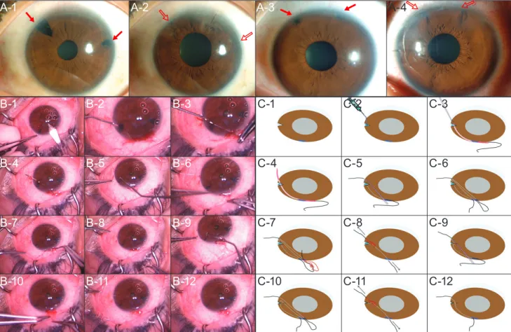
knot technique (Fig. 1B and 1C) [2-4]. The primary inci- sion was made in superior cornea (Fig. 1B-1 and 1C-1). A secondary incision was made near the iridotomy to allow for the injection of viscoelastics and insertion of the IOL manipulator. Sodium hyaluronate 1.4% (Healon GV; Phar- macia-Upjohn Ophthalmics, Kalamazoo, MI, USA) was injected to the anterior chamber and iridotomy site in a step-by-step manner to protect the lens (Fig. 1B-2 and 1C- 2). A CTC-6 long, curved needle was passed through the main incision with 10-0 polypropylene suture. The needle was use to elevate picked up one side of the iridotomy with counter-pressure provided by the IOL manipulator (Fig.
1B-3 and 1C-3) [5]. The needle was then passed through the other side of the iridotomy with the same support and fi- nally exited through the corneolimbal angle (Fig. 1B-4 and 1C-4). The IOL manipulator was introduced through the main incision, used to create a suture loop, and then re- tracted through the main incision (Fig. 1B-5 and 1B-6;
1C-5 and 1C-6).The free proximal end of the suture was passed through the loop three times (Fig. 1B-7 and 1C-7).
The first slipknot was gently slipped through into the ante- rior chamber, then engaged in situ around the iridotomy site as each free end of the suture was pulled (Fig. 1B-8 and 1C-8). This maneuver was repeated twice to secure the knot (Fig. 1B-9 to 1B-12, 1C-9 to 1C-12). Both ends of the knot were then cut using long Vannas scissors. Depending on the size of the iridotomy, it was secured using one to two additional slipknots. The viscoelastic material was re- moved by manual irrigation. In order to prevent AACG re- currence in the first patient, iridotomy size was reduced by 80%. Postoperatively, two patients reported that their symptoms disappeared completely (Fig. 1A-2 and 1A-4).
Their postoperative BCVAs were not decreased and IOPs were not elevated. No other postoperative complication was detected.
The use of peripheral iridoplasty to close symptomatic LPI without disturbing the lens represents a technical chal-
Fig. 1. Figures (A) presented preoperative and postoperative photographs of cases (case 1, A-1 and A-2; case 2, A-3 and A-4). Solid arrows indicate preoperative laser peripheral iridotomies (LPIs), and open arrows indicate closed LPIs. Figures (B) showed intraocular Siepser slipknot technique. Figures (C) presented schema of (B). Numbering is consistent with (B).
A-1
B-1
B-4
B-7
B-10
B-2
B-5
B-8
B-11
B-3 C-1
C-4
C-7
C-10
C-2
C-5
C-8
C-11
C-3
C-6
C-9
C-12 B-6
B-9
B-12
A-2 A-3 A-4
428
Korean J Ophthalmol Vol.28, No.5, 2014
lenge. Surgery can lead to iridodialysis, cyclodialysis, and subsequent bleeding [5]. The following steps prevented these complications in the two patients presented here.
First, we used a modified Siepser technique for creation of the knot in situ. This type of knot minimizes the force pulling on the iris from the direction of the corneolimbal angle. Second, we used an IOL manipulator to create a counter-force when the needle was passed through the iris.
Third, the viscoelastics is inserted in a step-wise manner.
This was performed to prevent iris prolapse through the primary incision, which would subsequently trigger a sharp spike in IOP. Finally, the needle was maneuvered extremely gently when passing horizontally through the anterior chamber to as to avoid contact with the lens. This is the first report to describe the adaptation of a modified Siepser slip- knot technique to treat symptomatic LPI in phakic eyes.
Eun Hye Jung
Department of Ophthalmology, Seoul National University Hospital, Seoul National University College of Medicine, Seoul, Korea
Mee Kum Kim, Won Ryang Wee
Department of Ophthalmology, Seoul National University Hospital, Seoul National University College of Medicine, Seoul;
Laboratory of Ocular Regenerative Medicine and Immunology, Seoul Artificial Eye Center, Seoul National University Hospital Biomedical Research Institute, Seoul, Korea
E-mail: [email protected]
Conflict of Interest
No potential conflict of interest relevant to this article was reported.
References
1. Spaeth GL, Idowu O, Seligsohn A, et al. The effects of iri- dotomy size and position on symptoms following laser pe- ripheral iridotomy. J Glaucoma 2005;14:364-7.
2. Lee EJ, Ahn JY, Wee WR, et al. In situ intraocular suture techniques for pupilloplasty and suspension of a subluxated intraocular lens. Ophthalmic Surg Lasers Imaging 2010;41:
266-71.
3. Osher RH, Snyder ME, Cionni RJ. Modification of the Siepser slip-knot technique. J Cataract Refract Surg 2005;
31:1098-100.
4. Siepser SB. The closed chamber slipping suture technique for iris repair. Ann Ophthalmol 1994;26:71-2.
5. Dunn SP, Stec L. Iris reconstruction. In: Macsai MS, editor.
Ophthalmic microsurgical suturing techniques. Berlin:
Springer; 2007. p. 71-83.
Ethmoidal Sinus Mucocele as a Cause of Acquired Brown Syndrome
Dear Editor,
A mucocele is composed of a respiratory epithelial lined mucus-containing lesion with accumulation of mucoid se- cretion. Sometimes, it produces remodeling of the bony structure and influences the adjacent tissues [1]. In this case, extension of the ethmoid sinus mucocele and remod- eling of the orbital structure were major causes of acquired Brown syndrome. Although various causes of acquired
Brown syndrome have been described, this is the first re- ported case of acquired Brown syndrome due to ethmoidal sinus mucocele in Korea.
A 69-year-old male presented to our institution with a newly-developed diplopia. There was no history of trauma or any medical illness. At initial examination, visual acuity was 20 / 30 in both eyes. The intraocular pressure was 17 mmHg in the right and 20 mmHg in the left eye. There was no relative afferent pupillary defect and color vision was normal.
A soft compressible tender mass measuring 2 × 2 mm on the superonasal aspect of the left upper lid extending from the trochlea to the medial canthal tendon was noted. A mo- tility examination showed an underelevation in adduction of the left eye mimicking Brown syndrome (Fig. 1A). A
Korean J Ophthalmol 2014;28(5):428-429 http://dx.doi.org/10.3341/kjo.2014.28.5.428