Chest Radiographic Findings of Novel Swine-Origin Influenza A (H1N1) Virus Infection in Children
1So Young Bae, M.D., Hyun Sook Hong, M.D., Yun-Woo Chang, M.D.2, Sang Hyun Paik, M.D., Seong-Jin Park, M.D., Jang-Gyu Cha, M.D., Hae Kyung Lee, M.D.
1Department of Radiology, Soonchunhyang University Bucheon Hospital, Korea
2Department of Radiology, Soonchunhyang University Hospital, Korea Received October 27, 2010 ; Accepted April 29, 2011
Address reprint requests to : Hyun Sook Hong, M.D., Department of Radiology, Soonchunhyang University Bucheon Hospital, 1174, Jung-dong, Wonmi- gu, Bucheon-si, Gyunggi-do 420-767, Korea.
Tel. 82-32-621-5851 Fax. 82-32-621-5874 E-mail: [email protected]
Purpose: To analyze chest radiographic findings in children infected with laboratory- confirmed novel swine-origin influenza A (H1N1) virus.
Materials and Methods: Three hundred seventy-two out of 2,014 children with labora- tory confirmed H1N1 infection and who also underwent a chest radiograph from September to November 2009 were enrolled in this study. Patients were divided into in-patients, out-patients, and patients with co-infections and further subdivided into with underlying disease and without underlying disease as well as age (<2 years old, 2-5 years, 5-10 years, 10-18 years old). The initial radiographs were evaluated for radi- ographic findings and the anatomic distribution of abnormalities.
Results: The initial radiographs were abnormal in 154 (41.39%) patients. The predom- inant radiographic findings were peribronchial wall opacity found in 85 (22.84%) pa- tients and hyperinflation observed in 69 (18.54%) patients. Further, 75 (71.42%) pa- tients exhibited central predominance and the right lower lung zone was also com- monly involved. There were statistically significant differences in the radiological find- ings between in-patient and out-patient groups. However, there were no significant dif- ferences in the radiographic findings between in-patients and the co-infection group with respect the presence of underlying disease and age.
Conclusion: Initial radiographs of children with laboratory confirmed H1N1 virus were abnormal in 41.39% of cases. The common radiographic findings included peri- bronchial opacities, hyperinflation, lower lung zonal distribution, and central predomi- nance.
Index words :Influenza A Virus, H1N1 Subtype Pneumonia
Children
Radiography, Thoracic
The first isolation of the swine influenza virus from a human occurred in 1974 (1), confirming the speculation that swine-origin influenza viruses could infect humans.
Since 1975, sporadic human infections with swine virus- es and a small outbreak have been documented (2-9).
The swine flu outbreak that occurred in Mexico in April 2009 (10) rapidly spread to many countries around the world. In June 2009, the World Health Organization (WHO) declared the emergence of a global pandemic, raising the alert level to phase 6 (pandemic phase), on the basis of documented human-to-human spread of in- fection in at least three countries from two WHO re- gions. In Korea, there have been 252 deaths through April, 2010 and among them, 54% of the patients have been younger than 19 years of age (www.cdc.go.kr).
Normal or prominent peribronchial opacities with hy- perinflation have been reported as common findings in children, whereas air space consolidation has been the most common finding in adults (11-13). To date, the de- scription of the radiologic manifestations of S-OIV has been limited in children. Therefore, the aim of this study was to analyze chest radiographic findings in chil- dren infected with laboratory-confirmed novel swine- origin influenza A (H1N1) virus (S-OIV).
Materials and Methods
This study was approved by the institutional review board with a waiver of informed consent. The study population included all patients less than 18 years of age with laboratory confirmed H1N1 from September 2009 to November 2009. Respiratory specimens were ana- lyzed by real-time reverse transcription polymerase chain reaction (RT-PCR) (Artus Infl./H1LC/RG RT-PCR kit, Qiagen, Hilden, Germany). A total of 2014 patients were identified, among whom, 375 underwent chest ra- diographs. The indications for chest radiographs were respiratory symptoms with high fever (>38 C), abnor- mal breathing sound on auscultation, or those requested by parents. Other than those with urinary tract infection (n = 2) and poor image quality (n = 1), a total of 372 pa- tients were enrolled in the study.
The common presenting symptoms of total laboratory confirmed H1N1 patients (n=2014) were fever (95.48%), cough (65.30%), and sore throat (20.41%). Further, 2.18% of patients had abdominal pain, 1.69% vomiting, and 0.55% had diarrhea.
On the initial chest radiographic images, lung parenchyma was classified as normal or abnormal. The
initial radiographs were evaluated for parenchymal le- sions (peribronchial wall opacity, hyperinflation, air space consolidation, atelectasis, or pneumomedi- astinum), anatomic distribution, (unilateral/bilateral, central/peripheral, diffuse, or upper/middle/lower lung zone), pleural effusion, and lymphadenopathy.
Radiologic findings were classified as peribronchial wall opacities with increased peribronchial cuffing with/
without some nodularity. Hyperinflation was evident on the chest radiographs in children with hyperlucency, depression and/or flattening of the diaphragms to more than 10 posterior ribs, increased anterior to posterior chest diameter, and an increased retrosternal space. Air space consolidation referred to a density conforming to the lobar or segmental boundaries with evidence of vol- ume expansion or minimal volume loss of the involved lobe and fluffy or homogeneously increased opacities with air- bronchograms. Atelectasis referred to densities conforming to lobar or segmental boundaries having ev- idence of volume loss. Pneumomediastinum was de- fined as the presence of one or more gas shadows within the mediastinum that did not correspond in position and contour with gas in the esophagus or tracheobronchial position. Pleural effusion was present under conditions of costophrenic angle blunting or fluid shifting on the decubitus view. Lymphadenopathy referred to lumpy round increased opacities at the outer edge of the hilum.
In cases with peribronchial opacities, a quantitative esti- mate was recorded as absent, mild, moderate, or severe, and it was regarded as a positive finding when occurring at a moderate degree. The anatomic distribution was characterized as unilateral or bilateral, with each lung divided into upper, middle, and lower lung zones. The pattern was classified as predominantly central, periph- eral, or diffuse. Further, the number of days of hospital- ization was assessed in patients with extensive disease involving three lung zones.
The 372 H1N1-infected patients were divided into three categories: in-patients (n=73), out-patients (n=286) who had H1N1 infection only and those pa- tients with co-infections (n=13); and further subdivided into with or without underlying disease and according to age (< 2 years old, 2 - 5 years, 5 - 10 years, and 10 - 18 years). We then evaluated the differences in radiolog- ic findings for each of the groups. Also we evaluated dif- ferences with respect to hospitalization in the in-patient and co-infection groups. The in-patient group included patients who were initially admitted to the emergency room or visited the out-patient clinic and were later ad-
mitted. Patients with co-infection included 11 with Mycoplasma pneumoniae, 1 with rhinovirus and aden- ovirus, and 1 with Epstein-Barr virus.
Eighty-six patients (25.09%) had chronic medical con- ditions including: asthma (n = 73), preterm delivery with low birth weight (n =12), a medicated seizure dis- order (n = 2), Wilson’s disease receiving penicilamine treatment (n = 1), tuberculosis (n = 1), and nephrotic syndrome (n = 1). Eighty-four of the 86 patients with chronic medical conditions were obtained with chest ra- diographs (16 in-patients, 65 out-patients, and 3 co-in- fected patients).
The posteroanterior/ anteroposterior (PA/AP) and lat- eral projection radiographs were performed with digital radiography equipment (Gold mountain medical, TDR4600-F80, Seoul, Korea). The images were re- viewed on a PACS viewer with a 2,560 2,048 pixel monitor (INFINITT, STAR PACS, v. 5081, Seoul, Korea). The chest radiographs were independently ana- lyzed by two pediatric radiologists (HS Hong, YW Chang) at different times, and the differences were then evaluated by consensus. The K-value was used for the inter-observer agreement rate. As such, K-values < 0 in- dicated that inter-observer agreement was less than the agreement by chance alone. K-values between 0.01- 0.20 indicated that, inter-observer agreement was slight:
0.21-0.40 indicated fair agreement, 0.41-0.60 indicated moderate agreement, 0.61-0.80 indicated good agree-
ment, and 0.81-0.99 indicated excellent agreement. The differences in chest radiographic abnormalities and anatomic distribution with respect to underlying dis-
Table 1. Chest Radiographic Findings, Pattern, and Anatomical Distribution of Enrolled Patients
Chest Radiographic Findings n=372 K-value Peribronchial opacity 85 (22.84%) 0.73
Hyperinflation 69 (18.54%) 0.74
Consolidation 030 0(8.06%)0 0.92
Pleural effusion 011 0(2.95%)0 0.94
Atelectasis 08 0(2.15%) 0.72
Pneumomediastinum, 01 0(0.27%) 1.00 Subcutaneous emphysema
Lymphadenopathy 00 0(0.00%) NA
Normal 218 (58.60%)0 0.75
Pattern n=105* K-value
Central 75 (71.42%) 0.73
Diffuse 30 (28.57%) 0.75
Peripheral 0 (0.00%) NA
Anatomical distribution n=105* K-value
RLL 71 (67.62%) 0.68
RUL 50 (47.61%) 0.72
RML 47 (44.76%) 0.76
LLL 44 (41.90%) 0.70
LUL 25 (23.80%) 0.61
LML 19 (18.09%) 0.57
Note.─NA: not applicable
*: Focal lung abnormality: From chest radiographic findings, with the exception of normal and hyperinflation (n=95; in-patient: 47, out-patient: 48)
Numbers in parentheses are percentages
A B
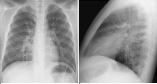
Fig. 1. A 10-year-old boy with laboratory confirmed S-OIV (H1N1) who presented with coughing.
Chest PA (A), right lateral view (B) on hospital admission showed peribronchial opacities in the RLL and RML. The distribution showed a central predominance as well as a presence in the lower and middle lung zone.
eases were analyzed using the Fisher’s exact test for cat- egorical data and the Mann-Whitney test for continuous data. Statistical analyses were performed with the STA- TA computer software (Stata, College station, TX., USA). A p-value < 0.05 was the threshold for statistical significance.
Results
Initial radiographs were abnormal in 154 of 372 (41.39%) patients. The anatomical distribution was char- acterized as being unilateral (71/105, 67.62%), or with bilateral involvement (34/105, 32.38%). The predomi- nant radiographic findings were peribronchial wall opacity (85/372; 22.84%) (Fig. 1), and hyperinflation (69/372; 18.54%). Moreover, the most common pattern was central (75/105; 71.42%), which also affected the right lower lung zone (71/105; 67.62%). Air space con- solidation was observed in 30 patients (30/372; 8.06%) (Fig. 2), while pleural effusion was observed in 11 pa- tients (11/372; 2.95%). None of the patients showed lymph node enlargement. Extensive disease involving three lung zones was observed in 44/372 (11.83%) pa- tients, resulting in a longer hospitalization time (mean:
5.54 days). However, the difference was not statistically significant (p > 0.05, Mann-Whitney test). The imaging findings have been summarized in Table 1. Inter- ob-
server agreement with regard to the radiologic findings was good to excellent.
The first available chest radiographs were abnormal in 54/73 (73.97%) patients that had been admitted to hospi- tal, 90/286 (31.47%) patients from the out-patient group, and 10/13 (76.92%) of the patients with co-infection (Table 2). Among the in-patients with abnormal radi- ographs, the predominant radiographic findings were peribronchial opacity (35/73, 47.94%), hyperinflation (21/73, 28.77%), and consolidation (19/73, 26.03%) in the in-patient group, while the common findings in the out-patient group were normal (196/286, 68.53%), with peribronchial wall opacities (45/286, 15.73%) and hyper- inflation (46/286, 16.08%) (Table 2). Each of the differ- ences in the imaging findings were statistically signifi- cant (p < 0.05), except for lymphadenopathy (Table 2).
For patients with co-infection, the predominant chest ra- diographic finding was air space consolidation (6/13, 46.15%). Further, the percent of cases with pleural effu- sion (3/13, 23.08%) was higher than in the in-patient group (8/73, 10.96%); however, there were no statistical- ly significant differences in the radiographic findings be- tween the in-patient group and the patient with co-infec- tions (p > 0.05) (Table 2). The median hospitalization duration for the in-patient group was 5.19 days (range 3~10 day), with a 6.00-day median (range 4~9 day) for patients with co-infections; this difference was not sta-
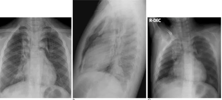
A B C
Fig. 2. A 7-year-old boy who presented with dyspnea and had a history of allergic rhinitis.
Chest PA (A), right lateral (B) and right decubitus view (C) on hospital admission showed consolidation with air-bronchogram in the BLL, RUL and increased peribronchial opacities in the right perihilar region, pneumomediastinum, subcutaneous emphysema, as well as fluid shifting in the dependent portion. The distribution of the lesion showed central predominance as well as diffuse in- volvement of the right lung.
tistically significant. There were no differences in the ra- diographic findings between the groups with or without underlying disease (p > 0.05). In addition, there were no statistically significant differences in the radiographic findings according to age. The most common radi- ographic finding, with the exception of a normal result, was peribronchial opacities in the under 2 years of age group (2.69%); peribronchial opacity and hyperinflation were common in the 2-18 years group. There were no parenchymal or pleural complications. All patients im- proved with regard to clinical symptoms and chest radi- ographic findings. The number of days to an improved chest radiograph was a median of 5.43 days (range, 2-17 days).
Discussion
Influenza A viruses are ubiquitous in nature. Pigs have been known to be infected with both avian and hu- man influenza viruses, enabling these viruses to interact and generate new forms (14). A new strain of triple-reas- sortant influenza A (H1N1) virus containing genes from swine, avian and human influenza viruses was identi- fied in Mexico in April 2009, and rapidly spread world- wide (10). Avian-like swine influenza A (H1N1) viruses can infect humans directly, without the need for a reas- sortment event; resulting in a serious disease threat.
Some of the characteristics of H1N1 viruses differ from the seasonal influenza virus, such as their affects on younger patients. The clinical outcomes of this pathogen have been reported as ranging from self-limited illness
to respiratory failure and death (10, 15). In the current study, most patients presented with flu- like symptoms such as fever, cough, sore throat, and sputum produc- tion. Abdominal symptoms were not common (4%) compared to other reports (25%) (15). The current study showed that about 60% of laboratory confirmed patients had normal radiographs. Among those with radiograph- ic abnormalities, the most common finding was peri- bronchial increased opacities, similar to other reports of H1N1 in children (13) and other viral pneumonia in chil- dren (14, 16-20). Parahilar peribronchial infiltrates, hy- perexpansion, segmental or lobar atelectasis, and hilar adenopathy have been common radiographic findings in the viral respiratory tract infections of children. The results of this study confirmed these findings (13, 16- 21).
Agarwal et al. (11) concluded that the most common abnormality in H1N1 adults was air-space disease (50%), with a predilection for lower and central lung preponderance in hospitalized patients requiring ad- vanced mechanical ventilation. In this study, anatomic distribution or central predominance was consistent with prior reports; however, the frequency of air space consolidation was lower. Lee et al. (13) concluded that patients with a mild and self- limited clinical course had normal radiographs or prominent peribronchial mark- ings with hyperinflation; a finding which was supported by this study. However, air space consolidation oc- curred with a high frequency in their study; with 57%
(12/21) of hospitalized children, and 86% (12/14) of chil- dren who required intensive care treatment.
Table 2. Differences in the Chest Radiographic Findings among in-patients, Out-patients, and Patients with Co-infection and the Duration of Hospital Stay between in-patients and Patients with Coinfections
Chest Radiographic Findings (n=372) In-patient (n=73) Out-patient (n=286) Co-infected Patient (n=13) p-value* p-value�
Peribronchial opacity 35 (47.94%) 45 (15.73%) 5 (38.46%) 0.00 0.56
Hyperinflation 21 (28.77%) 46 (16.08%) 2 (15.38%) 0.01 0.50
Consolidation 19 (26.03%) 07 0(2.45%) 6 (46.15%) 0.00 0.19
Pleural effusion 08 (10.96%) 01 0(0.35%) 3 (23.08%) 0.00 0.36
Atelectasis 04 0(5.48%) 01 0(0.35%) 3 (23.08%) 0.00 0.07
Pneumomediastinum,
Subcutaneous emphysema 01 0(1.37%) 00 0(0.00%) 00 0(0.00%)0 0.04 1.00
Lymphadenopathy 00 0(0.00%) 00 0(0.00%) 00 0(0.00%)0 NA NA
Normal 19 (26.02%) 196 (68.53%)0 3 (23.08%) 0.00 1.00
Duration of hospital stay (day) In-patients Co-infected patients p-value
Median 5.19 6.00 0.556
Range 3~10 4~9
Note.─NA: not applicable
Fisher’s exact test for categorical data, p < 0.05: indicates statistical significance
*: p-value between in-patients and out-patients
�: p-value between in-patients and co-infected patients
In contrast a report by Lee et al. (13), this study showed no differences in the radiographic findings be- tween patients admitted to the hospital and those with co-infections (Table 2). This discrepancy was likely due to the different study populations.
Again, in contrast to the report by Lee et al. (13), there were no differences in the radiographic findings be- tween a group with underlying disease and without un- derlying disease. It was presumed that patients with un- derlying disease would have had a more severe pattern of disease; however, there was no difference in radi- ographic findings among the groups. This may be ex- plained by the fact that the most common underlying disease was asthma, and that there were few additional risk factors in this study.
Mycoplasma pneumonia was the most common organ- ism affecting patients with co-infection, accountings for up to 30% of cases with pneumonia in school-aged chil- dren (22). The presence of upper respiratory tract viral infections is often complicated by more serious bacterial diseases. The influenza virus alters the host in a way that predisposes one to adherence, invasion, and induction of disease by pneumococcus. As such, Group A streptococ- cus, Staphylococcus aureus, and Streptococcus pneumo- niae have been found to be common offending organ- isms (23).
Children under the age of 5 (15) or 2 years of age (17) have been considered high risk groups. However, in this study, there were no differences in the duration of chest radiographic findings or hospital stays according to age.
Agarwal et al. (11) reported that patients with severe dis- ease were also at risk for developing a pulmonary em- bolism: a high frequency of obesity was also noted as a significant patient factor in prior studies. In this study, there were no patients that required mechanical ventila- tion, that were obese, or that of a developed a pul- monary embolism. In this study, there were also no pa- tients who needed admission to intensive care, or me- chanical ventilation and there were no deaths. These findings were probably due to the public and physician awareness of the H1N1 virus in the community), which led to prompt diagnosis and treatment. This result re- flected the lower mortality rate of H1N1 (0.017%) in Korea than seasonal influenza virus (0.1%) (www.cdc.go.kr) as well as a lower percentage observed in the USA (0.021%) (www.cdc.gov).
The limitations of this study stemmed from the fact that there were no patients with severe illness who also needed mechanical ventilation or who had developed
respiratory failure. Likely, this was likely due to early recognition, diagnosis, and treatment. In fact, only a lim- ited number of patients had risk factors such as asthma or, prematurity.
In conclusion, the initial radiographs were abnormal in 41.39% patients with H1N1 infection. Peribronchial wall opacities, hyperinflation, lower lung zonal distribu- tion, and central predominance were the common radi- ographic findings among children infected with H1N1 virus.
References
1. Smith TF, Burgert EO Jr, Dowdle WR, Noble GR, Campbell RJ, Van Scoy RE. Isolation of swine influenza virus from autopsy lung tissue of man. N Engl J Med 1976;294:708-710
2. Ito T, Couceiro JN, Kelm S, Baum LG, Krauss S, Castrucci MR, et al. Molecular basis for the generation in pigs of influenza A viruses with pandemic potential. J Virol 1998;72:7367-7373
3. De Jong JC, Paccaud MF, de Ronde-Verloop FM, Huffels NH, Verwei C, Weijers FF, et al. Isolation of swine-like influenza A (H1N1) viruses from man in switzerland and the netherlands. Ann Inst Pasteur Virol 1988;139:429-437
4. Goldfield M, Bartley JD, Pizutti W, Black HC, Altman R, Halperin WE. Influenza in New Jersey in 1976: isolations of influenza A/New Jersey/76 virus at Fort Dix. J Infect Dis 1977;136 Suppl:
S347-S355
5. Rota PA, Rocha EP, Harmon MW, Hinshaw VS, Sheerar MG, Kawaoka Y, et al. Laboratory characterization of a swine influenza virus isolated from a fatal case of human influenza. J Clin Microbiol 1989;27:1413-1416
6. Wells DL, Hopfensperger DJ, Arden NH. Swine influenza virus in- fections: transmission from ill pigs to humans at a wisconsin agri- cultural fair and subsequent probable person-person transmission.
JAMA 1991;265:478-481
7. Wentworth DE, Thompson BL, Xu X, Regnery HL, Cooley AJ, McGregor MW, et al. An influenza A (H1N1) virus, closely related to swine influenza virus, responsible for a fatal case of human in- fluenza. J Virol 1994;68:2051-2058
8. Gregory V, Bennett1 M, Thomas Y, Kaiser L, Wunderli W, Matter H, et al. Human infection by a swine influenza A (H1N1) virus in Switzerland Brief Report. Arch Virol 2003;148:793-802
9. Top FH, Russell PK. Swine influenza at Fort Dix, New Jersey (January-Febuary 1976). IV. Summary and speculation. J Infect Dis 1977;136 Suppl:S376-S380
10. Perez-Padilla R, de la Rosa-Zamboni D, Ponce de Leon S, Hernandez M, Quinones-Falconi F, Bautista E, et al. Pneumonia and respiratory failure from swine-origin influenza A (H1N1) in Mexico. N Engl J Med 2009;361:680-689
11. Agarwal PP, Cinti S, Kazerooni EA. Chest radiographic and CT findings in novel swine-origin influenza A (H1N1) virus (S-OIV) in- fection. AJR Am J Roentgenol 2009;193:1-6
12. Ajlan AM, Quiney B, Nicolaou S, Muller NL. Swine-Origin Influenza A (H1N1) Viral Infection: radiographic and CT findings.
AJR Am J Roentgenol 2009;193:1494-1499
13. Lee EY, McAdam AJ, Chaudry G, Fishman MP, Zurakowski D, Boiselle PM. Swine-Origin Influenza A (H1N1) viral infection in children: initial chest radiographic findings. Radiology 2010;254:
934-941
14. Webster RG, Bean WJ, Gorman OT, Chambers TM, Kawaoka Y.
Evolution and ecology of influenza A viruses. Microbiol Rev 1992;
56:152-179
15. Novel Swine-Origin Influenza A (H1N1) Virus Investigation Team, Dawood FS, Jain S, Finelli L, Shaw MW, Lindstrom S, et al.
Emergence of a novel swine-origin influenza A (H1N1) virus in hu- mans. N Engl J Med 2009;360:2605-2615
16. Condon VR. Pneumonia in children. J Thorac Imaging 1991;6:31- 44
17. Wildin SR, Chonmaitree T, Swischuk LE. Roentgenographic fea- tures of common pediatric viral respiratory tract infections. Am J Disc Child 1998;142:43-46
18. Griscom NT. Pneumonia in children and some of its variants.
Radiology 1988;167:297-302
19. Donnelly LF. Imaging in Immunocompetent children who have pneumonia. Radiol Clin N Am 2005;43:253-265
20. Bramson RT, Griscom NT, Cleveland RH. Interpretation of chest radiographs in infants with cough and fever. Radiology 2005;236:
22-29
21. Aherne W, Bird T, Court SDM, Gardner PA, McQillin J.
Pathological changes in virus infections of the lower respiratory tract in children. J Clin Path 1970;23:7-18
22. Broughton RA. Infections due to Mycoplasma pneumonia in child- hood. Pediatr lnftct Dis 1986;5:71-85
23. McCullers JA. Insights into the Interaction between influenza virus and pneumococcus. Clin Microbiol Rev 2006;19:571-582
대한영상의학회지 2011;64:615-621
신종인플루엔자 (H1N1) 바이러스에 감염된 소아환자의 흉부방사선소견1
1순천향대학교 의과대학 부천병원 영상의학과
2순천향대학교 의과대학 병원 영상의학과
배소영∙홍현숙∙장윤우2∙백상현∙박성진∙차장규∙이혜경
목적: 신종인플루엔자(H1N1) 바이러스에 감염된 소아환자의 흉부방사선소견을 알아보고자 한다.
대상과 방법: 2009년 9월부터 11월까지 H1N1 바이러스로 감염된 환자 2,014명 중, 흉부방사선 촬영을 시행한 372명의 환자를 대상으로 하였다. 환자를 3가지 그룹으로 나누었는데 입원환자, 외래환자, H1N1 바이러스 이외의 다른 균과 동시 감염된 환자로 나누었고, 또한 기저질환의 유무와 나이에 따라 환자를 나누었다. 처음 시행한 흉부방 사선촬영상의 영상소견, 양상, 이상 소견의 폐영역분포를 분석하였고, 기저질환이나 나이에 따른 흉부방사선소견의 차이도 분석하였다.
결과: 초기 흉부방사선촬영상에서 이상 소견은 154명(41.39%)의 환자에서 나타났다. 주된 영상소견은 기관지주위 음영(85; 22.84%)과 과팽창(69; 18.54%)이었고, 대부분이 폐의 중심부를 침범하였으며(75; 71.42%) 오른쪽 아 래폐영역을 침범하였다. H1N1 바이러스만 감염된 입원환자와 외래환자 사이에서 이러한 영상소견은 통계적으로 유의한 차이를 보였다. 그러나 H1N1 바이러스만 감염된 입원환자와 H1N1 바이러스 이외의 다른 균과 동시 감염 된 환자 사이의 영상소견과 기저질환의 유무, 혹은 나이에 따른 영상소견에 유의한 차이는 없었다.
결론: H1N1 바이러스에 감염된 소아환자에서 초기 흉부방사선촬영상 이상소견은 41.39%에서 보였고, 기관지주 위음영, 과팽창의 소견이 많았으며 대부분이 폐의 중심부, 아래 폐영역을 침범하였다.