https://doi.org/10.5468/ogs.2020.63.3.370 pISSN 2287-8572 · eISSN 2287-8580
Introduction
Female sexual dysfunction (FSD) is highly prevalent among women with pelvic floor dysfunction, such as those with stress urinary incontinence (SUI). SUI may have a negative effect on female sexual function [1-3]. The potential causes of FSD include the fear that incontinence may produce an embarrassing odor during penetration or intercourse [4,5]. A previous study suggested that women with SUI experienced
Effects of surface electrical stimulation during sitting on pelvic floor muscle function and sexual function in women with stress urinary incontinence
Ui-Jae Hwang, PhD, PT
1, Oh-Yun Kwon, PhD, PT
2, Min-Seok Lee, MD
3Department of Physical Therapy, 1Graduate School, 2College of Health Science, Laboratory of Kinetic Ergocise Based on Movement Analysis, Yonsei University, Wonju; 3Sophie-Marceau Women’s Clinic, Daegu, Korea
Objective
Dysfunction of the pelvic floor muscles (PFM) is associated with sexual dysfunction in women with stress urinary incontinence (SUI). The EasyK7 device was developed to stimulate the PFM by surface electrical stimulation during sitting (SESdS). We investigated the effects of SESdS on PFM function and sexual function in women with SUI.
Methods
Women with SUI were randomized into the SESdS and control groups. PFM function and sexual function were assessed using a perineometer and the pelvic organ prolapse–urinary incontinence sexual function questionnaire (PISQ), respectively. After 8 weeks, the groups were compared using either analysis of covariance with the baseline values as covariates or the paired Student’s t-test.
Results
The final analysis included 16 subjects from each group. There were significant differences between the SESdS and control groups after the intervention, as well as within the SESdS group between the pre- and post-intervention measurements. The P-values for the differences in PFM measurements between the groups, and between the pre- and post- intervention measurements within the SESdS group, were 0.001 and 0.004 for power, 0.015 and 0.011 for strength, and 0.012 and 0.034 for endurance, respectively. In addition, in the PISQ, there were significant differences between the groups and between the pre- and post-intervention measurements within the SESdS group in the partner-related domain (between groups: P=0.003; within SESdS group: P=0.024) and total score (between groups:
P<0.001; within SESdS group: P=0.001).
Conclusion
SESdS can improve PFM function and sexual function in women with SUI.
Trial Registrati
Clinical Research Information Service Identifier: KCT0003357
Keywords: Electrical stimulation; Pelvic floor; Sexual dysfunctions; Stress urinary incontinence
Received: 2019.08.21. Revised: 2019.10.21. Accepted: 2019.11.06.
Corresponding author: Min-Seok Lee, MD
Sophie-Marceau Women’s Clinic, 2395 Dalgubeol-daero, Suseong- gu, Daegu 42019, Korea
E-mail: [email protected] https://orcid.org/0000-0002-0028-8767
Articles published in Obstet Gynecol Sci are open-access, distributed under the terms of the Creative Commons Attribution Non-Commercial License (http://creativecommons.
org/licenses/by-nc/3.0/) which permits unrestricted non-commercial use, distribution, and reproduction in any medium, provided the original work is properly cited.
Copyright © 2020 Korean Society of Obstetrics and Gynecology
lower sexual satisfaction than those without urinary disorders [3].
SUI is caused by weak or damaged pelvic floor muscles (PFM) and connective tissues, that support the urethra. Prop- er functioning of the PFM is important for a woman’s plea- sure during vaginal intercourse and for the strength of grip felt by her partner [6]. During orgasm, involuntary rhythmic contractions are generated by the PFM, including the iliococ- cygeus and pubococcygeus muscles.
The aim of non-invasive treatment for FSD in women with SUI is to enhance the function of the PFM [7,8]. PFM train- ing (PFMT) using electrical stimulation (ES) may reduce urine leakage and improve female sexual function, as well as the strength and force of PFM contraction. Previous studies have shown that intravaginal ES, used as part of a rehabilitation program to treat SUI, may also be used to treat FSD [8-10].
Women with dysfunctional PFM showed improved sexual function after PFMT [8,11]. However, although various char- acteristics of the PFM (strength, power, and endurance) are functionally important, previous studies have focused only on the effect of ES on PFM strength [12,13].
The EasyK7 device (Alphamedic Co., Ltd., Daegu, Korea) was recently developed to improve the function of the PFM by surface ES during sitting (SESdS) via transcutaneous electrodes in contact with both the perivaginal and sacral regions. PFMT using ES is usually administered via a trans- vaginal or transanal electrode. This procedure is invasive, dependent on the degree of vaginal space, associated with hygiene problems and discomfort, leading to low levels of adherence [14]. Correia et al. [15] demonstrated that both surface ES (SES) and intravaginal ES can improve quality of life by enhancing the strength and force of contraction of the PFM. Green and Laycock [16] found that patients regard- ed intravaginal ES as too invasive. In contrast, the application of SES is more comfortable and potentially more acceptable to women with SUI [17].
Previous studies have investigated the effects of SES on patients in a supine or hook-lying position. This treatment is usually administered via transcutaneous electrodes in contact with the perivaginal or sacral region [15,18-20]. However, no study has investigated improvements in PFM function (muscle strength, power, and endurance) after SESdS. Therefore, the present study investigated the effects of SESdS on PFM func- tion and sexual function in women.
Materials and methods
1. Subjects and design
The present study was performed between September 2018 and December 2018 at an obstetrics and gynecology clinic in Seoul, South Korea. An investigator-blinded, parallel- group, randomized controlled trial was performed, including both control and SESdS groups (1:1). Based on pilot data gathered from 3 subjects in each group, G*Power software (ver. 3.1.3; University of Trier, Trier, Germany) [21] was used to calculate the sample size needed to achieve a power of 0.80 and an effect size of 0.917 with an α level of 0.05. The sample size calculation indicated that 6 more subjects must be recruited to each group. Subjects were recruited using advertisements that provided telephone contact details to women with SUI who may be interested in participating in the study. Visits were scheduled to determine the applicants’
suitability for the study, based on appropriate inclusion and
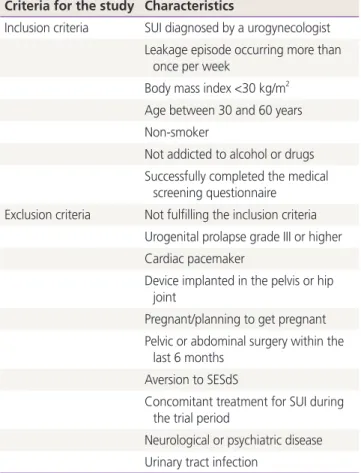
Table 1. Inclusion and exclusion criteria Criteria for the study Characteristics
Inclusion criteria SUI diagnosed by a urogynecologist Leakage episode occurring more than
once per week
Body mass index <30 kg/m
2Age between 30 and 60 years Non-smoker
Not addicted to alcohol or drugs Successfully completed the medical
screening questionnaire Exclusion criteria Not fulfilling the inclusion criteria
Urogenital prolapse grade III or higher Cardiac pacemaker
Device implanted in the pelvis or hip joint
Pregnant/planning to get pregnant Pelvic or abdominal surgery within the
last 6 months Aversion to SESdS
Concomitant treatment for SUI during the trial period
Neurological or psychiatric disease Urinary tract infection
SUI, stress urinary incontinence; SESdS, surface electrical stimulation
during sitting.
exclusion criteria. The severities of incontinence and FSD were determined in interviews.
The inclusion/exclusion criteria for the study are shown in Table 1. A total of 34 subjects who met these criteria were randomly separated into 2 groups (www.randomization.
com): a control and an SESdS group (Fig. 1).
2. Surface electrical stimulation during sitting
The EasyK7 is a SESdS device that stimulates the PFM and surrounding structures using 3 surface electrodes in contact with the perivaginal and sacral regions. In the present study, the surface electrodes were positioned near each partici- pant’s anus and sacrum to stimulate both the perivaginal and sacral regions, with the subject sitting on the EasyK7 device (Fig. 2). Subjects were asked to sit on the device to ensure that both electrodes made contact with the perivaginal and sacral regions. The amplitude used for stimulation was set to a comfortable level for each subject. The EasyK7 deliv-
ered biphasic and asymmetric impulses of 25 Hz at pulses of 11 seconds, with an 11-second rest period between pulses.
The mean intensities used were 19.37±6.29 mA (range, 2.5–30 mA). Each EasyK7 session was 15 minutes long.
3. Intervention
The subjects in the SESdS group were provided with an EasyK7 device and shown how to use and maintain the device correctly. These subjects were instructed to use the device for a single 15-minute session per day for 5–6 days per week, for a total of 8 weeks. In addition, the subjects were permitted to increase the EasyK7 stimulation amplitude within tolerable limits.
Control group subjects walked for more than 20 minutes in lieu of EasyK7 treatments. At the end of the 8-week inter- vention period, we provided an EasyK7 device as a reward to all subjects for participating in the study. Measurements were recorded in both groups before and after the 8-week inter-
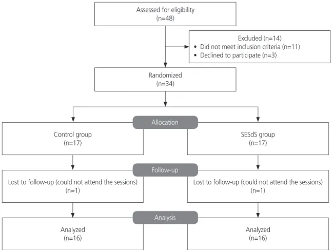
Control group (n=17)
Randomized (n=34)
Excluded (n=14)
• Did not meet inclusion criteria (n=11)
• Declined to participate (n=3) Assessed for eligibility
(n=48)
Lost to follow-up (could not attend the sessions) (n=1)
Analyzed (n=16)
SESdS group (n=17)
Lost to follow-up (could not attend the sessions) (n=1)
Analyzed (n=16) Allocation
Follow-up
Analysis
Fig. 1. Flow chart of patient selection into this randomized trial of transcutaneous electrical stimulation in women with stress urinary in-
continence. SESdS, surface electrical stimulation during sitting.
vention period. We measured PFM function using a perine- ometer and evaluated sexual function using a questionnaire.
4. Outcomes
Female sexual function was measured using the Korean version of the pelvic organ prolapse–urinary incontinence sexual function questionnaire (PISQ) [22]. The PISQ is a 31-item questionnaire with the responses based on a 5-point Likert scale [23]. It has been used to evaluate sexual func- tion in women with urinary incontinence and/or pelvic organ prolapse [24]. The PISQ has 3 distinct domains that cover behavioral/emotive (15 items), physical (10 items), and partner-related (6 items) aspects of female sexual function.
There is also an overall total score. The behavioral/emotive domain assesses sexual desire, frequency of sexual activity, and orgasm capability. The physical domain evaluates the ef- fect of urinary incontinence on sexual function. The partner- related domain evaluates the participant’s perception of her partner’s response to the effect of pelvic floor disorders on the couple’s sex life. Each domain score is calculated by add- ing the scores from the individual items in each domain. The total PISQ-31, physical domain, behavioral/emotive domain, and partner-related domain scores range from 0 to 125, 0 to 40, 0 to 61, and 0 to 24, respectively. In all domains, higher scores indicate better sexual function.
PFM function was evaluated using a vaginal pressure mea- surement device (VVP-3000 perineometer; QLMED, Ltd., Seongnam, Korea; length: 115 mm, active surface length:
66 mm, diameter: 24 mm), with each participant in the hook-lying position. A microprocessor with latex tubing was connected to the vaginal pressure probe and transmitted pressure readings when vaginal contractions compressed the probe. The baseline pressure value when the PFM was relaxed was set to 0 mmHg. Subjects were instructed to contract their PFM at maximum effort for 3 seconds. Next, they were asked to contract their PFM in an inward direction as much as possible, without contracting their abdominal or gluteal muscles [25]. The strength of the PFM was measured in mm Hg based on the difference between resting pressure and peak pressure. The mean value of 2 maximal voluntary contractions (MVCs) was recorded [26]. To measure muscle power, the subjects performed MVCs as rapidly as possible [27]. We defined the power of the PFM as the peak pressure divided by the time required to achieve MVC (mmHg/s). The time required to achieve MVC was measured from the start- ing point until peak pressure was achieved. We defined the endurance of the PFM as the mean force of vaginal contrac- tion for 10 seconds during a single attempt.
5. Statistical analyses
Kolmogorov–Smirnov Z-tests were used to confirm that the data were normally distributed. Analysis of covariance was used to compare variables between groups and within each group before and after the intervention. Baseline values were used as covariates. In addition, the paired Student’s t-test was used for comparisons before and after the intervention in each group. All data are expressed as mean±standard deviation. Effect sizes were calculated to estimate the sig- nificance of differences between groups. Each effect size (r) ranged from 0 (no effect) to 1 (complete effect). Effect sizes of 0 to <0.1, 0.1 to <0.3, 0.3 to <0.5, and ≥0.5 were cate- gorized as no effect, small effect, moderate effect, and large effect, respectively. All statistical analyses were performed using SPSS software (ver. 18.0; SPSS Inc., Chicago, IL, USA), and a P-value <0.05 was considered statistically significant.
Results
A total of 34 subjects with SUI were randomly separated into Fig. 2. Surface electrical stimulation during sitting using the Al-
phamedic EasyK7 device.
2 groups, each consisting of 17 subjects. However, due to time constraints, one subject from each group failed to com- plete the intervention period. Therefore, 32 subjects com- pleted the protocol and were included in the analysis (Table 2).
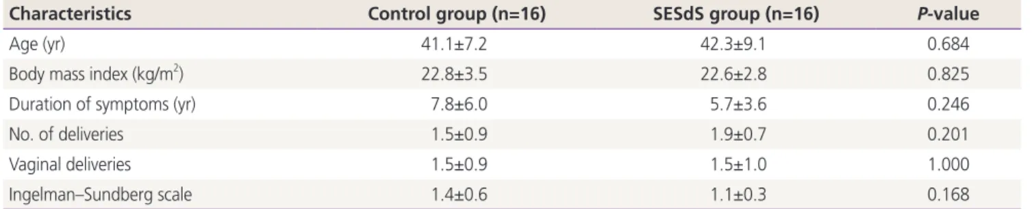
There were no significant differences in demographic char- acteristics or Ingelman–Sundberg scale values between the 2 groups.
Table 3 shows the SESdS-induced improvements in PFM function and PISQ parameters, determined by comparing the SESdS and control groups after the intervention period, as well as improvements within the SESdS group (i.e., the differ-
ence between measurements recorded before and after in- tervention). With regard to PFM function, there were signifi- cant differences between the groups, as well as between the pre- and post-intervention measurements within the SESdS group, in terms of power (between groups: P=0.001; within SESdS group: P=0.004), strength (between groups: P=0.015;
within SESdS group: P=0.011), and endurance (between groups: P=0.012; within SESdS group: P=0.034). In addi- tion, PFM function significantly increased after intervention within the SESdS group (strength: P=0.019; power: P=0.03;
endurance: P=0.018). However, there were no significant dif-
Table 2. Characteristics of the participants
Characteristics Control group (n=16) SESdS group (n=16) P-value
Age (yr) 41.1±7.2 42.3±9.1 0.684
Body mass index (kg/m
2) 22.8±3.5 22.6±2.8 0.825
Duration of symptoms (yr) 7.8±6.0 5.7±3.6 0.246
No. of deliveries 1.5±0.9 1.9±0.7 0.201
Vaginal deliveries 1.5±0.9 1.5±1.0 1.000
Ingelman–Sundberg scale 1.4±0.6 1.1±0.3 0.168
Data are shown as mean±standard deviation.
SESdS, surface electrical stimulation during sitting.
Table 3. Primary outcomes in each group pre- and post-intervention
Primary outcomes Pre-intervention Post-intervention Effect size Within P- value
Between P-value
PFM Power (mmHg/s) Control group 16.41±13.20 15.16±10.07 0.32 0.418 0.001
SESdS group
a)16.27±9.20 30.50±17.56 0.030
Strength (mmHg) Control group 18.70±10.07 19.02±9.40 0.19 0.557 0.015
SESdS group
a)20.21±9.09 26.60±11.28 0.019
Endurance (mmHg) Control group 13.72±7.96 13.27±7.35 0.20 0.704 0.012
SESdS group
a)14.49±7.23 20.46±10.34 0.018
PSIQ Behavioral/emotive score Control group 26.56±11.78 23.56±10.37 0.42 0.056 0.000
SESdS group
b)26.94±13.43 33.25±15.45 0.000
Physical score Control group 34.81±3.29 35.13±4.10 0.08 0.714 0.121
SESdS group
b)30.06±4.54 34.56±2.97 0.000
Partner-related score Control group 18.25±2.08 18.13±2.19 0.26 0.697 0.003
SESdS group
a)18.69±2.36 20.13±1.71 0.022
Total score Control group 79.63±14.29 76.81±12.10 0.54 0.140 0.000
SESdS group
b)75.69±16.42 87.69±16.76 0.000
Data are shown as mean±standard deviation.
PFM, pelvic floor muscles; PISQ, pelvic organ prolapse–urinary incontinence sexual function questionnaire; SESdS, surface electrical stimulation during sitting.
a)