This is an Open Access article distributed under the terms of the Creative Commons Attribution Non-Commercial License (http: //creativecommons.org/licenses/by- nc/4.0/) which permits unrestricted non-commercial use, distribution, and reproduction in any medium, provided the original work is properly cited.
Copyright: © 2019 Korean Journal of Agrcultural Science
https://doi.org/10.7744/kjoas.20190025
FOOD & CHEMISTRY
Protective effects of Cirsium japonicum var. maackii against amyloid beta-induced neurotoxicity in C6 glial cells
Ji Hyun Kim
1, Min Jeong Kim
1, Ji Myung Choi
1, Sanghyun Lee
2, Eun Ju Cho
1,*1
Department of Food Science and Nutrition & Kimchi Research Institute, Pusan National University, Busan 46241, Korea
2
Department of Plant Science and Technology, Chung-Ang University, Anseong 17546, Korea
*
Corresponding author: [email protected]
Abstract
Alzheimer’s disease (AD) is the most common neurodegenerative disease associated with age, and amyloid beta (Aβ) is known to cause Alzheimer’s disease. In the present study, we investigated the protective effects of Cirsium japonicum var. maackii extract and its fractions against Aβ-induced neurotoxicity in C6 glial cells. The cells treated with Aβ
25-35showed a decrease in cell viability and an increase in reactive oxygen species (ROS) production compared with the non-treated cells. However, the cells treated with the C. japonicum var.
maackii extract and its fractions increased the cell viability and inhibited the Aβ-induced ROS production. These results demonstrate the neuroprotective effects of C. japonicum var. maackii against Aβ. To further examine the protective mechanism, we measured inflammation and apoptosis related protein expressions. The cells treated with extract and fractions from C. japonicum var. maackii down-regulated inflammatory related proteins such as cyclooxygenase-2, interleukin (IL)-1β, and IL-6, and attenuated apoptosis related proteins including B-cell lymphoma-2 (Bcl-2) associated X protein/Bcl-2 ratio. In particular, the ethanol and ethylacetate fraction exhibited higher inhibitory effect against ROS production and apoptosis-related protein expressions among the extract and the other fractions. Therefore, this study demonstrated the protective effects of C. japonicum var. maackii extract and its fractions against Aβ-induced neurotoxicity in C6 glial cells through the regulation of oxidative stress, inflammation, and apoptosis, suggesting that it might have potential as a therapeutic for AD.
Keywords: amyloid beta, apoptosis, Cirsium japonicum var. maackii, inflammation, oxidative stress
Introduction
알츠하이머 질환(
Alzheimer
’s disease
)은 노화에 따른 대표적인 신경퇴행성질환으로 기억력 및 인 지능 손상을 일으킨다(Price and Morris
,1999
). 알츠하이머 질환의 발병 원인은 명확하게 규명되지는 않았지만, 뇌 내amyloid beta
(Aβ
) 단백질의 과발현이 주요 원인으로 보고되었다(Canevari et al
.,OPEN ACCESS
Accepted: May 28, 2019 Revised: May 23, 2019 Received: May 5, 2019 DOI:
Citation: Kim JH, Kim MJ, Choi JM, Lee S, Cho EJ. 2019. Protective effects of Cirsium japonicum var. maackii against amyloid beta-induced neurotoxicity in C6 glial cells. Korean Journal of Agricultural Science. https://doi.org/10.7744/
kjoas.20190025
2004
).Aβ
는39
-43
개의 아미노산으로 이루어진 물질로, 아밀로이드 전구 단백질인APP
(amyloid precursor protein
)로부터β
-,γ
-secretase
와 같은protease
에 의해 분해되어 생성되어진다(Butterfield et al
.,2013
). 국내·외 여러 연구에 의하면, 알츠하이 머 질환 환자의 뇌에서 과발현된Aβ
는 응집되어 뇌 내plaque
를 형성하게 되는데, 이는 알츠하이머 질환의 대표적인 주요 병인으로 알려져 있으며, 학습·기억력 손상과 밀접한 관련이 있는 것으로 보고되었다(Canevari et al
.,2004
;Butterfield et al
.,2013
).Aβ
의 과발현은 뇌를 구성하는 신경세포 및 신경교세포에서 산화적 스트레스, 염증반응 및 세포사멸의 원인이 된다 (Butterfield et al
.,2013
). 뇌에서Aβ
는 활성산소종(ROS
;reactive oxygen species
)을 증가시켜 생체를 구성하는 단백질,DNA
및RNA
의 산화와 지질과산화 등을 일으킨다(Cheignon et al
.,2018
). 특히, 이러한 산화적 스트레스는 염증반응을 유도하는데,cyclooxygenase
-2
(COX
-2
),inducible nitric oxygen synthase
(iNOS
)와 같은 염증 매개 인자를 활성화시키고,interleukin
(IL
)-1β
,IL
-6
와 같은 염증성cytokines
을 방출시킨다(Sawikr et al
.,2017
). 이와 같이Aβ
에 의해 유도된 산화적 스트레스 및 염증반응 은 뇌 내 세포사멸을 유도한다. 세포사멸 과정에서,Bcl
-2
-associated X protein
(Bax
) 단백질은pro
-apoptosis
인자로써 세포 사 멸을 유도하는 역할을 하는 반면B
-cell lymphoma
-2
(Bcl
-2
) 단백질은anti
-apoptosis
인자로 작용하여 세포사멸을 보호하는 역할을 하는데, 알츠하이머 질환에서Bcl
-2
단백질에 비해Bax
단백질 발현이 과발현되어 세포사멸이 유도되는 것으로 알 려져 있다(Obulesu and Lakshmi
,2014
). 또한,Aβ
가 과발현 된 알츠하이머 질환 동물 모델의 뇌에서 산화적 스트레스, 염증 반응, 세포사멸 관련 단백질 발현의 증가가 보고되어 있어(Ali et al
.,2017
;Ma et al
.,2018
),Aβ
로 인한 이들 작용 메커니즘 을 조절하는 것은 알츠하이머 질환의 예방 및 치료에 중요한 역할을 하는 것으로 생각된다.엉겅퀴(
Cirsium japonicum var
.maackii
)는 국화과(asteraceae
)의 다년생 초본으로, 한국, 중국, 일본 등에 자생하고 있으며, 항균, 항염, 해독, 이뇨작용 등으로 민간 및 한방에서 약용식물로써 널리 이용되는 식물이다. 이전 연구에 의하면, 엉겅퀴 는 항산화, 항염증, 항당뇨, 간 손상 보호 등 다양한 생리활성이 보고되었다(Liao et al
.,2010
;Mok et al
.,2011
;Wan et al
.,2014
). 또한, 엉겅퀴는 항산화 등의 생리활성이 우수한cirsimaritin
,acacetins
,luteolin
,linarin
,chlorogenic acid
등의 활성성분을 다량 함유하는 것으로 알려져 있다(Kim and Kim
,2003
;Liao et al
.,2010
;Shin et al
.,2017
). 특히 엉겅퀴에서 분리한luteolin
은 당뇨로 인지능이 손상된 동물모델에서 항산화, 항염증 및 신경보호 활성을 통해 인지능 개선 효능이 보고되었으며(Liu et
al
.,2013
), 엉겅퀴로부터 분리한kainic acid
는 신경세포에서ROS
소거와 항산화 효소 활성 증가를 통한 신경 세포 보호 효 과가 보고되었다(Han et al
.,2012
). 선행연구로써 엉겅퀴 추출물 및 분획물의H
2O
2 유도 산화적 손상에 대한 신경교세포 보 호 효과를 확인하였으나(Lee et al
.,2018
), 알츠하이머 질환의 주요 원인으로 알려진Aβ
로 유도된 신경독성에 대한 신경교 세포 보호 효과 및 작용 메커니즘 분석에 관한 연구는 부족한 실정이다. 따라서 본 연구에서는C6
신경교세포를 이용하여Aβ
로 유도된 신경독성에 대해 엉겅퀴 추출물 및 분획물의 신경교세포 보호 효능과 산화적 스트레스, 염증반응, 세포사멸 관련 인자 측정을 통해 관련 신경교세포 보호 작용기전을 규명하고자 한다.Materials and Methods
엉겅퀴의 추출물 및 분획물 조제
실험에 사용된 엉겅퀴(
Cirsium japonicum var
.maackii
) 봄 지상부는 임실생약(Imsil
,Korea
)에서 제공받아 실험재료로 사용 하였다. 세척 및 건조된 엉겅퀴 봄 지상부5
.71 kg
을ethanol
(EtOH
)로 환류냉각장치를 이용하여 추출하였고, 추출물667
.2
g
(수율11
.68
%)을 얻었다.EtOH
추출물은 유기용매를 이용하여 각각 분획하였고, 분획물로n
-hexane
(213
.6 g
, 수율3
.74
%),CHCl
3 (39
.0 g
, 수율0
.68
%),ethyl acetate
(EtOAc
,67
.6 g
, 수율1
.18
%),n
-butanol
(n
-BuOH
,47
.0 g
, 수율0
.82
%) 등 총4
개의 분 획물을 조제하여 실험재료로 사용하였다.세포 종류 및 시약
C6
신경교세포는 한국세포주은행(KCLB
,Korea Cell Line Bank
,Seoul
,Korea
)에서 구입하여 사용하였다. 세포 배양을 위 한Dulbecco
’s modified eagle medium
(DMEM
),fetal bovine serum
(FBS
),trypsin ethylenediaminetetraacetic acid
(EDTA
),100 units
/mL
의penicillin streptomycin
용액은Welgene
(Gyeongsan
,Korea
)에서 구입하여 사용하였다. 신경독성 유도를 위한Aβ
25-35는Sigma
(St
.Louis
,USA
)사의 제품을 구매하여 사용하였다. 세포 생존율 측정을 위한3
-(4
,5
-dimethylthiazol
-2
-yl
)-2
,5
-diphenyltetrazolium bromide
(MTT
)는Bio Basic
(Toronto
,Canada
)에서,dimethyl sulfoxide
(DMSO
)는Bio Pure
(Ontario
,Canada
) 에서 구매하였으며,ROS
측정을 위한dichlorofluorescein diacetate
(DCF
-DA
)는Sigma
(St
.Louis
,USA
)사 제품을 구매하여 사 용하였다.Western blot analysis
를 위한RIPA cell lysis buffer
는Elpis Biotech
(Daejeon
,Korea
)에서,Laemmli sample buffer
는Bio
-Rad
(Hercules
,USA
)에서 구입하였다.1
차 항체로 사용한IL
-1β
는Bioss Biotechnology
(Woburn
,Massachusetts
,USA
)에서,IL
-6
,Bax
,Bcl
-2
는Santa Cruz Biotechnology
(Dallas
,USA
)에서,β
-actin
,COX
-2
,2
차 항체로 사용한anti
-rabbit IgG HRP
-linked antibody
는Cell Signaling Technology
(Danvers
,USA
)에서 구입하여 실험에 사용하였다.세포 배양
C6
신경교세포는10
%의FBS
와1
%의penicillin
,streptomycin
이 포함된DMEM
배지에서37
℃,5
%CO
2incubator
에서 배 양하였다. 배양된 세포는2
-3
일 마다 한번 배양액을 바꾸어 주면서 배양하여 세포 분화가 최대에 도달했을 때,phosphate buffered saline
(PBS
,pH 7
.4
)로 세포를 세척 후,0
.05
%trypsin
과0
.02
%EDTA
혼합액으로 부착된 세포를 분리한 뒤, 원심분 리를 통해 집적시켰다. 집적된 세포는 골고루 분산되도록 세포와 배지를 잘 혼합하여 계대배양하면서 실험에 사용하였 다.세포 생존율 측정
세포가
confluence
상태가 되면96 well plate
에5
×10
4cells
/well
로seeding
하여4
시간37
℃에서 배양시켜 세포를 부착시킨 뒤, 엉겅퀴 추출물 및 분획물 시료를 희석하여 농도별로 각well
에 처리하였다.4
시간 뒤, 세포 손상을 유도하기 위해Aβ
25-35 (25 µM
)을 처리하여incubator
내에서 배양하였다.24
시간 뒤,5 mg
/mL MTT solution
을 각well
에 주입하여37
℃에서4
시간 동 안 배양하였고, 생성된 보라색의formazan
결정을DMSO
에 녹여540 nm
에서 흡광도를 측정하여 세포 생존율을 계산하였 다(Mosmann
,1983
).Reactive oxygen species (ROS) 소거능 측정
세포가
confluence
상태가 되면96 well black plate
에5
×10
4cells
/well
로seeding
하여4
시간37
℃에서 배양하여 세포가 잘 부착되면 엉겅퀴 추출물 및 분획물 시료를 농도별로 희석하여 각well
에 처리하여4
시간 배양한 뒤,Aβ
25-35 (25 µM
)을 처리 하여 배양하였다.24
시간 뒤,80 µM DCF
-DA solution
을 각well
에 주입하여37
℃에서30
분 동안 재배양하였고,FLUOstar OPTIMA
(BMG labtech
.,Ortenberg
,Germany
)excitation
-480 nm
,emission
-535 nm
를 이용하여 형광도를 측정하여ROS
소거능 을 계산하였다(Wang and Joseph
,1999
).Western blot analysis
세포에
RIPA buffer
를 첨가하여4
℃에서1
시간 동안 반응시킨 뒤,12
,000 rpm
에서30
분간 원심분리하여 상층액의 단백질 을 분리하였다. 단백질 정량 시약인Bio
-Rad protein assay kit
(Bio
-Rad
,Richmond
,USA
)를 이용하여 정량하고,sample buffer
와 혼합하여sample
을 제작하였다. 동량의sample
을10
% 또는13
%의sodium dodecyl sulphate
(SDS
)-polyacrylamide gel
을 이용하여 전기영동으로 분리한 후,
polyvinylidene difluoride
(PVDF
)membrane
에transfer
한 뒤,5
%skim milk
에서1
시간 동안blocking
하였다. 이 후,1
차항체와4
℃에서overnight
반응 시킨 뒤2
차 항체와 실온에서1
시간 반응시켰다.PBS
-T
로 세척 후enhanced chemiluminescence
(ECL
)solution
과 반응시킨 후Chemiliuminescence image system
(Davinch
-Chemi
TM,Davinchi
-K
,Seoul
,Korea
)를 이용하여 특정 단백질 발현을 확인하였다.통계 분석
실험 결과는 평균 ± 표준편차 (
n
=3
)로 나타내었고,Statistical Package for the Social Sciences
(SPSS
) 프로그램(version 20
.0
,Chicago
,USA
)을 이용하여 각 실험결과들로부터analysis of variance
(ANOVA
)를 구한 뒤,Duncan
’s multiple Range test
및Student
’s t
-test
를 이용하여p
<0
.05
수준에서 각 군의 평균간의 유의성을 검정하였다.Results and Discussion
알츠하이머 질환의 주요 병리학적 원인으로 알려진
Aβ
는 뇌 내 신경독성을 일으켜 산화적 스트레스, 염증반응, 세포사 멸을 유도하며, 이는 결국 기억력 및 인지능 손상을 나타낸다(Canevari et al
.,2004
;Butterfield et al
.,2013
).Aβ
는 뇌의 신경세 포 뿐만 아니라 신경전달물질 분비와 신호전달에 관여하는 신경교세포의 손상을 유도하여 알츠하이머 질환에 영향을 미 치는 것으로 보고되었다(Block and Hong
,2005
). 뇌에서 신경교세포는 시냅스 가소성, 성장인자 분비 등의 조절을 통해 신 경세포를 보호하는 역할을 한다(Travis
,1994
;Farfara et al
.,2008
). 그러나,Aβ
로 손상이 유도된 신경교세포는 •OH
,O
2- 등의ROS
생성으로 인한 산화적 스트레스를 일으킨다(Tanaka et al
.,1999
). 이는 염증매개인자, 염증성cytokine
분비 등을 통해 염증반응을 유도 할 뿐만 아니라, 신경교세포 사멸을 통해 신경교세포 기능의 손상을 일으켜AD
를 악화시킨다(Farfara et
al
.,2008
).Aβ
25-35는full length Aβ
의 절단된 형태로써, 보다 작은 크기로 인해 세포막을 통과하기 쉬울 뿐만 아니라full length
Aβ
와 유사한 독성을 나타내어 국내외in vivo
및in vitro
연구에 널리 사용되고 있다(Mattson et al
.,1997
;Lau et al
.,2007
). 또한,
Aβ
25-35를 동물의 뇌 내 주입 시, 뇌 내 산화적 스트레스, 염증반응, 세포사멸이 유도되었으며, 이로 인해 기억력 및 인지능 손상의 임상적 특징을 나타냄을 확인하였다(
Fang and Liu
,2006
;Lu et al
.,2009
;Kim et al
.,2015
). 따라서, 본 연구에서Aβ
25-35를 이용하여 신경교세포 손상을 유도한 모델에서 엉겅퀴 추출물 및 분획물의 신경교세포 보호 효과를 확인하고자하였다.
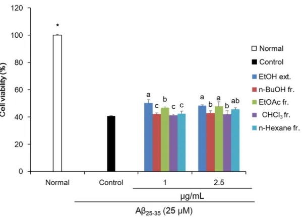
엉겅퀴 추출물 및 분획물의
Aβ
25-35로 유도된 신경독성으로부터C6
신경교세포 보호 효과를MTT assay
를 통해 확인하였 다.MTT assay
는 세포 생존율 측정에 주로 사용되는assay
로, 탈수소 효소 작용에 의한mitochondria
활성을 이용하는 측정 법이다(Mosmann
,1983
).C6
신경교세포에 아무것도 처리하지 않은normal
군의 세포생존율을100
% 라고 했을 때,Aβ
25-35를25 µM
의 농도로 처리한 경우40
.56
%로 유의적으로 세포 생존율 감소를 나타내어,Aβ
25-35로 인한C6
신경교세포 손상을 확인하였다(Fig
.1
). 반면, 엉겅퀴 추출물 및 분획물을 처리한 군에서Aβ
25-35만을 처리한control
군에 비해 유의적으로 세포 생존율 증가를 나타내었다. 특히,EtOH
및EtOAc
분획물을2
.5 µg
/mL
의 농도로 처리 시, 각각48
.37
,47
.84
%의 수치를 나 타내어 다른 추출물 및 분획물에 비해 우수한 신경교세포 보호 효과를 확인하였다.Aβ
25-35로 신경독성을 유도한C6
신경교세포에서 엉겅퀴 추출물 및 분획물의 산화적 스트레스 개선 효과를 확인하기 위해
DCF
-DA assay
를 이용하여ROS
생성량을 측정하였다.DCF
-DA assay
는DCFH
가ROS
에 의해 산화되어 강한 형광을 나 타내는DCF
로 전환되는 원리를 이용한 측정법으로, 세포 내ROS
생성 측정에 널리 이용되는 방법이다(Rastogi et al
.,2010
).ROS
생성을 측정한 결과, 시간이 지남에 따라 모든 군에서fluorescence
수치 증가를 나타내어ROS
생성량이 증가하는 것 을 확인하였으며, 특히Aβ
25-35를 처리한control
군은normal
군에 비해ROS
생성량이 더욱 많이 증가하여Aβ
25-35로 인한 산화 적 스트레스가 유도되었음을 알 수 있었다(Fig
.2A
).60
분 기준으로ROS
생성량을 측정한 결과,normal
군100
% 대비control
군은
108
.36
%로ROS
생성이 증가함을 확인하였다. 반면, 엉겅퀴 추출물 및 분획물을1
,2
.5 µg
/mL
의 농도로 처리했을 때, 모든 군에서control
군에 비해 낮은 수치를 나타내었다(Fig
.2B
). 특히 엉겅퀴 추출물 및 분획물을2
.5 µg
/mL
의 농도로 처리 시EtOH
및EtOAc
분획물에서 각각102
.42
,102
.53
%의 낮은 수치를 나타내어,EtOH
및EtOAc
분획물이 다른 추출물 및 분 획물에 비해 유의적으로 우수한ROS
소거능을 나타냄을 확인하였다. 또한, 이전 연구에서 엉겅퀴EtOAc
분획물은EtOH
,n
-BuOH
,CHCl
3,n
-hexane
과 같은 다른 추출물 및 분획물에 비해in vitro
에서DPPH
, ·OH
,O
2- 라디칼 소거능이 우수하였으 며,aldose reductase
억제 활성이 가장 우수한 것으로 보고되었다(Lee et al
.,2017
;Lee et al
.,2018
).뇌에서
Aβ
로 인한 신경독성은ROS
생성량 증가를 통한 산화적 스트레스 뿐만 아니라nuclear factor
-κB
(NF
-κB
)pathway
활성화를 통한 염증반응을 유도한다(Kempuraj et al
.,2016
).NF
-κB
는 알츠하이머 질환에 관여하는 염증반응 작용 기전으 로, 세포의 손상 시 활성화되어COX
-2
,iNOS
와 같은 염증 매개 인자를 발현시킨다(Kempuraj et al
.,2016
). 이와 같이 발현 된 염증 매개 인자는IL
-6
,IL
-1β
,tumor necrosis factor
-α
와 같은 염증성cytokines
를 과발현시켜 세포 손상을 유도한다(Apelt and Schliebs
,2001
). 또한,Aβ
가 과발현된 뇌 조직에서 염증 매개 인자 및 염증성cytokines
이 과발현되어 염증반응이 유도 됨이 보고되었다(Halliday et al
.,2000
). 본 연구에서Aβ
25-35로 인한 신경교세포에서의 염증반응에 대해 엉겅퀴 추출물 및 분 획물의 염증반응 개선 작용 기전을 규명하기 위해COX
-2
,IL
-1β
,IL
-6
와 같은 염증반응 관련 단백질 발현을 측정하였다(Fig
.3
).Aβ
25-35만을 처리한control
군의 경우 아무것도 처리하지 않은normal
군에 비해 유의적으로COX
-2
,IL
-1β
,IL
-6
단백질 발현이 증가하여 염증반응이 유도되었음을 확인하였다. 반면 엉겅퀴 추출물 및 분획물을
2
.5 µg
/mL
의 농도로 처리하였을 때,control
군에 비해 유의적으로 모든 군에서 이들 단백질 발현이 감소함을 확인하였다. 따라서 엉겅퀴 추출물 및 분획물은
Aβ
25-35로 유도된 신경교세포의 염증반응 개선 효과가 있는 것으로 생각된다.Fig. 1. Effect of Cirsium japonicum var. maackii extract (ext.) and fractions (fr.) on cell viability in
Aβ25-35-treated C6 glial cells. Values are means ± SD (n = 3). a - c: Means with the different letters
are significantly different (p < 0.05) by Duncan’s multiple range test among extract- and fractions- treated groups. *p < 0.05 compared with the control group by Student’s t-test.
Aβ
로 인한 신경독성은 산화적 스트레스와 염증반응을 유도하여, 결국 뇌 내 신경세포 및 신경교세포의 사멸을 유도한 다(Yang et al
.,2004
;Han et al
.,2017
).Bcl family
는 세포사멸에 관여하는 중요한 작용 기전으로, 뇌에서Bcl family
발현 조절 은 기억력 및 인지능 손상을 유도하여 알츠하이머 질환의 원인이 되는 것으로 보고되었다(Kitamura et al
.,1998
).Bax
단백Fig. 2. Effect of Cirsium japonicum var. maackii extract (ext.) and fractions (fr.) on reactive oxygen species
(ROS) production in Aβ25-35-treated C6 glial cells. Time course of change in intensity of ROS fluorescence during 1 h (A) and production of ROS at 1 h (B). Values are means ± SD (n = 3). NS, non-significance. a, b:Means with the different letters are significantly different (p < 0.05) by Duncan’s multiple range test among
질은 세포사멸을 촉진하는
pro
-apoptosis
인자로써Aβ
와 같은 신경독성으로 인한 세포 손상 시 과발현되며, 이는cytochrome C
및caspase
를 활성화 시켜 세포사멸을 유도한다(Kitamura et al
.,1998
;Obulesu and Lakshmi
,2014
). 반면,Bcl
-2
단백질은 세 포사멸을 보호 하는 역할을 하는anti
-apotosis
인자로써 작용한다(Ferreiro et al
.,2007
).Aβ
25-35로 세포사멸을 유도한 신경교 세포에서 엉겅퀴 추출물 및 분획물의 세포사멸 보호 작용 기전을 확인하기 위해 세포사멸과 관련된 인자인Bax
와Bcl
-2
단백질 발현을 확인하였다(Fig
.4
).Aβ
25-35만을 처리한control
군의 경우 아무것도 처리하지 않은normal
군에 비해 유의적으 로Bax
/Bcl
-2
단백질 발현 비율이 증가하여Aβ
25-35로 인한 세포사멸이 유도되었음을 알 수 있었다. 반면2
.5 µg
/mL
의 농도 로 엉겅퀴MeOH
추출물 및EtOAc
,CHCl
3,Hx
분획물을 각각 처리 시,control
군에 비해Bax
/Bcl
-2
단백질 발현 비율이 개선 되었으며, 특히EtOAc
분획물의 경우 다른 추출물 및 분획물에 비해 가장 낮은Bax
/Bcl
-2
수치를 나타내어 세포사멸 보호 효과가 우수한 것으로 사료된다. 엉겅퀴EtOAc
분획물에는radical
소거능이 우수한 항산화 물질로cirsimaritin
,hispidulin
,cirsimarin
,luteolin
등과 같은flavonoid
계열의 활성성분을 함유하는 것으로 보고되었다(Jung et al
.,2009
;Lee et al
.,2017
). 뿐 만 아니라,hispidulin
은 인지능 손상 동물 모델에서Aβ
축적 억제, 염증 반응 및apoptosis
억제를 통한 신경 보호 효과가 보 고되었다(Huang et al
.,2018
). 또한,luteolin
은Aβ
유도 인지능 손상 동물 모델에서 항산화 및cholinergic function
조절을 통한 인지능 개선 효과와mitogen
-activated protein kinases signaling
조절을 통한 신경보호 효과 등이 보고되었다(Cheng et al
.,2010
;Yu et al
.,2015
). 따라서, 본 연구에서 우수한 신경교세포 보호 효능을 보인 엉겅퀴EtOAc
분획물은 이러한 활성성분의 작 용으로 다른 추출물 및 분획물에 비해 우수한 신경교세포 보호 효능을 나타낸 것으로 사료된다.Fig. 3. Effect of Cirsium japonicum var. maackii extract (ext.) and fractions (fr.) on inflammation-related
protein expressions in Aβ25-35-treated C6 glial cells. Protein bands intensity of COX-2, IL-1β, and IL-6 (A) and quantitative analysis of COX-2 (B), IL-1β (C), and IL-6 (D). Values are means ± SD (n = 3). a - g: Means with the different letters are significantly different (p < 0.05) by Duncan’s multiple range test.Conclusion
본 연구에서는 엉겅퀴 추출물 및 분획물의
Aβ
유도 신경독성에 대한 신경교세포 보호 효과와 그 작용기전에 관하여 연 구하였다. 엉겅퀴 추출물 및 분획물은Aβ
25-35로 손상이 유도된C6
신경교세포에서 세포 생존율 증가와ROS
생성을 감소 시켜 신경교세포 보호 효과를 나타내었다. 신경교세포 보호 작용기전 확인을 위해 염증과 세포사멸 관련 단백질 발현을Fig. 4. Effect of Cirsium japonicum var. maackii extract (ext.) and fractions (fr.) on apoptosis-related protein
expressions in Aβ25-35-treated C6 glial cells. Values are means ± SD (n = 3). a - g: Means with the different letters are significantly different (p < 0.05) by Duncan’s multiple range test.확인하였을 때, 엉겅퀴 추출물 및 분획물은
COX
-2
,IL
-1β
,IL
-6
와 같은 염증 관련 인자의 억제와Bax
,Bcl
-2
와 같이 세포사 멸에 관여하는 인자 조절을 통해 항염증 및 항세포사멸 효과를 나타내었다. 특히, 여러 추출물 및 분획물 중EtOAc
분획 물에서 가장 우수하게ROS
소거능과 세포사멸 관련 단백질 발현 조절을 나타냄을 확인하여,EtOAc
분획물이Aβ
25-35로 인 한 신경교세포의 손상에 대한 보호 효과가 우수한 것으로 생각된다. 이상의 결과에서 엉겅퀴는Aβ
25-35로 인한 신경독성에 대해 산화적 스트레스, 염증반응, 세포사멸 조절을 통해 신경교세포 보호 효과를 나타내는 것으로 사료된다.Acknowledgements
이 논문은