Development of a Nuclease Protection Assay With Sandwich Hybridization (NPA-SH) to Monitor Heterosigma akashiwo
Mingyeong Kang1.2, Mirye Park3, Kang Eun Kim1,2 and Taek-Kyun Lee1,2*
1Ecological Risk Research Division, Korea Institute of Ocean Science and Technology, Geoje 53201, Korea
2Ocean Science, University of Science and Technology, Daejeon 34113, Korea
3Bioresources Culture Collection Division, Nakdonggang National Institute of Biological Resources, Sangju 37242, Korea Received November 1, 2019 /Revised December 31, 2019 /Accepted January 2, 2020
Heterosigma akashiwo is a globally distributed raphidophyte that forms blooms and causes significant losses to the aquaculture industry in many coastal countries. The development of a fast and sensitive detection method is therefore required to facilitate the appropriate warning of harmful algal blooms.
In this study, a nuclease protection integrated with sandwich hybridization (NPA-SH) assay was de- veloped to both qualitatively and quantitatively detect H. akashiwo. The NPA, capture and signal probes were designed by nucleotide sequencing of H. akashiwo. The applicability of NPA-SH was eval- uated using cultured H. akashiwo cells and field samples collected at Goseong Bay, Korea. The results show that this method has good applicability and effectiveness in analyzing cultured cells and field samples. A linear regression equation for the quantitative analysis of H. akashiwo was obtained, and the lower detection limit of the assay was 1×10⁴ cells/ml. There was no statistically significant differ- ence in the results of H. akashiwo quantitation using NPA-SH compared to those obtained using a microscope. These results indicate that NPA-SH can be a good alternative to the traditional micro- scopic method used to monitor H. akashiwo.
Key words : Algal blooms, field monitoring, HABs, Heterosigma akashiwo, NPA-SH
*Corresponding author
*Tel : +82-55-639-8630, Fax : +82-55-639-8509
*E-mail : [email protected]
This is an Open-Access article distributed under the terms of the Creative Commons Attribution Non-Commercial License (http://creativecommons.org/licenses/by-nc/3.0) which permits unrestricted non-commercial use, distribution, and reproduction in any medium, provided the original work is properly cited.
Journal of Life Science 2020 Vol. 30. No. 1. 26~31 DOI : https://doi.org/10.5352/JLS.2020.30.1.26
Introduction
Harmful algal blooms (HABs) caused by toxic species have a negative effect on fisheries, human health, tourism and aquatic ecosystems. Heterosigma akashiwo (Hada) (Chro- mophyta: Raphidophyceae) is a eurythermal and euryhaline flagellated golden-brown marine microalga with a global distribution [1]. This harmful microalga has been associated with blooms that kill fish and shellfish on the coasts of many countries. Therefore, H. akashiwo monitoring is needed to op- timize the early warning of HABs and fish loss [7, 16].
Like many microalgal species, H. akashiwo is not easy to identify by optical microscopy because of its high morpho- logical diversity [2, 16]. H. akashiwo cells are small (8-25 μm in length and 6-15 μm in width), fragile, and vary in shape depending on various sea conditions [2, 14]. Also, the mor- phological features, size and color of H. akashiwo could be
changed when exposed to fixative [4]. Therefore, substantial experience and expertise are needed to microscopically iden- tify natural Heterosigma species. Consequently, a fast and ac- curate detection method is required for effective monitoring of H. akashiwo. Molecular probes have replaced classical mi- croscopy for identification at the species level [12]. Accord- ingly, various detection methods, such as sandwich hybrid- ization assay (SHA), fluorescence in situ hybridization (FISH), DNA array, quantitative real-time PCR (qRT-PCR), and loop-mediated isothermal amplification (LAMP), have been applied for H. akashiwo [4, 5, 11, 12, 16].
H. akashiwo-specific SHA methods and comparative stud- ies with qPCR for environmental samples have been re- ported [5, 8, 12]. However, SHA has been estimated to have low species specificity, sensitivity and reproducibility due to unstable reactivity [6]. To overcome these disadvantages, development of a nuclease protection (NPA-SH) assay based on sandwich hybridization was required. Previous re- searchers have developed NPA-SH methods to detect envi- ronmental samples of various HAB species. The NPA-SH methods upgrade specificity and sensitivity. Also, It can be checked the result visually, and reduce the time for prepara- tion and hybridization [9]. Until now, NPA-SH methods for Prorocentrum minimum, P. micans, P. donghaiense, Skeletonema
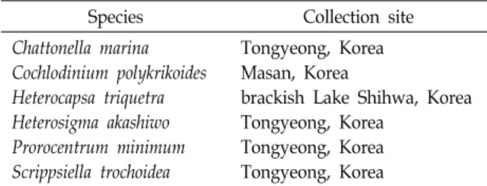
Table 1. Algal species
Species Collection site
Chattonella marina Cochlodinium polykrikoides Heterocapsa triquetra Heterosigma akashiwo Prorocentrum minimum Scrippsiella trochoidea
Tongyeong, Korea Masan, Korea
brackish Lake Shihwa, Korea Tongyeong, Korea
Tongyeong, Korea Tongyeong, Korea
costatum, Phaeocystis globosa, Cochlodinium polykrikoides, and Heterocapsa triquetra [3, 9, 10, 15, 17] were developed. Howev- er, NPA-SH probes for H. akashiwo detection have not yet been developed.
In this study, we developed a species-specific NPA-SH probe set for the rapid and accurate detection of H. akashiwo and performed qualitative and quantitative analysis. The species specificity and sensitivity of the developed probes were analyzed, and H. akashiwo abundance was monitored by applying the probes to environmental samples.
Materials and Methods
Algal cultures
Six species of HAB-causing marine microalgae: Chattonella marina, Cochlodinium polykrikoides, Heterocapsa triquetra, Heter- osigma akashiwo, Prorocentrum minimum, and Scrippsiella tro- choidea, were obtained from the Library of Marine Samples, KIOST, Korea (Table 1). Microalgae were cultured axenically in f/2 medium at 20℃ under a 12 hr light–dark cycle with a photon flux density of 4,000 lx.
Primer design and RT-PCR
RNA was extracted using the modified method of Venugopalan and Kapoor [13]. Primer design, RT- PCR and LSU rRNA sequencing were performed for H. akashiwo [9, 10]. Primers for RT-PCR were designed using Oligo 6.0 in the most conserved regions of LSU rRNA (forward 5'-CGGAGGAAAAGAAACTAAC-3', reverse 5'-AGCTACT AGATGGTTCGAT-3') [18]. The primers were evaluated us- ing Oligo software and chemically synthesized by Bioneer Corporation (Daejeon, Korea). RT-PCR amplification, clon- ing and sequencing were performed using primers and com- mercial kit (One-Step RNA PCR Kit; TaKaRa, Biotechnology, Seoul, Korea). All PCR products were separated by 1% agar- ose gel electrophoresis and the DNA bands were purified by using MEGA-spin™ Agarose Gel DNA Extraction Kit (Intron, Korea). The PCR products were cloned into pGEM-
T-Easy vector (Promega, Madison, WI, USA) and trans- formed into E. coli DH5α competent cells. Cloned LSU rRNA genes were sequenced by Bioneer Corporation (Daejeon, Korea) and LSU rRNA sequences for H. akashiwo were obtained.
Probe design and synthesis
The LSU rDNA sequence of H. akashiwo was identified by searching with BLASTn (http://www.ncbi.nlm.nih.gov/
Blast.cgi). To design H. akashiwo-specific NPA-SH probes, the nucleotide sequences of the six microalgae used in this study, as well as other microalgae belonging to the same genus, were analyzed. The specificity of NPA probes was verified using Clustal W in MEGA6, and capture probe and signal probes were designed (Table 2). The capture probe was complementary to the 3'-end of the NPA probe and the 5'-end was labeled with biotin. The signal probe was com- plementary to the 5'-end of the NPA probe and the 3'-end was labeled with fluorescein. All designed probes were chemically synthesized by Bioneer Corporation (Daejeon, Korea).
NPA-SH assay
NPA-SH was performed to detect H. akashiwo qual- itatively and quantitatively [3, 9]. Capture probe immobiliza- tion, microalgae collection and lysis, protection with S1 nu- clease and sandwich hybridization with the capture probe were carried out. After that, the absorbance was measured at 450 nm and 620 nm using a plate reader (FLUOstar, BMG Thermo Fisher Scientific Inc., USA) and the A450 nm/A620 nm ratio was calculated.
Specificity
Five species of microalgae (Table 1): Chattonella marina, Cochlodinium polykrikoides, Heterocapsa triquetra, Prorocentrum minimum and Scrippsiella trochoidea were selected to test the specificity of the assay for H. akashiwo. All six species are common in Korean coastal waters. The cultured microalgae were collected from the stationary growth phase, lysed, and the NPA-SH assay was performed. The experiment was car- ried out in triplicate. The probe specificity for H. akashiwo was evaluated by comparing the absorbance values of H.
akashiwo with other microalgae.
Calibration curve
For the quantitative NPA-SH assay, calibration curves
A B
Fig. 1. Specificity of the sandwich hybridization integrated with nuclease protection assay (NPA-SH) probes for H. akashiwo. A.
Comparison of NPA-SH assay on the six species of microalgae. Negative control is the microalgae mix with all species except H. akashiwo. B. Comparison of NPA-SH assay on the samples with and without H. akashiwo. Data are expressed as means
± SE and obtained from triplicate experiments. ** denotes a significant difference compared with the controls (p<0.01).
were constructed. H. akashiwo cells in the stationary growth phase were collected, serially diluted, and analyzed using both NPA-SH and microscopy. The experiment was carried out in four replicates. The absorbance value of NPA-SH and the number of cells obtained from the microscope were plot- ted to establish a calibration curve. The correlation between absorbance value and cell number was calculated.
Cultured and field sample tests
To evaluate the applicability of NPA-SH to the field sam- ples, natural sea water was collected at Goseong Bay, Korea contained with H. akashiwo cells. The samples were evenly divided into two parts. One was analyzed with the NPA-SH probe for H. akashiwo, while the other was counted using a microscope. All experiments were repeated three times.
For NPA-SH analysis of mixed cells, samples of pure cul- tured C. marina, C. polykrikoides, H. triquetra, P. minimum, S.
trochoidea and H. akashiwo were collected in similar cell counts and RNA was extracted. The extracts were mixed with H. akashiwo NPA probes and analyzed using NPA-SH.
Each assay was performed in triplicate. NPA-SH was ap- plied to monitor the abundance of H. akashiwo in field samples. Natural seawater was sampled from January to December 2014 near Tongyeong, Korea. Samples were col- lected by microfiltration with a 0.45 μm Millipore membrane and stored at -70℃ until NPA-SH analysis.
Statistical analysis
The NPA-SH assay was performed with at least 3 repli-
cates per experiment. All data are expressed as means ± SE.
Differences between the control and each experimental group were analyzed using Student's t-test. One-way ANOVA (Tukey's multiple comparison test) was applied to evaluate the difference between the control and each experimental group. Statistical significance was assigned to p<0.05 and p<0.01.
Result and Discussion
Probe specificity
For the results of NPA-SH analysis of mixed microalgal species, the A450 nm/A620 nm value of each sample was expressed as a relative value to the negative control group (Fig. 1A). The absorbance value (1.402) for H. akashiwo was clearly higher than that for other microalgae (<0.98) (p<0.01).
In addition, when analyzing samples with (+mix) or without (-mix) H. akashiwo, the NPA-SH absorbance value was found only in the sample including H. akashiwo (+mix) (Fig. 1B, p<0.01). These results indicate that the NPA-SH oligonucleo- tide probe can distinguish H. akashiwo from five other micro- algae.
Probe sensitivity
H. akashiwo samples were collected, serially diluted, and compared using the NPA-SH method and microscopic cell counting method. The absorbance value of NPA-SH was plotted against the number of cells determined microscopi- cally (Fig. 2A). NPA-SH was performed at concentrations
A B
Fig. 2. Absorbance value (A) and regression curve (B) for the detection of H. akashiwo by NPA-SH assay. Data are expressed as means ± SE and obtained from triplicate experiments.
A B
Fig. 4. Sampling locations near Tongyeong, in the South Sea of Korea. A. The red square shows the location of Tongyeong. B.
Expansion of Tongyeong region. The red circle shows the sampling location.
Fig. 3. Comparison of NPA-SH vs. microscopy assay. Data are expressed as means ± SE obtained from triplicate experi- ments.
between 1×101 and 1×10⁸ cells/ml and a standard curve was established between 1×10⁴ and 1×10⁸ cells/ml. The fitted lin- ear regression equation was y = 0.3509x + 0.4504 and R2
= 0.9571 (Fig. 2B), where x is the cell density (number of cells per ml of seawater) and y is the absorbance value (A450 nm/A620 nm). The linearity of the regression equation in- dicates that it can be used to transform NPA-SH absorbance values to cell numbers. The results show that the detection limit of NPA-SH is 1×10⁴ cells/ml, which is lower than that required for a red tide alert (3.0×10⁴ cells/ml) by the Korea Ministry of Oceans and Fisheries in 2015.
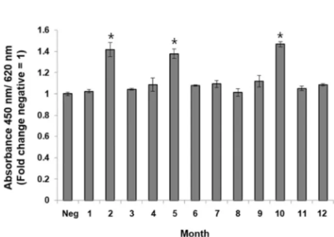
Fig. 5. Application of NPA-SH probes for detection of annual field samples from Tongyeong, Korea. Data are ex- pressed as means ± SE and obtained from triplicate experiments. Neg is a negative control using natural sea- water without phytoplankton. * denotes a significant dif- ference compared with the controls (p<0.05).
Comparison of NPA-SH and microscopic cell counts The results obtained from NPA-SH and microscopic anal- ysis of H. akashiwo cells in natural sea water were compared (Fig. 3). The number of H. akashiwo cells estimated with NPA-SH and the number of cells counted using a micro- scope were similar (Fig. 3). There was no statistically sig- nificant difference (p<0.05) and the mean deviation between the two methods was 5.4%. These results indicate that the substances present in natural seawater do not affect the qual- itative and quantitative NPA-SH detection of H. akashiwo.
NPA-SH Probe testing in field samples
Finally, the NPA-SH detection method was applied to monitor the annual occurrence of H. akashiwo. Each 100 ml of the site samples were collected three times a month in Goseong Bay, Korea (Fig. 4) in 2014 and analyzed using NPA-SH probes capable of detecting H. akashiwo. When the occurrence of microalgae was analyzed for one year, H. aka- shiwo appeared in February, May, and October, and no stat- istically significant (p<0.05) microalgae were observed in the other months (Fig. 5). Based on the standard regression equation, the numbers of H. akashiwo cells in February, May, and October were 2,752, 2,641, and 2,900 cells/ml, respec- tively. Taken together, these results suggest that the NPA- SH probe for H. akashiwo detection in this study is very use- ful for in situ monitoring in marine environments.
Acknowledgements
This research was supported by the Bio & Medical
Technology Development Program of the National Research Foundation (NRF), funded by the Ministry of Science and ICT (MSIT) (NRF-2017M3A9E4072753).
The Conflict of Interest Statement
The authors declare that they have no conflicts of interest with the contents of this article.
References
1. Blanco, E. P., Hagström, J., Salomon, P. S. and Granéli, E.
2013. Detection of Heterosigma akashiwo (Hada) using specific RNA probes: variability of RNA content with environmental conditions. Harmful Algae 24, 80-88.
2. Bowers, H. A., Tomas, C., Tengs, T., Kempton, J. W., Lewitus, A. J. and Oldach, D. W. 2006. Raphidophyte [Chadefaud ex Silva] systematics and rapid identification: sequence anal- yses and real-time PCR assays. J. Phycol. 42, 1333-1348.
3. Cai, Q., Li, R., Zhen, Y., Mi, T. and Yu, Z. 2006. Detection of two Prorocentrum species using sandwich hybridization integrated with nuclease protection assay. Harmful Algae 5, 300-309.
4. Chen, G. F., Wang, G. C., Zhang, C. Y., Zhang, B. Y., Wang, X. K. and Zhou, B. C. 2008. Development of rRNA and rDNA-targeted probes for fluorescence in situ hybridization to detect Heterosigma akashiwo (Raphidophyceae). J. Exp.
Mar. Biol. Ecol. 355, 66-75.
5. Doll, C., Main, C. R., Bianco, C., Coyne, K. J. and Greenfield, D. I. 2014. Comparison of sandwich hybridization assay and quantitative PCR for the quantification of live and preserved cultures of Heterosigma akashiwo (Raphidophyceae). Limnol.
Oceanogr. Meth. 12, 230-243.
6. Ebenezer, V., Medlin, L. K. and Ki, J. S. 2012. Molecular detection, quantification, and diversity evaluation of micro- algae. Mar. Biotechnol. 14, 129-142.
7. Keppler, C. J., Hoguet, J., Smith, K., Ringwood, A. H. and Lewitus, A. J. 2005. Sublethal effects of the toxic alga Heterosigma akashiwo on the southeastern oyster (Crassostrea virginica). Harmful Algae 4, 275-285.
8. Main, C. R., Greenfield, D. I., Doll, C., Wang, Y., Whereat, E. B., Mortensen, R., Pettay, D. T. and Coyne, K. J. 2018.
Critical comparison of molecular methods for detection and enumeration of the harmful algal species, Heterosigma aka- shiwo, in environmental water samples. J. Appl. Phycol. 30, 2425-2434.
9. Park, M., Park, S. Y., Hwang, J., Jung, S. W., Lee, J., Chang, M. and Lee, T. K. 2018. Integration of the nuclease protection assay with sandwich hybridization (NPA-SH) for sensitive detection of Heterocapsa triquetra. Acta Oceano. Sin. 37, 107- 112.
10. Suh, S. S., Park, M., Hwang, J., Kil, E. J., Lee, S. and Lee, T. K. 2015. Detection of the dinoflagellate, Cochlodinium poly- krikoides, that forms algal blooms using sandwich hybrid-
초록:
Heterosigma akashiwo
를 모니터하기 위한 뉴클레아제 보호 분석이 통합된 샌드위치 혼성 (NPA-SH)의 개발강민경1,2․박미례3․김강은1,2․이택견1,2*
(1한국해양과학기술원 생태위해성 연구부, 2과학기술 연합대학원 대학교 해양과학, 3국립낙동강생물자원관 미생물실)
Heterosigma akashiwo는 전세계적으로 분포된 침편모조류이며, 대발생을 형성하여 많은 나라의 양식산업에 커다 란 손실을 유발시킨다. 따라서 빠르고 민감한 검출방법을 개발하는 것은 유해조류 대발생에 대한 적절한 경보를 위해서 필요하다. 이 연구에서는 H. akashiwo를 정성 및 정량적으로 검출하기 위하여 뉴클레아제 보호 분석이 통 합된 샌드위치 혼성(NPA-SH)을 개발하였다. NPA-SH 프로브는 여섯 종의 미세조류 핵산 서열을 이용하여 디자 인 후, 특이성을 확인하여 capture 프로브와 signal 프로브를 선정하였다. 배양시료와 현장시료를 이용하여 NPA-SH의 적용성을 평가한 결과, NPA-SH의 좋은 적용성과 효과를 확인하였다. H.akashiwo의 정량분석을 위한 선형회귀식을 확인하였으며, 최소 검출한계는 1×10⁴ cells/ml이었다. NPA-SH를 사용하여 얻은 H. akashiwo의 정 량결과와 현미경을 사용하여 얻은 결과 사이에는 통계학적으로 유의한 차이는 없었다. NPA-SH 분석은 환경시료 에 잘 적용되었다. 이러한 결과는 NPA-SH가 H. akashiwo의 모니터링에 사용되어 왔던 전통적인 현미경적 방법에 대한 좋은 대안이 될 수 있음을 보여준다.
ization integrated with nuclease protection assay. Biotechnol.
Lett. 38, 57-63.
11. Tyrrell, J. V., Bergquist, P. R., Bergquist, P. L. and Scholin, C. A. 2001. Detection and enumeration of Heterosigma aka- shiwo and Fibrocapsa japonica (Raphidophyceae) using rRNA- targeted oligonucleotide probes. Phycologia 40, 457-467.
12. Tyrrell, J. V., Connell, L. B. and Scholin, C. A. 2002. Monitor- ing for Heterosigma akashiwo using a sandwich hybridization assay. Harmful Algae 1, 205-214.
13. Venugopalan, C. and Kapoor, H. C. 1997. Single step iso- lation of plant RNA. Phytochemistry 46, 1303-1305.
14. Woelfl, S. and Whitton, B. A. 2000. Sampling, preservation and quantification of biological samples from highly acidic environments (pH≤3). Hydrobiologia 433, 173-180.
15. Yu, Z., Tiezhu, M. and Zhigang, Y. 2008. Detection of Phaeo-
cystis globosa using sandwich hybridization integrated with nuclease protection assay (NPA-SH). J. Environ. Sci. 20, 1481- 1486.
16. Zhang, C., Wang, Y., Guo, C., Chen, G., Kan, G., Cai, P.
and Zhou, J. 2018. Comparison of loop-mediated isothermal amplification with hyperbranched rolling circle amplifica- tion as a simple detection method for Heterosigma akashiwo.
Harmful Algae 73, 1-11.
17. Zhen, Y., Mi, T. and Yu, Z. 2009. Detection of several harm- ful algal species by sandwich hybridization integrated with a nuclease protection assay. Harmful Algae 8, 651-657.
18. Zhen, Y., Yu, Z., Cai, Q., Mi, T. and Li, R. 2007. Detection of two diatoms using sandwich hybridization integrated with nuclease protection assay (NPA-SH). Hydrobiologia 575, 1-11.