Change in Lactobacillus brevis GS1022 and Pediococcus inopinatus GS316 in Gajami Sikhae Fermentation
Soo-Jeong Lim1, Eun-Yeong Bae1, Min-Kyeong Seol1, Young-je Cho1, Hee-Young Jung2 and Byung-Oh Kim1*
1School of Food Science, Kyungpook National University, Daegu 41566, Korea
2School of Applied Biosciences, Kyungpook National University, Daegu 41566, Korea Received March 16, 2020 /Revised May 4, 2020 /Accepted May 4, 2020
Lactic acid bacteria are widely known to prevent and treat intestinal health conditions, heart disease, depression, and obesity. In Korea, such bacteria are commonly consumed through various fermented foods, although most are isolated from kimchi, and research on the lactic acid bacteria in fermented seafood is insufficient. This study was therefore conducted to observe changes in bacterial flora ac- cording to the culture date of lactic acid bacteria in the fermentation of traditional Korean Gajami Sikhae produced in Pohang and to isolate the bacteria of probiotic value. The bacteria were periodi- cally isolated and identified from date of preparation to 50 days after preparation to investigate which Lactobacillus are involved in Gajami Sikhae. As fermentation progressed, it was confirmed that Pediococcus sp. and Lactobacillus sp. participate predominantly in the early and later periods of fermen- tation, respectively. During the entire fermentation period, 170 isolates were screened, and the follow- ing five species were found to be involved: Pediococcus pentosaceus, Pediococcus inopinatus, Leuconostoc mesenteroides, Lactobacillus brevis, and Lactobacillus plantarum. Five strains of these species were selected through acid and bile tolerance tests, and their coaggregation, autoaggregation, hydrophobicity, anti- bacterial, and antioxidant activities were then evaluated. As a result, it is thought that L. brevis GS1022, which has excellent digestive fluid resistance, and P. inopinatus GS316, which has excellent cohesiveness, may be useful as probiotic strains.
Key words : Antimicrobial activity, Gajami Sikhae, lactic acid bacteria, probiotics
*Corresponding author
*Tel : +82-53-950-7756, Fax : +82-53-950-6772
*E-mail : [email protected]
This is an Open-Access article distributed under the terms of the Creative Commons Attribution Non-Commercial License (http://creativecommons.org/licenses/by-nc/3.0) which permits unrestricted non-commercial use, distribution, and reproduction in any medium, provided the original work is properly cited.
Introduction
Flounder is widely used as a food item in various prod- ucts, such as dried products, raw fish, and frozen food, be- cause it is abundant in nutrients such as proteins and un- saturated fatty acids [12]. Sikhae is prepared by dehydrating seafood such as squid, flounder, and pollack with salt and fermenting them with grains, red pepper powder, and vegetables. Organic acids produced by lactic acid bacteria (LAB) prohibit decay of the food, and yeast provides a tex- ture and flavor that makes the food suitable for consumption [23]. As fermentation progresses, the fishbone becomes ten- der and the unsavory smell of raw marine products dis- appears.
A previous study assessed the microbiological and phys- icochemical characteristics of flounder [11]. Because its fer- mentation principle and ingredients are similar to those of kimchi, sikhae has similar physiological characteristics. The anticancer effects of LAB, fibrin, as well as the blood pres- sure-lowering effects of γ-aminobutyric acid (GABA), have been previously reported [17]. Lee [16] reported that Weissella sp. isolated from kimchi demonstrated antibacterial and anticancer effects.
According to Guarner and Schaafsma [10], probiotics is a generic term for living organisms that are beneficial for the body when consumed in moderate amounts. Probiotics generally comprise LAB, including some Bacillus sp., as well as Saccharomyces, and have diverse health functions [5].
Probiotics have functions such as relieving lactose intoler- ance and allergic reactions, immunomodulation, decreasing blood cholesterol levels, suppressing colorectal cancer, alle- viating atopic dermatitis, and alleviating Crohn’s disease [24]. However, essential conditions must be met for these probiotics to perform these functions: safety and stability in the human body, possibility of colonization, epithelial cell
adhesion, self-aggregation, and stability in the production process must be ensured [1]. Because LAB are tolerant to low pH conditions and bile acid, they are considered high- value probiotics [13]. LAB are generally recognized as safe and can produce organic acids by metabolizing sugar. LAB also suppress constipation and prevent harmful bacteria in the intestines by inhibiting intestinal decay [29]. Additionally, probiotics have beneficial effects, such as reduction of blood cholesterol levels, promotion of anticancer activities, and en- hancement of immunity, when ingested in appropriate amounts [6]. Because many LAB isolated from kimchi are probiotic, it is expected that LAB isolated from Gajami Sikhae will also be probiotic. The purpose of this study was to investigate the types and dominant species of LAB in- volved in the fermentation of Gajami Sikhae. LAB were iso- lated every five days during the fermentation process, and changes in the microflora were examined. The most basic function of a probiotic is perhaps the ability to survive in the digestive tract, which was evaluated using acid solution, bile acid solution, artificial gastric fluid, and artificial small intestine fluid. Acid tolerance was evaluated by measuring the survival rate of the isolated LAB with different adapta- tion times for each solution. In addition, autoaggregation and coaggregation abilities were evaluated to determine the beneficial effect of intestinal colonization. We also evaluated the function of these bacteria to exclude the formation of harmful bacterial colonies. Antioxidant activities, cell surface hydrophobicity, and antimicrobial activities were evaluated to assess the potential of the isolated bacterial strains as probiotics. Morphological, physiological, and molecular biol- ogy techniques were used to identify strains exhibiting high probiotic activity.
Materials and Methods
Isolation and preservation of LAB
LAB strains were isolated from a home-made Gajami Sikhae sample produced in Pohang, Gyeongsangbuk-do province, South Korea. The sample was stored at 10℃ for 50 days from the day of preparation. To determine the types of LAB involved in the fermentation process, 30 g of sikhae and 30 ml of sterilized, distilled water were mixed, homo- genized, and centrifuged at 3,100× g for 10 min (1248R, GYROZEN Co., Ltd., Korea). The supernatant was diluted and inoculated on Lactobacilli de Man, Rogosa and Sharpe (MRS) (Difco, USA) agar medium. After incubation at 37℃
for 24 hr, colonies were first selected based on shape, color, and size. Each of the selected colonies was separated by sin- gle-colony isolation. The total glycerol concentration was ad- justed to approximately 20%, and samples were stored at -80℃. The isolation of LAB was conducted every 5 days for a total of 50 days. For secondary selection of isolated LAB, MRS agar medium supplemented with 0.1%(w/v) CaCO3
was used, and strains that produced a clear zone of ≥35 mm were selected.
Identification of LAB
Morphological characterization
Prior to molecular biological identification, Gram staining was performed. First, selected LAB were cultured for 12-16 hr in CaCO3-supplemented MRS medium and stained with a Gram staining kit (Difco, USA). The staining results were viewed under a microscope to determine the cell wall struc- ture and shape and size of the strain. (Data not shown.)
Molecular biological identification
16S rDNA sequencing was performed on the initially se- lected strains to identify the change of LAB during Gajami Sikhae fermentation. The phenol–chloroform method [13]
was used to isolate the chromosomal DNA of the strains.
For this, 2 μl of 10× Taq buffer, 1.6 μl of 2.5 mM dNTPs, 1 μl of primers (forward and reverse), 20 ng of template DNA, and 0.2 U Taq polymerase (LPS solution, Korea) were mixed to prepare a reaction mixture for polymerase chain reaction (PCR). After initial denaturation at 95℃ for 5 min, denaturation at 95℃ for 1 min, annealing at 55℃ for 30 s, and extension at 72℃ for 1 min were repeated 35 times, fol- lowed by a final extension at 72℃ for 5 min. Amplification products were identified using 1.5% agarose gel electro- phoresis. The primers used in the PCR reaction were pre- pared according to Petri [20], and are shown in Table 1.
Biochemical identification
Isolated LAB were subcultured on MRS broth, and the availability of 49 carbon sources was confirmed using a Api 50 CHL kit (Biomérieux, France). The Api web program (http://apiweb.biomerieux.com) was used to read standard species and confirm the results.
Evaluation of the probiotic characteristics of the isolated LAB
Evaluation of acid tolerance
Table 1. Primers used in this study
Species Sequence Target size (bp)
L. lactis sp. F
R
TTG CAT GGA ATG AGC GGA AAC
TAT CCT CCC ATT GAT AAA CCA GCG 248
L. brevis sp. F
R
GGA AGA TCA AGA ATA TCG GTG
GCG TCT CTA ATT CAC TGA GC 1361
L. buchneri sp. F
R
CTA TCT TTA ACC GCA TTG CCG
GAC ACG CTT CTC ATG ATT GTC 1007
L. curvatus sp. F
R
CCA GAT CCA TCA GAA GAT ACG
GCT AAC TTA CCA CTA ACG ACC 480
L. hilgardii sp. F
R
TTC CTT GGT AAT GTG CTT GC
AAT GGC AAT CGC AAT GGA CG 684
L. plantarm sp. F
R
GAA GAT TTG CCC ATC GGT G
CGT TTG ATG GTA GCG TTG C 1113
Lc. mesenteroides sp. F
R
GTG GTC ATG GGT CTT AGC
GGA TCA AGA CTA GCC AAT GG 886
O. oeni sp. F
R
GGT AGA TTA ACC CGC GAC G
GGA ATC GGT AGC ATC CTG 1588
P. acidilactici sp. F
R
ATG ATG GAC AGA CTC CCT G
CGA GCT GCG TAG ATA TGT C 776
P. damnosus sp. F
R
GTC TAA ACT GGT GGT TAA ACG
ATC GCA CCT GGT TCA ATG C 470
P. inopinatus sp. F
R
CTA TCC TTA CAA TGT GCA TCG
TGG TGC GTC AGT AAA TGT AAG 567
P. parvurus sp. F
R
GCA TGA ATC ACT TTT CGC TC
CAA AGA TTG TGA CCC AGT TG 331
P. pentosaceus sp. F
R
GGG AAC GGT TTT AGT TTT ATA CG
CTA AGA GCG GTG ATG ATA AG 396
W. paramesenteroides sp. F R
GCT GAT GAA CCC ATA CCT C
GAC CTG ATT CGC TCG TTG 641
To evaluate the acid tolerance of the isolated LAB, the method described by Tokatlı [29] was used. LAB cultured at 37℃ for 24 hr were centrifuged at 6,000× g for 15 min to collect the cells. The cells were washed twice with phos- phate-buffered saline (PBS) and resuspended in PBS, with the adjusted to 2.5. The suspension was incubated at 37℃
for 4 hr, and the suspensions at 0 and 4 hr of culture were diluted with sterilized saline and cultured on MRS agar. The survival rate of LAB was calculated as shown in equation (1).
Evaluation of bile acid tolerance
To evaluate the bile acid tolerance of isolated LAB, the method described by Tokatlı [29] was used. Using the same preparations as those for evaluating acid tolerance, cells were washed twice and inoculated in 1% MRS broth contain- ing 0.3%(w/v) oxgall (Difco, USA). The inoculum was in-
cubated at 37°℃ for 4 hr, and the cultures at 0 and 4 hr were diluted with sterile saline and cultured on MRS agar.
The survival rate of LAB was calculated as shown in equa- tion (1).
%
survival= logCFU of viable cells survited
×100 (1) logCFU of initial viable cells inoculated
Autoaggregation and coaggregation assays
The method described by Tareb [26] was used to inves- tigate the autoaggregation of LAB and coaggregation with pathogenic bacteria. The following indicator strains were used: Candida albicans ATCC 10231, cultured at 28℃-30℃ us- ing Yeast extract Peptone Dextrose medium; Escherichia coli KCTC 2571, cultured at 37℃ using Luria Bertani (LB) me- dium; Helicobacter pylori HPKCTC B0150, cultured at 37℃
using brucella medium containing 10%(v/v) fetal bovine se- rum; Staphylococcus aureus KCTC 1916, cultured at 35℃-40
℃ using LB medium, and Listeria monocytogenes KCTC 13064, cultured at 30℃–37℃ using listeria enrichment medium.
The LAB and pathogens were cultured under optimal cul- ture conditions, washed twice with PBS, and mixed in the same buffer, and the optical density (OD)600 value was ad- justed to 0.3. The OD600 was measured at 0, 1, 2, 3, 4, 5, 20, and 24 hr while each suspension was kept at 25℃.
Autoaggregation (%) was calculated using the equation (1
− ODtime / ODT0) ×100. ODtime is the OD600 value for each hour (0, 1, 2, 3, 4, 5, 20, or 24), and ODT0 is represented by the OD600 value at 0 hr. To investigate coaggregation, LAB and pathogens were cultured and suspended under the same conditions as that used in the autoaggregation test. The OD600 was measured at 0, 1, 2, 3, 4, 5, 20, and 24 hr after mixing the LAB and the pathogen suspension at a ratio of 1:1. The coaggregation (%) was calculated using the equation [(ODPatho + ODLb) / 2 − (ODmix) / (ODPatho + ODLb) ×100], where ODPatho and ODLb were the OD600 values of each strain and ODmix was the value of the mixed-strain culture medium.
The indicator strain C. albicans ATCC 10231 was procured from American Type Culture Collection (ATCC). Escherichia coli KCTC 2571, Listeria monocytogenes KCTC 13064, and Staphylococcus aureus KCTC 1916 were procured from Korean Agricultural Culture Collection. Helicobacter pylori HPKCTC B0150 was procured from the microbiology class of Gyeong- sang National University.
Antimicrobial activity assay
The antimicrobial activities of the isolates were de- termined using the disc diffusion method [4]. The indicator strain was the same as that used in the autoaggregation and coaggregation test. Each indicator strain was cultivated un- der optimal culture conditions and washed twice with PBS, the OD600 value was adjusted to 0.1 using the same buffer, and 1% was inoculated on Mueller Hinton (Difco, USA) agar medium. LAB culture supernatants (60 μl) were dispensed into of sterilized paper discs (8 mm, Toyo Roshi Kaisha, Ltd., Japan) and incubated at 30℃ for 24 hr. Ampicillin and am- photericin B were used as positive controls. The anti- microbial activity was measured by the size of the inhibition zone in mm.
Evaluation of cell surface hydrophobicity
To indirectly confirm the adhesion ability of the LAB, the cell surface hydrophobicity was evaluated using the method described by Doyle [7]. LAB incubated at 37℃ for 18 hr were centrifuged (5,000× g, 15 min) to collect the cells. After wash-
ing twice with PBS, a suspension having OD600 = 1.0 was prepared using PBS. The same quantity of ethyl acetate, chloroform, and xylene was added to the suspension, and the mixture was allowed to stand for 30 min at room temper- ature after vortexing for 1 min. The OD600 value of the sus- pension and the separated layer were measured, and the hy- drophobicity (%) was calculated according to the following formula: hydrophobicity (%) = [(absorbance of sample at 600 nm − absorbance of solvent at 600 nm) / absorbance of sol- vent at 600 nm ×100].
2,2-Diphenyl-1-picrylhydrazyl (DPPH) free radical scavenging ability
The isolated LAB were cultured, and the supernatants were then subjected to centrifugation (3,100× g, 10 min). The supernatants were filtered using a syringe filter (0.45 μm, echromscience, Korea). The DPPH free radical scavenging activity was measured according to the method of Brand- Williams [2]. A 1-ml volume of DPPH solution (200 μM in methanol) was mixed with 800 μl of LAB culture super- natant, and the reaction was terminated at room temperature for 30 min. After the reaction was terminated, the absorbance was measured using a spectrophotometer (Libra S22, Biochrom Ltd., England) at 517 nm. Distilled water was used as a control, and butylated hydroxytoluene (BHT, Sigma Co., USA), a synthetic antioxidant, was used as a positive control.
DPPH radical scavenging activity (%) was calculated accord- ing to the equation [1 − (absorbance of sample at 517 nm / absorbance of solvent at 517 nm) ×100].
Statistical analysis
All values are expressed as mean ± standard deviation.
Data were analyzed using one-way analysis of variance us- ing Statistical Package for Social Science, version 23 (SPSS;
Chicago, USA) for Windows. The differences among groups were assessed using Duncan’s multiple range test. Statistical significance was considered at p<0.05.
Results and Discussion
Isolation and identification of LAB
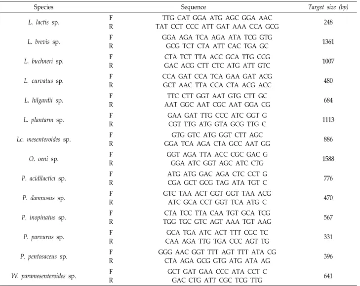
A total of 170 LAB strains were isolated over 50 days to assess the LAB involved in the fermentation of Gajami Sikhae. 16S rDNA analysis showed that P. inopinatus, P. pen- tosaceus, Lc. mesenteroides, L. brevis, and L. plantarum were present in sikhae over the 50 days of fermentation. Pediococ- cus spp. were dominant at the beginning of fermentation
Fig. 1. Changes in lactic acid bacteria during Gajami Sikhae fermentation.
Fig. 2. Survival rates of lactic acid bacteria after 4 hr at a pH of 2.5.
(0-20 days), and Lactobacillus spp. were dominant at the mid- dle stage (21-50 days) (Fig. 1). When LAB were cultured in CaCO3-supplemented medium, 29 strains were selected based on the size of the clear zone. According to a study on the distribution of LAB in kimchi, Lc. mesenteroides, P.
cerevisiae, L. plantarum, and L. brevis were dominant [22], and the distribution of LAB in sikhae showed that Lc. mesenter- oides and L. plantarum were dominant [15]. In the present study, LAB were isolated from the production date to day 50 after production, and the dominant strains were different from those identified in previous studies.
Probiotic activity
Evaluation of acid tolerance
LAB are ingested via the oral cavity and pass through
the stomach, where various enzymes are present. They fi- nally reach the small intestine through the bile duodenum.
Thus, LAB should be resistant to acid to survive at the low pH of the stomach, which contains gastric acid. Acid toler- ance tests showed that of 29 strains selected through acid production, 17 exhibited a survival rate of 20% or more at pH 2.5 (Fig. 2). Eight strains showing a high survival rate of 60% or more were selected, and their tolerance to bile acid was measured.
Evaluation of tolerance to bile acid
LAB must pass through the stomach and are thus exposed to bile secreted from the gallbladder. To survive in the ex- treme intestinal environment, LAB must display bile acid tolerance. Many LAB have a high survival rate in the in-
Fig. 3. Survival rates of lactic acid bacteria after 4 hr in 0.3% (w/v) oxgall.
Table 2. Autoaggregation percentages for probiotic strains of Gajami Sikhae origin after incubation
Probiotic strains % of autoaggregation (25℃)
2 hr 4 hr 20 hr 24 hr
Pathogenic strains C. albicans ATCC 10231 E. coli KCTC 2571 H. pylori HPKCTC B0150 L. monocytogenes KCTC 13064 S. aureus KCTC 1916
6.28±0.17d 3.42±0.37c 3.45±0.36c 2.47±0.28ab 3.39±0.64c
15.39±0.34e 10.82±0.31bc 12.54±0.80cd 10.26±0.35b 12.20±1.92bc
70.08±0.81h 51.10±0.92g 47.61±0.89f 21.73±1.23b 17.41±0.25a
89.43±1.54g 61.61±1.39f 50.27±1.61e 34.46±2.14c 19.07±2.70a Probiotic strains
P. inopinatus GS316 P. pentosaceus GS53 L. plantarum GS86 Lc. mesenteroides GS919 L. brevis GS1022 L. plantarum GS1025
5.87±0.77d 2.09±0.95a 3.45±0.36c 2.47±0.28ab 3.99±0.64c 3.06±0.14bc
14.33±1.66de 6.79±0.71a 11.17±0.76bc 7.73±0.75a 11.05±2.13bc 11.12±0.78bc
47.45±1.41f 20.18±1.27b 31.37±1.59d 24.33±0.83c 31.57±0.67d 33.44±1.36e
58.84±1.49f 26.14±1.22b 37.20±2.31c 28.24±1.07b 37.64±1.95c 41.99±1.74d
(a-h)
Significant differences (p<0.05) among all bacteria strains tested at same time. The data are expressed as the mean ± SD (n=3).
testinal environment, and resistance to bile has been re- ported to be due to a bile acid-degrading enzyme [8].
Gilliland [9] reported that cholesterol-degrading strains have a strong resistance to bile salts and bile acid-degrading activity. Six of the eight strains with high resistance to acid also had a high survival rate of >70% in the 0.3%(w/v) ox- gall-supplemented medium (Fig. 3). Six strains with choles- terol degradation potential were used in a simulated diges- tive fluid tolerance test.
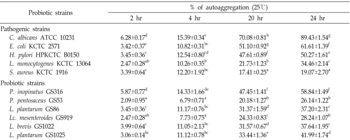
Autoaggregation and coaggregation assays
For the LAB used as probiotics to function continuously in the body, they must have the ability to attach to the in- testinal cells. The autoaggregation ability is a method to in- directly confirm the cell adhesion ability. It is generally un-
derstood that strains with a high aggregation ability also have a high cell adhesion ability. In this study, 11 strains showed a aggregation ability of 19%–89% after 24 hr (Table 2). Among these strains, C. albicans ATCC 10231 showed the most effective autoaggregation ability. Among the LAB, P.
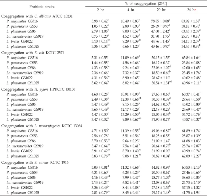
inopinatus GS316 showed a higher autoaggregation rate and better adhesion ability than the other strains. The ability to coaggregate with pathogenic strains prevents the clustering of disease-causing microorganisms. Therefore, LAB strains can prevent infection by competitively binding to pathogenic microorganisms and intestinal cells. The coaggregation abil- ities of five pathogenic strains and six LAB were inves- tigated, and P. inopinatus GS316 showed the highest coag- gregation ability (>60%), outperforming all other experi- mental groups (Table 3). L. plantarum GS86 and L. plantarum
Table 3. Coaggregation of probiotic and pathogenic strains as determined by spectrophotometry
Probiotic strains % of coaggregation (25℃)
2 hr 4 hr 20 hr 24 hr
Coaggregation with C. albicans ATCC 10231 P. inopinatus GS316
P. pentosaceus GS53 L. plantarum GS86 Lc. mesenteroides GS919 L. brevis GS1022 L. plantarum GS1025
3.98 ± 0.42c 1.05 ± 0.22a 2.79 ± 1.06b 0.75 ± 0.20a 3.10 ± 0.14bc 3.36 ± 0.34bc
10.49 ± 0.83e 2.80 ± 0.93a 9.00 ± 0.57d 4.52 ± 0.39b 9.29 ± 0.39de 6.66 ± 1.20c
78.85 ± 0.88e 26.69 ± 0.97a 47.60 ± 2.42d 31.90 ± 1.75b 44.76 ± 1.45c 43.46 ± 0.97c
83.92 ± 1.88e 38.18 ± 0.70c 63.43 ± 2.09d 25.75 ± 0.85a 34.15 ± 2.07b 34.46 ± 0.76b Coaggregation with E. coli KCTC 2571
P. inopinatus GS316 P. pentosaceus GS53 L. plantarum GS86 Lc. mesenteroides GS919 L. brevis GS1022 L. plantarum GS1025
5.31 ± 0.55c 1.44 ± 0.55a 4.33 ± 0.58bc 2.36 ± 0.66a 4.31 ± 0.56bc 3.62 ± 0.64b
11.09 ± 0.69d 4.56 ± 0.66a 9.24 ± 0.60c 7.32 ± 0.37b 8.90 ± 0.65c 8.82 ± 0.64c
50.15 ± 1.53e 16.12 ± 0.32a 32.06 ± 1.02d 18.50 ± 0.60b 28.67 ± 1.10c 30.54 ± 1.31cd
65.84 ± 1.64c 23.84 ± 0.88a 42.00 ± 1.65b 23.45 ± 1.76a 40.02 ± 2.48b 40.96 ± 2.11b Coaggregation with H. pylori HPKCTC B0150
P. inopinatus GS316 P. pentosaceus GS53 L. plantarum GS86 Lc. mesenteroides GS919 L. brevis GS1022 L. plantarum GS1025
4.60 ± 0.26c 2.49 ± 0.36a 3.47 ± 0.49b 3.65 ± 0.49b 4.47 ± 0.30c 3.47 ± 0.32b
10.91 ± 0.90b 12.38 ± 0.66cd 9.15 ± 0.26a 12.17 ± 0.29c 13.29 ± 0.50d 9.89 ± 0.67ab
27.65 ± 0.66c 30.35 ± 0.39d 24.62 ± 0.50b 22.18 ± 0.29a 25.05 ± 0.36b 31.90 ± 0.73e
60.37 ± 0.41f 27.64 ± 0.50b 45.02 ± 0.80e 23.69 ± 0.47a 34.72 ± 0.76c 40.57 ± 0.37d Coaggregation with L. monocytogenes KCTC 13064
P. inopinatus GS316 P. pentosaceus GS53 L. plantarum GS86 Lc. mesenteroides GS919 L. brevis GS1022 L. plantarum GS1025
4.71 ± 1.50b 2.56 ± 0.78a 3.70 ± 0.53ab 3.47 ± 0.64ab 3.91 ± 0.42ab 3.83 ± 0.76ab
11.39 ± 0.55d 5.51 ± 0.56a 9.64 ± 0.23c 7.54 ± 0.41b 8.70 ± 1.40bc 9.08 ± 1.21bc
49.06 ± 0.87d 18.25 ± 0.55a 31.09 ± 1.47c 20.64 ± 0.73b 31.99 ± 0.90c 30.82 ± 0.94c
61.89 ± 1.74c 25.87 ± 1.39a 41.88 ± 1.03b 25.74 ± 2.07a 40.99 ± 0.74b 42.89 ± 2.27b Coaggregation with S. aureus KCTC 1916
P. inopinatus GS316 P. pentosaceus GS53 L. plantarum GS86 Lc. mesenteroides GS919 L. brevis GS1022 L. plantarum GS1025
5.03 ± 0.81d 6.31 ± 0.60e 4.16 ± 0.43cd 2.13 ± 0.24a 3.36 ± 0.49bc 2.81 ± 0.79ab
11.32 ± 0.66c 6.28 ± 0.23a 7.99 ± 0.47b 6.52 ± 0.41a 8.44 ± 0.88b 8.45 ± 0.44b
44.82 ± 0.96c 20.50 ± 0.62a 28.77 ± 1.05b 22.08 ± 0.69a 27.18 ± 1.55b 29.17 ± 1.48b
60.53 ± 2.13d 27.46 ± 0.65a 38.63 ± 0.85b 24.73 ± 2.10a 37.15 ± 1.32b 41.75 ± 1.94c
(a-f)
Significant differences (p<0.05) among all bacteria strains tested at same time. The data are expressed as the mean ± SD (n=3).
GS1025 showed >35% activity in all experimental groups.
Evaluation of cell surface hydrophobicity
High hydrophobicity among the cell surface properties of microorganisms is strongly related to their ability to adhere to the intestinal cell surface [27]. L. rhamnosus GG, one of the representative probiotic LAB, exhibited a hydrophobicity of 53.3% in a study by Todorov [28]. The six LAB selected in this study showed a hydrophobicity of approximately 25% for ethyl acetate, 70% for chloroform, and 95% for xy- lene (Fig. 4). According to Perez [19], bacterial cell surface hydrophobicity >85% becomes beneficial for adhering to epi-
thelial cells. All the six LAB showed high hydrophobicity to xylene at approximately 90%, suggesting that they have high epithelial cell adhesion ability.
Antimicrobial activity assay
According to Reid [21], LAB inhibit pathogenic micro- organisms via various mechanisms, often causing anti- microbial action through metabolites such as lactic acid, hy- drogen peroxide, and bacteriocin. In the present study, the antimicrobial activities of LAB isolated from Gajami Sikhae were measured. To measure the antimicrobial activity ex- hibited by substances other than bacteriocin, the pH of the
Fig. 4. Cellular hydrophobicity of lactic acid bacteria. Data are expressed as the mean ± SD (n=3). Means with different letters (a-e) above the bars for the same strain are significantly different at p<0.05 by Duncan’s multiple range test.
Table 4. Antimicrobial activity of probiotic strains present in Gajami Sikhae
Indicator strain
Inhibition zone (mm) C. albicans
ATCC 10231
E. coli KCTC 2571
H. pylori HPKCTC B0150
L. monocytogenes KCTC 13064
S. aureus KCTC 1916 Probiotic strains
P. inopinatus GS316 P. pentosaceus GS53 L. plantarum GS86 Lc. mesenteroides GS919 L. brevis GS1022 L. plantarum GS1025
ND ND ND ND ND ND
23.1 ± 0.36d 24.4 ± 0.60e 20.7 ± 0.57bc 10.5 ± 0.76a 21.2 ± 0.76c
ND
16.4 ± 0.60c 16.1 ± 0.31bc 16.3 ± 0.70bc 15.3 ± 0.61ab 18.2 ± 0.70d 14.8 ± 0.50a
12.1 ± 0.36a ND 15.9 ± 0.60b
ND 18.8 ± 0.31c
ND
11.1 ± 0.42a 10.7 ± 0.53a 15.9 ± 0.71b 10.3 ± 0.64a 15.9 ± 0.83b
ND Probiotic strains
Ampicillin* Amphotericin B✝
ND 11.9 ± 0.40
19.7 ± 0.25b ND
31.9 ± 0.40e ND
34.0 ± 0.20d ND
32.7 ± 0.35c ND The data are expressed as the mean ± SD (n=3). ND; Not Detected.
Means with differed letters (a-e) with the value in the same pathogenic strains are significantly different at p<0.05 by Duncan’s multiple range test.
*Ampicillin concentration: 10 μg/ml.
✝Amphotericin B concentration: 50 μg/ml.
culture was not controlled. No LAB showed antimicrobial activity against C. albicans ATCC 10231. All strains except Lc. mesenteroides GS919 and L. brevis GS1025 showed higher antimicrobial activity than ampicillin 10 μg/ml against E.
coli KCTC 2571. All LAB showed lower antimicrobial activity against H. pylori HPKCTC B0150, L. monocytogenes KCTC 13604, and S. aureus KCTC 1916 than ampicillin 10 μg/ml (Table 4).
Measurement of DPPH free radical scavenging ability Oxidative stress causes harmful biochemical reactions and various vascular diseases, mutations, cancer, and aging in living organisms [3]. Recently, studies on the antioxidant ac- tivity measurement using natural products have been ac- tively performed. Spices such as herbs, oregano, and cinna- mon have a high antioxidant ability, similar to those of me- dicinal materials [25]. LAB producing superoxide dismutase (SOD), such as L. lactis and L. plantarum, were found to have
Fig. 5. DPPH radical scavenging activity of lactic acid bacteria. Data are expressed as the mean ± SD (n=3). Means with different letters (a-e) above the bars for the same strains are significantly different at p<0.05 by Duncan’s multiple range test.
effective antioxidant activity as well as improved colitis. To investigate the antioxidant activity of isolated LAB, DPPH radical scavenging activity of LAB culture was assessed. All six LAB showed over 75% activity (Fig. 5), demonstrating that all the LAB have antioxidant activity similar to that of BHT 200 μg/ml (77.6%), a synthetic antioxidant.
The Conflict of Interest Statement
The authors declare that they have no conflicts of interest with the contents of this article.
References
1. Bang, J. H., Shin, H. J., Choi, H. J., Kim, D. W., Ahn, C.
S., Jeong, Y. K. and Joo, W. H. 2012. Probiotic potential of Lactobacillus isolates. J. Life Sci. 22, 251-258.
2. Brand-Williams, W., Cuvelier, M. E. and Berset, C. 1995. Use of a free radical method to evaluate antioxidant activity.
Lebensm. Wiss. Technol. 28, 25-30.
3. Castro, L. and Freeman, B. A. 2001. Reactive oxygen species in human health and disease. Nutrition 17, 163-165.
4. Chang, Y. H., Kim, J. K., Kim, H. J., Yoon, J. H., Kim, W.
Y., Choi, Y. W., Kim, Y. B. and Park, Y. H. 1999. Characteris- tics of Lactobacillus reuterii BSA - 131 isolated from swine intestine. Kor. J. Appl. Microbiol. Biotechnol. 27, 23-27.
5. Choi, H. J., Kim, D. W. and Joo, W. H. 2014. Characteristics of Paenibacillus sp. BCNU 5016 as a novel probiotic. J. Life Sci. 24, 161-166.
6. Choi, Y. J., Kim, S. W., Jang, J. K., Choi, Y. J., Park, Y. S., Park, H., Sim, G. S., Lee, H. S. and Chung, M. S. 2009.
Development of fermented functional onion juice using lac- tic acid bacteria. Food Engineering Progress 13, 1-7.
7. Doyle, R. J. and Rosenberg, M. 1995. Measurement of micro- bial adhesion to hydrophobic substrates. Methods Enzymol.
253, 542-550.
8. Gilliland, S. E., Staley, T. E. and Bush, L. J. 1984. Importance of bile tolerance of Lactobacillus acidophilus used as a dietary adjunct. J. Dairy Sci. 67, 3045-3051.
9. Gilliland, S. E. and Walker, D. K. 1990. Factors to consider when selecting a culture of Lactobacillus acidophilus as a diet- ery adjunct to produce a hypocholesterolemic effect in humans. J. Dairy Sci. 73, 905-911.
10. Guarner, F. and Schaafsma, G. J. 1998. Probiotics. Int. J. Food Microbiol. 39, 237-238.
11. Han, D. W., Han, H. J., Kim, D. G., Im, M. J. and Cho, S.
Y. 2013. Quality characterization of commercial flounder Verasper moseri Jordan et Gillberu sikhae. Kor. J. Fish Aquat.
Sci. 46, 696-701.
12. Hwang, S. H. and Youn, K. S. 2008. Stability and quality characteristics of squid liver oil during refining process. Food Eng. Prog. 40, 284-288.
13. Isolauri, E., Salminen, S. and Ouwehand, A. C. 2004.
Microbial-gut interactions in health and diseases. Probiotics.
Best Pract. Res. Clin. Gastroenterol. 18, 299-313.
14. Javadi, A., Shamaei, M., Mohammadi, Z. L., Pourabdollah, M., Dorudinia, A., Seyedmehdi, S. M. and Karimi, S. 2014.
Qualification study of two genomic DNA extraction meth- ods in different clinical samples. Tanaffos 13, 41-47.
15. Lee, C. H. 1997. Lactic acid fermented foods and their bene- fits in asia. Food Control 8, 259-269.
16. Lee, K. H. and Lee, J. H. 2011. Isolation of Leuconostoc and Weissella species inhibiting the growth of lactic acid bacteria sakei from kimchi. Kor. J. Microbiol. Biotechnol. 39, 175-181.
17. Lee, S. H. 2011. Production of γ-aminobutyric acid by lactic acid bacteria brevis GD-16 Isolated from kimchi.
Unpublished master's thesis, Kyungpook National University, Daegu, Korea.
18. Perez, M. K., Paulson, H. L., Pendse, S. J., Saionz, S. J.,
초록:가자미 식해 발효에서
Lactobacillus brevis
GS1022과Pediococcus inopinatus
GS316 의 균총 변화 연구임수정1․배은영1․설민경1․조영제1․정희영2․김병오1*
(1경북대학교 식품공학부, 2경북대학교 응용생명과학부)
유산균은 장 건강, 심장질환, 우울증, 비만 등을 예방하고 치료할 수 있다고 널리 알려져 있고, 특히 한국은 다 양한 발효식품에서 유산균을 섭취할 수 있다. 하지만 국내 발효식품에서 분리된 유산균에 대한 연구는 대부분 김치에 한정되어 있기 때문에 대표적인 어류 전통 발효식품인 식해류의 유산균에 대한 연구는 미비한 실정이다.
따라서 본 연구는 한국의 전통발효식품인 가자미 식해의 발효에 관여하는 유산균의 배양일에 따른 균총의 변화를 관찰하고, 가자미 식해로부터 프로바이오틱스로 이용 가치가 있는 균을 분리하기 위해 시행되었다. 포항의 가정집 에서 제조된 가자미 식해에서 유산균을 제조일로부터 50일까지 5일 간격으로 분리하고 동정하였고, 동정 결과 P. pentosaceus, P. inopinatus, Lc. mesenteroides, L. brevis, L. plantarum 5개 균주가 발효에 관여함을 알 수 있었다.
발효가 진행됨에 따라 초기 발효에는 Pediococcus sp.가, 중기 이후부터는 Lactobacillus sp.가 우세하게 발효에 관여 하는 것을 확인할 수 있었다. 분리한 유산균 중 우수한 프로바이오틱 활성을 가지는 균주를 알아보기 위하여 산 내성, 담즙산 내성 시험을 통해 균주를 선별한 뒤 응집능, 표면 소수성, 항균 활성, 항산화 활성 등을 평가하였다.
그 결과 본 연구의 가자미 식해 분리 균주 중 최종적으로 선택된 소화액 내성이 뛰어난 L. brevis GS1022와 응집능 이 뛰어난 P. inopinatus GS316은 프로바이오틱 균주로 이용할 가치가 충분하다고 사료된다.
Bonini, N. M. and Pittman, R. N. 1998. Recruitment and the role of nuclear localization in polyglutamine-mediated aggregation. J. Cell Biol. 143, 1457-1470.
19. Petri, A., Pfannebecker, J., Frohlich, H. and Konig, H. 2013.
Fast identification of wine related lactic acid bacteria by multiplex PCR. Food Microbiol. 33, 48-54.
20. Reid, G., McGroarty, J. A., Angotti, R. and Cook, R. L. 1988.
Lactobacillus inhibitor production against Escherichia coli and coaggregation ability with uropathogens. Can. J. Microbiol.
34, 344-351.
21. Rhee, S. J., Lee, J. E. and Lee, C. H. 2011. Importance of lactic acid bacteria in asian fermented foods. Microbial. Cell Factories 10, S5.
22. Shin, S. M. 1978 A study on the regional characteristics of Korean chotkal. Kor. J. Diet. Cult. 2, 149-161.
23. Shin, H. J., Choi, H. J., Kim, D. W., Ahn, C. S., Lee, Y. G., Jeong, Y. K. and Joo, W. H. 2012. Probiotic potential of Pedio- coccus pentosaceus BCNU 9070. J. Life Sci. 22, 1194-1200.
24. Su, L., Yin, J. J., Charles, D., Zhou, K., Moore, J. and Yu, L. 2007. Total phenolic contents, chelating capacities, and radical-scavenging properties of black peppercorn, nutmeg, rosehip, cinnamon and oregano leaf. Food Chem. 100, 990-
997.
25. Tareb, R., Bernardeau, M., Guequen, M. and Vernoux, J. P.
2013. In vitro characterization of aggregation and adhesion properties of viable and heat-killed forms of two probiotic Lactobacillus strains and interaction with foodborne zoonotic bacteria, especially Campylobacter jejuni. J. Med. Microbiol. 62, 637-649.
26. Todorov, S. D., Botes, M., Guigas, C., Schillinger, U., Wild, I., Wachsman, M. B., Holzapfel, W. H. and Dicks, L. M. 2008.
Boza, a natural source of probiotic lactic acid bacteria. J.
Appl. Microbiol. 104, 465-477.
27. Todorov, S. D. and Dicks, L. M. T. 2010. Characterization and optimization of bacteriocin from Lactobacillus plantarum isolated from fermented beef. Meat Sci. 84, 334-343.
28. Tokatlı, M., Gülgör, G., Elmacı, S. B., İşleyen, N. A. and Özçelik, F. 2015. In vitro properties of potential probiotic indegenous lactic acid bacteria originating from traditional pickles. Biomed. Res. Int. 2015, 1-8.
29. Yoon, S. S., Park, Y. S. and Choi, H. J. 2013. Genetics and research revolutions in the lactic acid bacteria: focused on probiotics and immunomodulation. Curr. Top. LAB Probiot- ics 1, 1-9.