Activation of Pro-Apoptotic Multidomain Bcl-2 Family Member Bak and Mitochondria-Dependent Caspase Cascade are Involved in p-Coumaric Acid-Induced Apoptosis in Human Jurkat T Cells
Je Won Lee and Young Ho Kim*
Laboratory of Immunobiology, School of Life Science and Biotechnology, College of Natural Sciences, Kyungpook National University, Daegu 702-701, Korea
Received November 11, 2011 /Revised December 8, 2011 /Accepted December 92011
The apoptogenic effect ofp-coumaric acid, a phenolic acid found in various edible plants, on human acute leukemia Jurkat T cells was investigated. Exposure of Jurkat T cells top-coumaric acid (50-150 μM) caused cytotoxicity and TdT-mediated dUTP nick-end labeling (TUNEL)-positive apoptotic DNA fragmentation along with Bak activation, Δψm loss, activation of caspase-9, -3, -7, and -8, and PARP degradation in a dose-dependent manner. However,these apoptotic events were completely abrogated in Jurkat T cells overexpressing Bcl-2.Under these conditions, necrosis was not accompanied.
Pretreatment of the cells with the pan-caspase inhibitor (z-VAD-fmk) could prevent p-coumaric acid-induced sub-G1 peak representing apoptotic cells, whereas it failed to block Δψm loss, indicating that the activation of caspase cascade was prerequisite for p-coumaric acid-induced apoptosis as a downstream event of Δψm loss. FADD- and caspase-8-positive wild-type Jurkat T cell clone A3, FADD-deficient Jurkat T cell clone I2.1, and caspase-8-deficient Jurkat T cell clone I9.2 exhibited sim- ilar susceptibilities to the cytotoxicity ofp-coumaric acid, excluding an involvement of Fas/FasL sys- tem in triggering the apoptosis. The apoptogenic activity ofp-coumaric acid is more potent in malig- nant Jurkat T cells than in normal human peripheral T cells. Together, these results demonstrated that p-coumaric acid-induced apoptogenic activity in Jurkat T cellswas mediated by Bak activation, Δψm loss, and subsequent activation of multiple caspases such as caspase-9, -3, -7, and-8, and PARP degra- dation, which could be regulated by anti-apoptotic protein Bcl-2.
Key words : Apoptosis, cytotoxicity,p-coumaric acid, caspase cascade, leukemia Jurkat T cells, Bcl-2
*Corresponding author
*Tel:+82-53-950-5378, Fax:+82-53-955-5522
*E-mail : [email protected]
Introduction
Apoptosis is the process of programmed cell death that can be triggered by a variety of pathological and physio- logical stimuli. Morphologically, it is characterized by cell shrinkage, chromatin condensation, membrane blebbing, chromosomal DNA fragmentation and forming mem- brane-bound apoptotic bodies [12]. While apoptosis is known to play an important role in the regulation of homeo- stasis in multicellular organism, an imbalance between cell proliferation and apoptotic cell death may cause tumor for- mation [28]. In addition, apoptotic cell death has been pro- posed as an efficient mechanism by which malignant tumor cells can be removed upon treatment with chemotherapeutic drugs, in that the induction of apoptosis in tumor cells re- sults in their own destruction into apoptotic bodies which can be cleared by surrounding cells without accompanying
a local damaging inflammatory response [13]. Diverse cyto- toxic approaches including anticancer drugs, ionizing radia- tion, immunotherapy, and suicide gene therapy, which are currently used for treatment of tumor cells, are known to be predominantly mediated through triggering apoptosis program [17]. However, all these cytotoxic treatments often have significant limitations due to their side effects on nor- mal cells and tissues [13]. In this context, the development of pharmacologically safe anticancer agents, whose apopto- genic activity can be more confined to tumor cells rather than normal cells, has been required.
Hydroxycinnamic acids, including p-coumaric, caffeic, ferulic, and sinapic acids, are a major class of phenolic com- pounds found in edible plants and their products such as cereals, coffee, peanuts, fruits, and vegetables [29,34].
Whereas hydroxycinnamic acids are usually found as esters of either glycosides or organic acid, or are bound to protein and other cell wall polymers, only a small number of them are found as free acids in nature [14]. In addition, hydrox- ycinnamic acids are known to possess a number of im-
portant health benefits [3,4,15,25,33]. In particular, it has been shown thatp-coumaric acid can exert various beneficial effects which include antioxidation [9], antimicrobial activity [36], cancer chemoprevention with inhibiting cancer cell growth [16], and antimelanogenesis [2]. However, little is known about the mechanism responsible for the antitumor activity of p-coumaric acid.
In the present study, we have investigated whether the cytotoxicity ofp-coumaric acid toward human acute leuke- mia Jurkat T cells is attributable to induction of apoptotic cell death. To elucidate the apoptotic mechanism,p-coumaric acid-induced apoptotic events of Jurkat T cells transfected with the vector (JT/Neo) have been compared with those of Jurkat T cells transfected with the Bcl-2 gene (JT/Bcl-2).
The inhibitory effects of the pan-caspase-inhibitor z-VAD- fmk onp-coumaric acid-induced apoptotic cell death have also been analyzed. The results show thatp-coumaric acid induces apoptosis of Jurkat T cells through Bak activation, mitochondrial damages, leading to mitochondrial membrane potential (Δψm) loss and subsequent activation of caspase cascade including caspase-9, -3, -7, and -8, which can be blocked by overexpression of Bcl-2. As compared with ma- lignant Jurkat T cells, normal human T cells appear to be more refractory to the apoptogenic activity of p-coumaric acid.
Materials and Methods Reagents, antibodies, cells, and culture medium
The ECL Western blotting kit was purchased from Amersham (Arlington Heights, IL, USA), and Immobilon-P membrane was obtained from Millipore Corporation (Bedford, MA, USA). Anti-cytochromec was purchased from Pharmingen (San Diego, CA, USA), and anti-caspase-3, an- ti-Bid, anti- poly (ADP-ribose) polymerase (PARP), anti-Bid, ant-Bax, anti-Bcl-2, anti-Bcl-xL, and anti-β-actin were pur- chased from Santa Cruz Biotechnology (Santa Cruz, CA, USA). Anti-caspase-8, and anti-caspase-9, anti-caspase-7, an- ti-Bad, and anti-Bid were from Cell Signaling (Beverly, MA, USA). Anti-Bak (Ab-1) and anti-Bax (6A7) were obtained from Calbiochem (San Diego, CA, USA). p-Coumaric acid, phytohemagglutinin A (PHA), and 3,3'dihexyloxacarbocyanine iodide (DiOC6) were purchased from Sigma (St. Louis, MO, USA). The broad-range caspase inhibitor z-VAD-fmk was obtained from Calbiochem (San Diego, CA, USA). Annexin V-FITC apoptosis kit was purchased from Clontech (Takara
Bio Inc., Shiga, Japan). FADD-positive wild-type Jurkat T cell clone A3, FADD-deficient Jurkat T cell clone I2.1, and ca- pas-8-deficient Jurkat T cell clone I9.2 were purchased from ATCC (Manassas, VA, USA). Jurkat T cell clone transfected with vector (JT/Neo), and Jurkat T cell clone transfected with Bcl-2 gene (JT/Bcl-2) were used in this experiment.
Jurkat T cells and human peripheral T cells were maintained in RPMI 1640 (Hyclone, Gaithersburg, MD, USA) containing 10% fetal bovine serum, 20 mM HEPES (pH 7.2), 5×10⁻⁵M β-mercaptoethanol, and 100 μg/ml gentamycin. For the cul- ture of both JT/Neo and JT/Bcl-2 cells, G418 (A.G. Scientific Inc., San Diego, CA, USA) was added to the RPMI 1640 me- dium at a concentration of 200 μg/ml.
Cytotoxicity assay
The cytotoxic effect ofp-coumaric acid on Jurkat T cell was analyzed by MTT assay reflecting the cell viability as previously described [19]. For MTT assay, Jurkat T cells transfected with vector JT/Neo or Bcl-2 gene (5×10⁴/well) were added to serial dilutions ofp-coumaric acid in 96-well plates. At 63 hr after incubation, 50 μg of MTT solution (1.1 mg/ml) was added to each well and incubated for an addi- tional 4 hr. After centrifugation, the supernatant was re- moved from each well and then 150 μl of DMSO was added to dissolve the colored formazan crystal produced from MTT. OD values of the solutions were measured at 540 nm by a plate reader.
TdT-mediated dUTP nick-end labeling (TUNEL) assay
Jurkat T cells treated with apigeninidin were adhered on- to glass cover slips pretreated with 2% amino- propyltriethoxysilane for 30 min in a humidified chamber as previously described [21]. The cells were then subjected to fluorescence-terminal dUTP nick-end labeling (TUNEL) using an In Situ Cell Death Detection Kit (Roche). Thereafter, the cells were mounted with propidium iodide (PI) on slides to label nuclei and then examined under a confocal laser scanning microscope.
Flow cytometric analysis
Cell cycle progression of Jurkat T cells following exposure top-coumaric acid was analyzed by flow cytometry as de- scribed elsewhere [18]. The extent of necrosis was detected with an Annexin V-FITC apoptosis kit (Clontech, Takara Bio Inc., Shiga, Japan). The cells (5×10⁵) were washed with 1X
binding buffer and then incubated with Annexin V-FITC and propidium iodide (PI) for 15 min before being analyzed by flow cytometry according to the manufacturer's instructions.
p-Coumaric acid-induced alteration in the mitochondrial membrane potential (Δψm) was measured by flow cy- tometry after cells were stained with 50 nM 3,3'dihexyloxacarbocyanine iodide (DiOC6) for 15 min at 37°C as previously described [43]. Activation of Bak and Bax following treatment withp-coumaric acid was measured by flow cytometry as previously described [35]. Briefly, cells (1×10⁶) were washed with PBS and fixed in PBS/1.0% paraf- ormaldehyde on ice for 30 min. Cells were then washed three times in PBS/1% FBS. Staining with con- formation-specific antibodies against Bak (Ab-1) and Bax (6A7) was performed with a proper dilution of individual antibodies in 100 μl staining buffer (PBS, 500 μg/ml dig- itonin). Then, cells were washed and resuspended in 100 μl staining buffer containing Alexafluor 488-labeled goat an- ti-mouse IgG. The conformational changes of Bak and Bax were measured by flow cytometry.
Preparation of cell lysates and Western blot analysis
The cell lysates were prepared by suspending 5×10⁶Jurkat T cell in 250 μl of lysis buffer (137 mM NaCl, 15 mM EGTA, 1 mM sodium orthovanadate, 15 mM MgCl2, 0.1% Triton X-100, 25 mM MOPS, 5.0 μg/ml proteinase inhibitor E-64, and pH 7.2). The cells were disrupted by sonication and ex- tracted at 4oC for 30 min. An equivalent amount of protein lysate (20-30 μg) was denatured with SDS sample buffer, and subjected to electrophoresis on a 10% SDS gradient poly- acrylamide gel with MOPS buffer. The proteins were electro- transferred to Immobilon-P membranes and then probed with individual antibodies. Detection of each protein was carried out with and ECL Western blotting kit according to the manufacturer's instructions.
Isolation and activation of human peripheral T cells
To prepare human peripheral blood mononuclear cells (PBMC), heparinized blood obtained from healthy labo- ratory personnel by venipuncture was centrifuged at 800×
g for 20 min over HISTOPAQUE-1077 (Sigma Chemical, St.
Louis, MO, USA), according to manufacturer's instructions.
This protocol was approved by the Ethics Committee of the Kyungpook National University, Daegu, Korea. Isolation of
T cells from PBMC was performed using a human T cell enrichment column kit (R and D System, Minneapolis, MN, USA). For activation of the peripheral T cells, the isolated peripheral T cells at a density of 2×10⁶/ml were incubated with phytohemagglutinin A (PHA) at a concentration of 1.0 μg/ml for 72 hr. To induce the interleukin-2 (IL-2)-dependent proliferation of T cells, the PHA-activated T cells (1×10⁵/well) were cultured with 50 units (U) of recombinant IL-2 in 96-well plates.
Results and Discussion
Comparison of effect Bcl-2 overexpression on p-coumaric acid-mediated cytotoxicity and apoptotic DNA fragmentation in Jurkat T cells transfected with vector (JT/Neo) and Jurkat T cells transfected with Bcl-2 gene (JT/Bcl-2)
Previous studies have demonstrated that cytochrome c re- lease from mitochondria and subsequent activation of cas- pase cascade are often involved in chemotherapeutic agent-induced apoptotic signaling pathway [27,37]. It has al- so been demonstrated that anti-apoptotic protein Bcl-2 can protect cells from apoptotic cell death induced by diverse stimuli including chemotherapeutic agents, via blocking mi- tochondrial damage which leads to mitochondrial mem- brane potential (Δψm) loss and cytochrome c release [22,42].
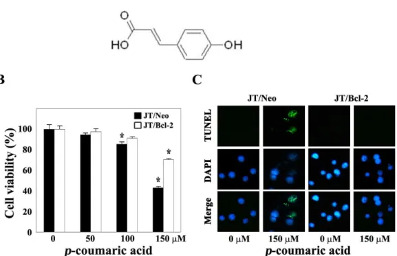
To examine whether apoptotic cell death is involved in the cytotoxicity ofp-coumaric acid, in this context, we decided to compare the cytotoxic effect ofp-coumaric acid on JT/Neo and JT/Bcl-2 cells. When JT/Neo cells were treated with p-coumaric acid (50-150 μM) for 60 hr, the cell viability, which was determined by MTT assay, appeared to decline significantly in a dose-dependent manner (Fig. 1B). Although the viability of JT/Neo cells was not affected upon treatment with 50 μMp-coumaric acid, the cell viabilities declined to the levels of 85.4% and 43.0% following exposure top-cou- maric acid at concentrations of 100 μM and 150 μM, respectively. Under these conditions, however, p-coumaric acid-induced cytotoxicity was significantly reduced in JT/Bcl-2 cells overexpressing Bcl-2, demonstrating sup- pressive effect of Bcl-2 on the cytotoxicity ofp-coumaric acid.
Since current results raised the possibility that the apoptotic cell death, which is sensitive to the anti-apoptotic action of Bcl-2, was mainly attributable to the cytotoxicity ofp-couma- ric acid, we further investigated whether the apoptotic DNA fragmentation was induced in JT/Neo cells following ex-
Fig. 1. Chemical structure ofp-coumaric aicd (A), and effect ofp-coumaric acid on cell viability (B), and DNA fragmentation (C) in Jurkat T cell clone transfected with vector (JT/Neo) and Jurkat T cell clone transfected with Bcl-2 gene (JT/Bcl-2). For cell viability analysis, continuously growing JT/Neo cells or JT/Bcl-2 cells (5×10⁴/well) were incubated with indicated concen- trations ofp-coumaric acid in a 96-well plate for 60 hr and further incubated with MTT for 4 hr. The cells were sequentially processed to assess the colored formazan crystal produced from MTT as an index of cell viability. Each value is expressed as mean±SD (n=3 with three replicates per independent experiment). *p<0.05 compared with control. Equivalent cultures were prepared and cells were harvested to analyze apoptotic DNA fragmentation by TUNEL assay. A representative study is shown and two additional experiments yielded similar results.
posure top-coumaric acid, and whether the induced apop- totic DNA fragmentation was abrogated in JT/Bcl-2 cells overexpressing Bcl-2. As shown in Fig. 1C, whereasp-cou- maric acid-treated JT/Neo cells clearly showed TUNEL-pos- itive nuclei compared to control cells,p-coumaric acid-treat- ed JT/Bcl-2 cells failed to show TUNEL-positive cells. These results indicated that the cytotoxicity ofp-coumaric acid is caused by induced apoptosis, which could be negatively regulated by anti-apoptotic protein Bcl-2, in Jurkat T cells.
Flow cytometric analysis of p-coumaric acid- induced apoptotic cells by either propidium iodide staining or by FITC-conjugated Annexin V staining
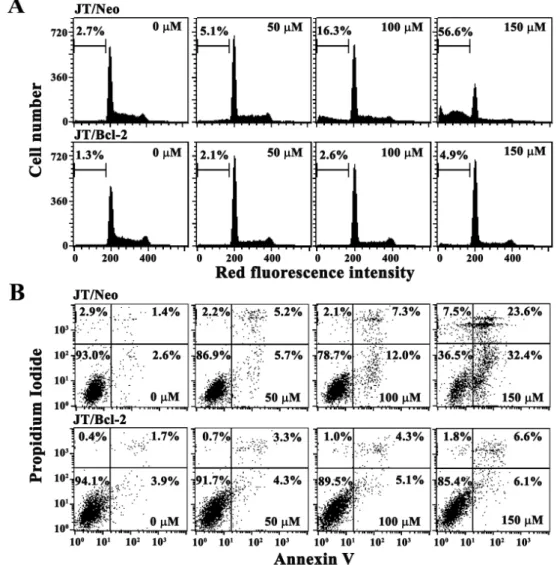
In order to confirm a dose-dependent enhancement in the level of apoptosis by p-coumaric acid in JT/Neo cells and its abrogation in JT/Bcl-2 cells, p-coumaric acid-induced apoptotic sub-G1peak representing apoptotic cells were in- vestigated in JT/Neo and JT/Bcl-2 cells following treatment with p-coumaric acid (50-150 μM) for 60 hr. As shown in
Fig. 2A, JT/Neo cells untreated or JT/Neo cells treated with 50 μMp-coumaric acid exhibited a barely detectable apop- totic sub-G1peak, it increased to the level of 16.3% and 56.6%
in the presence of 100 μM and 150 μM p-coumaric acid, respectively. Under the same conditions,p-coumaric acid-in- duced sub-G1 peak was not detected in JT/Neo cells.
In order to examine whether necrosis was accompanied by p-coumaric acid-induced apoptosis in JT/Neo cells, the cells treated withp-coumaric acid (50-150 μM) for 60 hr were analyzed by Annexin V staining. In accordance with the in- duced apoptotic sub-G1peak, the treatment of JT/Neo cells with 50 μMp-coumaric acid caused no enhancement in the levels of either early apoptotic cells stained only with Annexin V-FITC, or late apoptotic cells stained with both Annexin V-FITC and propidium iodide (PI) (Fig. 2B). Under these conditions, while the apoptotic changes appeared to be more apparent when the cells were treated with 150 μM than with 100 μMp-coumaric acid, the necrotic cells stained only with PI were barely detected. The levels of neither
Fig. 2. Changes in cell cycle distribution (A) and apoptotic cell death (B) in JT/Neo and JT/Bcl-2 cells following exposure to various concentrations ofp-coumaric acid for 60 hr. Both JT/Neo cells and JT/Bcl-2 cells were incubated at a density of 2×10⁵/ml with various concentrations ofp-coumaric acid for 60 hr. The analysis of cell cycle distribution was performed on an equal number of cells (2×10⁴) by flow cytometry after staining of DNA by PI.p-Coumaric acid-induced mitochondrial membrane potential (Δψm) loss was measured after staining of cells with DiOC6, and the apoptotic and necrotic cells were evaluated after staining with Annexin V-PI.
apoptotic nor necrotic cells, however, were enhanced in JT/Bcl-2 cells. Consequently, these results demonstrated that p-coumaric acid (100-150 μM) could induce apoptotic cell death in a dose-dependent manner, and confirmed that the cytotoxic effect exerted byp-coumaric acid on Jurkat T cells was mainly due to apoptosis, but not to necrosis.
Involvement of mitochondrial membrane potential (Δψm) loss and mitochondria-dependent activation of caspase cascade in p-coumaric acid-induced apoptosis
To investigate the death-signaling pathway underlying p-coumaric acid-induced apoptosis, the change in mitochon-
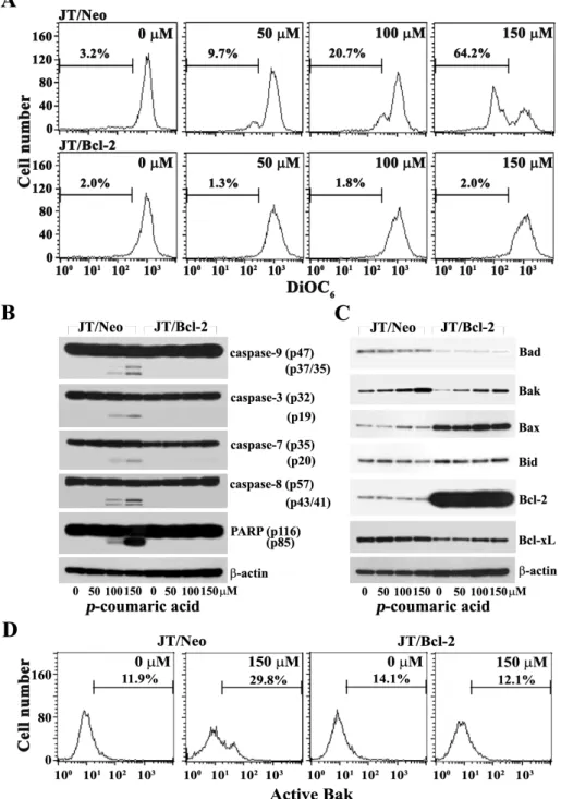
drial membrane potential (Δψm) of JT/Neo cells and JT/Bcl-2 cells following exposure top-coumaric acid (0, 50, 100, and 150 μM) was measured by flow cytometry using 3,3'dihexyloxacarbocyanine iodide (DiOC6) staining.
Although there was barely detectable Δψm loss in con- tinuously growing JT/Neo cells as well as JT/Neo cells treat- ed with 50 μM p-coumaric acid, 20.7% and 64.2% of the cells exhibited Δψm loss in the presence of 100 μM and 150 μM p-coumaric acid, respectively (Fig. 3A). This demonstrated thatp-coumaric acid (100-150 μM) could disrupt Δψm in a dose-dependent manner. At the same time, however,p-cou- maric acid failed to disrupt Δψm in JT/Bcl-2 cells. Since Δψ m loss is known to be one of the initial intracellular changes
Fig. 3. Change in mitochondrial membrane potential (Δψm) (A), Western blot analysis of activation of caspase-9, -3, -7, and -8, cleavage of PARP and β-actin (B), and pro-apoptotic Bcl-2 family members (Bad, Bak, Bax and Bid), anti-apoptotic Bcl-2 family members (Bcl-2 and Bcl-xL) and β-actin (C), and flow cytometric analysis of Bak activation (D) in JT/Neo and JT/Bcl-2 cells after treatment with p-coumaric acid. Both JT/Neo and JT/Bcl-2 cells were incubated at a density of 2×10⁵/ml with indicated concentrations of p-coumaric acid for 60 hr, and prepared for the cell lysates. Equivalent amounts of cell lysates were electrophoresed on 4-12% SDS gradient polyacrylamide gels and electrotransferred to Immobilon-P membrane. Western blot analysis and flow cytometric analysis of Bak activation were performed. A representative study is shown and two additional experiments yielded similar results.
that are accompanied by apoptotic cell death [40,41], these results suggested that Δψm disruption was associated with p-coumaric acid-induced apoptosis in JT/Neo cells. These re-
sults also indicated that the Δψm loss was mediated by a conserved apoptogenic mechanism, which could be targeted by the anti-apoptotic role of Bcl-2.
Since several studies have reported that Δψm loss pre- cedes the release of cytochrome c release into the cytosol, leading to activation of capase-9 and -3 [23,24,39], it was like- ly that Δψm loss and subsequent induction of mitochon- dria-dependent caspase cascade activation, which could be blocked by overexpression of Bcl-2, might play an essential role inp-coumaric acid-induced apoptosis of Jurkat T cells.
To test this prediction, it was investigated in both JT/Neo and JT/Bcl-2 cells, by Western blot analysis, whether the in- duced apoptosis by p-coumaric acid was accompanied by mitochondrial cytochrome c release and subsequent activa- tion of caspase cascade. In accordance with the Δψm loss, the caspase-9 activation that proceeded through proteolytic cleavage of inactive proenzyme (47 kDa) to active forms (37/35 kDa) was detected (Fig. 3B). The cleavage of procas- pase-3 (32 kDa) into active form (17 kDa) as well as the cleavage of procaspase-7 (35 kDa) into active form (20 kDa) was also detected. The activation of caspase-8 through pro- teolytic cleavage of proenzyme (57 kDa) into active forms (43/41 kDa) was significantly enhanced. In JT/Neo cells af- ter treatment with p-coumaric acid, the PARP degradation was detected along with the activation of caspase-3.
However, these apoptotic events were completely abrogated in JT/Bcl-2 cells.
Previously, it has been reported that the pro-apoptotic multidomain Bcl-2 family members (Bax and Bak) mediate permeabilization of the mitochondrial outer membrane (MOM), whereas anti-apoptotic Bcl-2 family members (Bcl-2, Bcl-xL and Mcl-1) prevent cytochrome c efflux triggered by Bak or Bax via either directly or inactivating the BH3-only pro-apoptotic Bcl-2 family members (Bad, Bid, Bim and Puma) [5,8]. It has also been shown that alteration in the expression ratio of Bak to Bcl-2 and/or Bax to Bcl-2, resulting in an enhancement in the ratio of Bak to Bcl-2 and/or Bax to Bcl-2, is often required for provoking the activation of Bak and/or Bax during the mitochondria damage-mediated apoptotic cell death induced by chemotherapeutic agents [1,5,7,11]. To examine the upstream pro-apoptotic events that mediatep-coumaric acid-induced Δψm loss, the expression levels of Bcl-2 family proteins, such as the pro-apoptotic Bcl-2 family members (Bad, Bak, Bax and Bid) and the an- ti-apoptotic Bcl-2 family members (Bcl-2 and Bcl-xL), were compared by Western blot analysis between JT/Neo and JT/Bcl-2 cells after treatment with p-coumaric acid. As shown in Fig. 3C, the expression level of pro-apoptotic mul-
tidomain Bcl-2 family member Bak was enhanced in JT/Neo and JT/Bcl-2 cells following exposure to p-coumaric acid, whereas those of Bad, Bax, Bcl-2, and Bcl-xL remained con- stant in both cells. In addition, the level of Bid protein (22 kDa), which was previously cleaved by active caspase-8 to generate the truncated Bid (tBid, 15 kDa) causing Δψm loss [26], appeared to slightly decline in JT/Neo cells treated with 150 μMp-coumaric acid, whereas this apoptotic change was completely abrogated in JT/Bcl-2 cells. These results suggested that the upregulation of in the level of Bak might be associated with p-coumaric acid-mediated disruption of Δψm loss, possibly via causing the activation of Bak. In order to confirm that thep-coumaric acid-induced apoptosis is ac- companied by the activation of pro-apoptotic multidomain Bcl-2 family members (Bak and Bax), which is known to be upstream of Δψm loss [5-7], the activation of Bak and Bax in JT/Neo and JT/Bcl-2 cells treated with 150 μMp-coumaric acid was analyzed by flow cytometry using the con- formation-specific anti-Bak (Ab-1) or anti-Bax (6A7). As shown in Fig. 3D, the activation of Bak was detected in JT/Neo cells, but not in JT/Bcl-2 cells overexpressing Bcl-2.
Under the same conditions, the activation of Bax was not observed in JT/Neo and JT/Bcl-2 cells (data not shown). In order to examine whether the activation of caspase cascade was critical forp-coumaric acid-induced apoptosis, we inves- tigated the effect of the pan-caspase inhibitor (z-VAD-fmk) [38] onp-coumaric acid-induced apoptotic events in JT/Neo cells. After JT/Neo cells were pretreated with z-VAD-fmk for 2 hr, the cells were exposed to 150 μMp-coumaric acid for 48 hr. Although the apoptotic sub-G1 peak was barely detectable in continuously growing JT/Neo cells, it in- creased to the level of 28.6% in the presence of 150 μMp-cou- maric acid for 48 hr (Fig. 4A). Thep-coumaric acid-induced sub-G1peak was diminished to the basal level by pretreat- ment with z-VAD-fmk, whereas the p-coumaric acid-in- duced Δψm loss was not abrogated by z-VAD-fmk (Fig. 4B).
These results demonstrated that the Δψm loss was an up- stream event of the caspase cascade activation which was a prerequisite for p-coumaric acid-induced apoptotic cell death. Consequently, current results indicated thatp-couma- ric acid-induced apoptosis was mediated by Bak activation, Δψm loss, and subsequent activation of multiple caspases including caspase-9, -3, -7 and -8, leading to PARP degrada- tion, which could be blocked by Bcl-2.
Fig. 4. Apoptotic change in cell cycle distribution (A) and Δψm loss (B) in Jurkat T cells (JT/Neo) after treatment withp-coumaric acid (150 μM) in the presence of the pan-caspase inhibitor z-VAD-fmk at concentration of 30 μM and 50 μM. JT/Neo cells (2×10⁵/ml) was pretreated with z-VAD-fmk for 2 hr and then treated with 150 μMp-coumaric acid for 48 hr. The analysis of cell cycle distribution was performed on an equal number of cells (2×10⁴) by flow cytometry after staining of DNA by propidium iodide. For the analysis of Δψm loss, the cells (~5×10⁵cells) were stained with DiOC6for 10 min at 37 °C. The percentage of red and green fluorescence was estimated by flow cytometry.
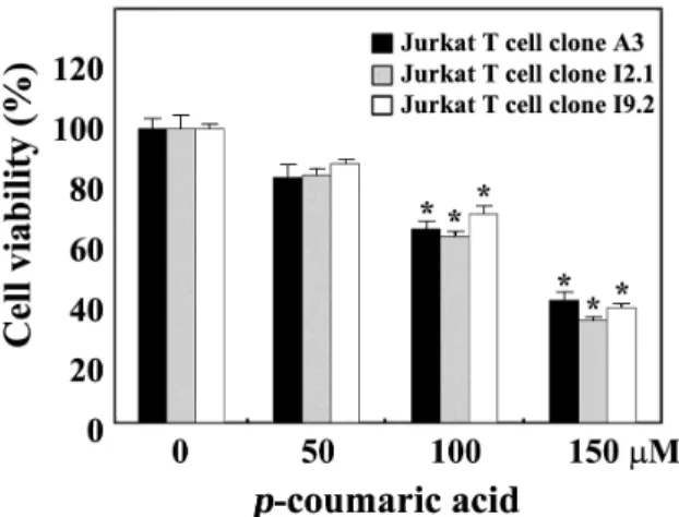
Comparison of cytotoxic effect of p-coumaric acid on FADD- and caspase-8 positive wild-type Jurkat T cell clone A3, FADD-deficient Jurkat T cell clone I2.1, and caspase-8-deficient Jurkat T cell clone I9.2
As a potential mechanism underlying the apoptosis pro- voked by antineoplastic drugs, upregulation of FasL and/or Fas expression has been implicated [10,30,31]. In order to further examine an involvement of Fas/FasL system in p-coumaric acid-induced apoptosis, we compared cytotoxic effect ofp-coumaric acid on FADD- and caspase-8-positive wild-type Jurkat T cells (clone A3) with that on FADD-defi- cient Jurkat T cells (clone I2.1) and caspase-8-deficient Jurkat T cells (clone I9.2), both of which were previously refractory to Fas-mediated apoptosis [20]. As shown in Fig. 5, irre- spective of the FADD deficiency, these Jurkat T cell clones exhibited essentially similar sensitivity to the cytotoxicity of p-coumaric acid. These results confirmed that the p-coumaric acid-induced apoptosis of Jurkat T cells was not provoked by the interaction of Fas with FasL. In addition, these results exclude the possibility that the p-coumaric acid-induced apoptosis of Jurkat T cells was initiated by endoplasmic re-
Fig. 5. Effect of p-coumaric acid on cell viability in wild-type Jurkat T cells (clone A3), FADD-deficient Jurkat T cells (clone I2.1) and caspase-8-deficient Jurkat T cells (clone I9.2). A3 cells, I2.1 cells, or I9.2 cells were incubated at a density 5×10⁴ cells per well with indicated concen- trations ofp-coumaric acid in a 96-well plate for 60 hr and further incubated with MTT for 4 hr with MTT to assess the cell viability. Each value is expressed as mean±SD (n=3 with three replicates per independent ex- periment). *p<0.05 compared to control.
ticulum (ER) stress-mediated activation of caspase-8 and re- sultant cleavage of Bid into tBid.
Cytotoxic effect of p-coumaric acid on human peripheral T cells
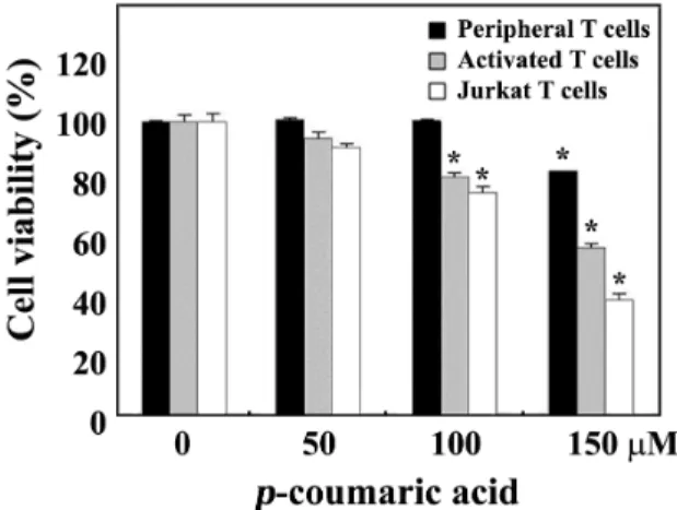
Sincep-coumaric acid possessed cytotoxicity toward ma- lignant Jurkat T cells, it was of interest to investigate wheth- er the drug is less toxic to normal T cells. In this context, we have investigated the cytotoxic effects ofp-coumaric acid on the viability of human resting peripheral T cells or the interleukin-2 (IL-2)-dependent proliferation of activated T cells, which were obtained by the stimulation of human pe- ripheral T cells with 1.0 μg/ml phytohemagglutinin A (PHA) for 72 hr. When the individual cells were incubated with various concentrations ofp-coumaric acid in a 96-well plate for 60 hr and then cell viability was measured by the MTT assay, the viability of unstimulated peripheral T cells as well as the IL-2-dependent proliferation of activated T cells was not markedly affected in the presence of 50-150 μMp-couma- ric acid (Fig. 6). However, the IL-2-dependent proliferation of activated T cells was more sensitive to the cytotoxicity ofp-coumaric acid than resting T cells and exhibited a via- bility of 56.8% at a concentration of 150 μM. Under these conditions, the viability of Jurkat T cells (clone A3) was re- duced to the level of 99.0%, 88.1%, and 37.3% at concen- trations of 50 μM, 100 μM, and 150 μM p-coumaric acid, respectively. These results indicated that malignant leuke- mia Jurkat T cells, as compared to normal T cells, were more sensitive to the apoptogenic activity of p-coumaric acid.
In conclusion, these results demonstrate thatp-coumaric acid, a naturally occurring phenolic acid found in a variety of edible plants including cereals, coffee, fruits, and vegeta- bles, induces apoptotic DNA fragmentation of human acute T cell leukemia Jurkat cells via Bak activation, mitochondrial membrane potential (Δψm) loss, activation of caspase-9, -3, -7, and -8, and resultant cleavage of PARP. Involvement of the extrinsic apoptotic pathway that is triggered by Fas/FasL system in p-coumaric acid-induced apoptosis can be ex- cluded, since the sensitivity of wild-type Jurkat T cell clone A3 to the cytotoxicity ofp-coumaric acid is similar to that of FADD-deficient Jurkat T cell clone I2.1 or caspase-8-defi- cient Jurkat T cells clone I9.2. Current results also indicate that the apoptogenic activity ofp-coumaric acid is more po- tent in malignant Jurkat T cells than in normal human periph- eral T cells. These findings will extend our understanding on the potency of p-coumaric acid as an antitumor agent.
Fig. 6. Effect of p-coumaric acid on human peripheral T cells unstimulated, IL-2-dependent proliferation of PHA-acti- vated T cells, and proliferation of Jurkat T cells.
Peripheral T cells (2×10⁵cells/well) were incubated with various concentrations ofp-coumaric acid for 60 hr, and the cells were further incubated with MTT for 4 hr to assess the cell viability. To induce IL-2-dependent pro- liferation of activated T cells, human peripheral mono- nuclear cells were activated with PHA (1.0 μg/ml) for 72 hr, and then the activated T cells were harvested and incubated with various concentrations of p-coumaric acid at a density of 1×10⁵/well as well as 50 U/ml of recombinant human IL-2 in 96-well plates. For treatment of Jurkat T cell clone A3 withp-coumaric acid, the cell density was 5×10⁴/well. Each value is expressed as mean±SD (n=3 with three replicates per independent ex- periment). *p<0.05 compared to control.
Acknowledgements
The authors thank Dr. Dennis Taub (Gerontology Research Center, NIA/NIH) for providing Jurkat T cell clones JT/Neo and JT/Bcl-2. This work was carried out with the support of “Cooperative Research Program for Agriculture Science & Technology Development (Project No.
PJ006638)” Rural Development Administration, Republic of Korea.
References
1. Adams, J. M. and S. Cory. 2007. Bcl-2-regulated apoptosis:
mechanism and therapeutic potential.Curr. Opin. Immunol.
19, 488-496.
2. An, S. M., S. I. Lee, S. W. Choi, S. W. Moon, and Y. C.
Boo. 2008. p-Coumaric acid, a constituent of Sasa quel- paertensisNakai, inhibits cellular melanogenesis stimulated by α-melanocyte stimulating hormone.Br. J. Dermatol.159, 292-299.
3. Chan, R. I., R. H. San, and H. F. Stich. 1986. Mechanism
of inhibition of N-methyl-N'-nitrosoguanidine-induced mu- tagenesis by phenolic compounds. Cancer Lett. 31, 27-34.
4. Chen, J. H., Y. Shao, M. T. Huang, C. K. Chin, and C. T.
Ho. 1996. Inhibitory effect of caffeic acid phenethyl ester on human leukemia HL-60 cells. Cancer Lett.108, 211-214.
5. Chipuk, J. E. and D. R. Green. 2008. How do BCL-2 proteins induce mitochondrial outer membrane permeabilization?
Trends Cell Biol. 18, 157-164.
6. Chipuk, J. E., L. Bouchier-Hayes, and D. R. Green. 2006.
Mitochondrial outer membrane permeabilization during apoptosis: the innocent bystander scenario.Cell Death Differ. 13, 1396-1402.
7. Chipuk, J. E., T. Moldoveanu, F. Llambi, M. J. Parsons, and D. R. Green. 2010. The BCL-2 family reunion. Mol. Cell37, 299-310.
8. Czabotar, P. E., P. M. Colman, and D. C. Huang. 2009. Bax activation by Bim? Cell Death Differ. 16, 1187-1191.
9. Ferguson, L. R., S. T. Zhu, and P. J. Harris. 2005. Antioxidant and antigenotoxic effects of plant cell wall hydroxycinnamic acids in cultured HT-29 cells. Mol. Nutr. Food Res. 49, 585-693.
10. Friesen, C., I. Herr, P. H. Krammer, and K. M. Debatin. 1996.
Involvement of the CD95 (APO-1/FAS) receptor/ligand system in drug-induced apoptosis in leukemia cells. Nat.
Med. 2, 574-578.
11. Gross, A., J. M. McDonnell, and S. J. Korsmeyer. 1999. BCL-2 family members and the mitochondria in apoptosis.Genes Dev. 13, 1899-1911.
12. Hacker, G. 2000. The morphology of apoptosis.Cell Tissue Res. 301, 5-17.
13. Hannun, Y. A. 1997. Apoptosis and the Dilemma of cancer chemotherapy. Blood89, 1845-1853.
14. Herrmann, K. 1989. Occurrence and content of hydroxycin- namic and hydroxybenzoic acid compounds in foods.Cri.
Rev. Food Sci. Nutr. 28, 315-347.
15. Huang, M. T., R. C. Smart, C. Q. Wong, and A. H. Conney.
1988. Inhibitory effect of curcumin, chlorogenic acid, caffeic acid, and ferulic acid on tumor promotion in mouse skin by 12-O-tetradecanoylphorbol-13-acetate. Cancer Res. 48, 5941-5946.
16. Hudson, E. A., P. A. Dinh, T. Kokubun, M. S. Simmonds, and A. Gescher. 2000. Characterization of potentially chemo- preventive phenols in extracts of brown rice that inhibit the growth of human breast and colon cancer cells. Cancer Epidemiol. Biomarkers Prev. 9, 1163-1170.
17. Johnstone, R. W, A. A. Ruefli, and S. W. Lowe. 2002.
Apoptosis: A link between cancer genetics and chemotherapy. Cell 108, 153-164.
18. Jun, D. Y., H. S. Park, J. S. Kim, J. S. Kim, W. Park, B. H.
Song, H. S. Kim, D. Taub, and Y. H. Kim. 2008. 17α-Estradiol arrests cell cycle progression at G2/M and induces apoptotic cell death in human acute leukemia Jurkat T cells.Toxicol.
Appl. Pharmacol. 231, 401-412.
19. Jun, D. Y, J. S. Kim, H. S. Park, C. R. Han, Z. Fang, M.
H. Woo, I. K. Rhee, and Y. H. Kim. 2007. Apoptogenic activ- ity of auraptene ofZanthoxylum schinifoliumtoward human
acute leukemia Jurkat T cells is associated with ER stress-mediated caspase-8 activation that stimulates mi- tochondria-dependent or -independent caspase cascade.
Carcinogenesis 28, 1303-1313.
20. Juo, P., S. Woo, C. J. Kuo, P. Signorelli, H. P. Biemann, Y.
A. Hannun, and J. Blenis. 1999. FADD is required for multi- ple signaling events downstream of the receptor Fas. Cell Growth Differ. 10, 797-804.
21. Kim, Y. H., J. J. Proust, M. J. Buchholz, F. J. Chrest, and A. A. Nordin. 1992. Expression of the murine homologue of the cell cycle control protein p34cdc2 in T lymphocytes.
J. Immunol. 149, 17-23.
22. Kluck, R. M., E. Bossy-Wetzel, D. R. Green, and D. D.
Newmeyer. 1997. The release of cytochrome c from mi- tochondria: a primary site for Bcl-2 regulation of apoptosis.
Science275, 1132-1136.
23. Kroemer, G., and J. C. Reed. 2000. Mitochondrial control of cell death. Nat. Med. 6, 513–519.
24. Kroemer, G., L. Galluzzi, and C. Brenner. 2007. Mitochondrial membrane permeabilization in cell death. Physiol. Rev.87, 99-163.
25. Laranjinha, J, O. Vierira, L. Almeida, and V. Madeira. 1996.
Inhibition of metmyoglobin/H2O2- dependent low density lipoprotein lipid peroxidation by naturally occurring phe- nolic acids. Biochem. Pharmacol.51, 395-402.
26. Li, H., H. Zhu, C. Xu, and J. Yuan. 1998. Cleavage of Bid by caspase 8 mediates the mitochondrial damage in the Fas pathway of apoptosis. Cell 94, 491-501.
27. Li, P., D. Nijhawan, I. Budihardjo, S. M. Srinivasula, M.
Ahmad, E. S. Alnemri, and X. Wang. 1997. Cytochrome c and dATP-dependent formation of Apaf-1/caspase-9 com- plex initiates an apoptotic protease cascade.Cell91, 479-489.
28. Lowe, S. W. and A. W. Lin. 2000. Apoptosis in cancer.
Carcinogenesis 21, 485-495.
29. Mathew, S. and T. E. Abraham. 2004. Ferulic acid: an anti- oxidant found naturally in plant cell walls and feruloyl es- terases involved in its release and their applications. Cri.
Rev. Biotechnol. 24, 59-83.
30. Muller, M., S. Strand, H. Hug, E. M. Heinemann, H.
Walczak, W. J. Hofmann, W. Stremmel, P. H. Krammer, and P. R. Galle. 1997. Drug-induced apoptotsis in hepatoma cells is mediated by the CD95 (APO-1/Fas) receptor/ligand sys- tem and involves activation of wild-type p53. J. Clin. Invest.
99, 403-413.
31. Nagarkatti, N. and B. A. Davis. 2003. Tamoxifen induces apoptosis in Fas+tumor cells by upregulating the expression of Fas ligand. Cancer Chemother. Pharmacol. 51, 284-290.
33. Nardini, M., M. D’Aquino, G. Tomassi, V. Gentili, M. Di Felice, and C. Scaccini. 1995. Inhibition of human low-den- sity-lipoprotein oxidation by caffeic acid and other hydrox- ycinnamic acid derivatives.Free Radic. Biol. Med.19, 541-552.
34. Nystrom, L., M. Makinen, A. M. Lampi, and V. Piironen.
2005. Antioxidant activity of steryl ferulate extracts from rye and wheat bran. J. Agric. Food Chem. 53, 2503-2510.
35. Park, H. S., D. Y. Jun, C. R. Han, H. J. Woo, and Y. H.
Kim. 2011. Proteasome inhibitor MG132-induced apoptosis
초록:p-Coumaric acid에 의해 유도되는 인체 Jurkat T 세포의 에폽토시스 기전 이제원․김영호*
(경북대학교 자연과학대학 생명과학부)
다양한 식용식물에 함유되어 있는 것으로 알려진 phenolic acids의 일종인p-coumaric acid의 항암활성을 규명 하고자, 인체 급성백혈병 T 세포주인 Jurkat T 세포에 대한 p-coumaric acid의 에폽토시스 유도기전을 조사하였 다. Jurkat T 세포를 p-coumaric acid (50-150 μM)로 처리한 결과, 세포독성, 에폽토시스-관련 DNA fragmenta- tion, 및 pro-apoptotic multidomain Bcl-2 family member인 Bak의 활성화, Δψm loss, caspase-9, -3, -7, 및 -8의 활성화, 그리고 PARP 분해 등의 여러 에폽토시스-관련 생화학적 현상들이 농도의존적으로 나타났다. 그러나 이 러한 에폽토시스-관련 생화학적 현상들은 Jurkat T 세포에 anti-apoptotic Bcl-2 단백질을 과발현할 경우에는 나타 나지 않았다. 또한 p-coumaric acid처리에 의해 유도되는 Jurkat T 세포의 에폽토시스에는 necrosis가 수반되지 않는 것으로 확인되었다. Jurkat T 세포를 pan-caspase inhibitor인 z-VAD-fmk를 전처리할 경우,p-coumaric acid 처리에 의해 유도되는 apoptotic sub-G1peak는 차단되어 나타나지 않았으나 Δψm loss는 여전히 나타났는데, 이 는 p-coumaric acid처리에 의한 에폽토시스의 유도에 caspase cascade 활성화가 필수적이며Δψm loss의 down- stream 현상임을 나타낸다. 한편, FADD 및caspase-8을 함께 발현하는 Jurkat T 세포주 A3, FADD-결손 Jurkat T 세포주 I2.1, 그리고 caspase-8-결손 Jurkat T 세포주 I9.2의 p-coumaric acid의 세포독성에 대한 감수성은 서로
유사하게 나타났는데, 이는 p-coumaric acid처리에 의한 에폽토시스의 유도가 Fas와 FasL간의 상호작용에 의해
개시되지 않음을 시사한다.p-Coumaric acid의 세포독성은 Jurkat T 세포에 비해 인체 정상 말초혈액 T 세포에서 훨씬 낮게 나타났다. 이러한 결과들은p-coumaric acid 처리에 의해 유도되는 Jurkat T 세포의 에폽토시스가 Bak 활성화, Δψm loss, caspase-9, -3, -7, 및 -8로 이루어진 caspase cascade의 활성화, 그리고 PARP 분해에 의해 유도 되며, 또한 anti-apoptotic 단백질인 Bcl-2의 과발현에 의해서 음성적으로 조절됨을 나타낸다.
via ER stress-mediated apoptotic pathway and its potentia- tion by protein tyrosine kinase p56lck in human Jurkat T cells. Biochem. Pharmacol. 82, 1110-1125.
36. Park, S. K. and J. C. Park. 1994. Antimicrobial activity of extracts and coumaric acid isolated fromArtemisia princeps var. orientalis. Kor. J. Biotechnol. Bioeng. 5, 506-511.
37. Saleh, A., S. M. Srinivasula, S. Acharya, R. Fishel, and E.
S. Alnemri. 1999. Cytochrome c and dATP-mediated oligo- merization of Apaf-1 is a prerequisite for procaspase-9 activation. J. Biol. Chem.274, 17941-17945.
38. Slee, E. A., H. Zhu, S. C. Chow, M. MacFarlane, D. W.
Nicholson, and G. M. Cohen. 1996. Benzyloxycarbonyl- Val-Ala-Asp (OMe) fluoromethylketone (z-VAD-fmk) in- hibits apoptosis by blocking the processing CPP32.Biochem.
J. 315, 21-24.
39. Tada-Oikawa, S., S. Oikawa, and S. Kawanishi. 1998. Role of ultraviolet A-induced oxidative DNA damage in apopto- sis via loss of mitochondrial membrane potential and cas- pase-3 activation, Biochem. Biophys. Res. Commun. 247, 693
-696.
40. Tait, S. W. and D. R. Green. 2010. Mitochondria and cell death: outer membrane permeabilization and beyond.Nat.
Rev. Mol. Cell Biol. 11, 621-632.
41. Waterhouse, N. J., J. C. Goldstein, O. von Ahsen, M. Schuler, D. D. Newmeyer, and D. R. Green. 2001. Cytochrome c maintains mitochondrial transmembrane potential and ATP generation after outer mitochondrial membrane per- meabilization during the apoptotic process. J. Cell Biol.153, 319-328.
42. Yang, J., X. Liu, K. Bhalla, C. N. Kim, A. M. Ibrado, J. Cai, T. I. Peng, D. P. Jones, and X. Wang. 1997. Prevention of apoptosis by Bcl-2: release of cytochrome c from mitochon- dria blocked. Science275, 1129-1132.
43. Zamzami, N., P. Marchetti, M. Castedo, C. Zanin, J. L.
Vayssiere, P. X. Petit, and G. Kroemer. 1995. Reduction in mitochondrial potential constitutes an early irreversible step of programmed lymphocyte death in vivo.J. Exp. Med.181, 1661-1672.