• 검색 결과가 없습니다.

Relation of apolipoprotein E polymorphism to clinically diagnosed Alzheimer's disease in the Korean population

N/A
N/A
Protected

Academic year: 2021

Share "Relation of apolipoprotein E polymorphism to clinically diagnosed Alzheimer's disease in the Korean population"

Copied!
6
0
0

로드 중.... (전체 텍스트 보기)

전체 글

(1)

Regular Article

Relation of apolipoprotein E polymorphism to clinically diagnosed Alzheimer’s disease in the Korean population

HEE-CHEOL KIM, md, ms,

1

DAE-KWANG KIM, md, p h d ,

2

IN-JANG CHOI, p h d ,

2

KYUNG-HEE KANG, ms,

2

SANG-DO YI, md, p h d ,

3

JONGHAN PARK, md, ma, dmsc

4

AND YOUNG-NAM PARK, md, p h d

1

Departments of

1

Psychiatry,

2

Anatomy,

3

Neurology, Institute for Medical Genetics, Keimyung University School of Medicine and

4

Department of Psychiatry, Catholic University of Taegu-Hyosung School of Medicine,Taegu, Korea

Abstract The gene for human apolipoprotein E (APOE) is found on the long arm of chromosome 19 (19q13.2) and exists in three common allelic forms, e2, e3, and e4. The APOE e4 allele is over- represented in Alzheimer’s disease (AD) and is accepted as a genetic risk factor. Some studies reported a protective effect of the APOE e2 allele for AD. However, there are some ethnic vari- ations in the proportion of different APOE alleles and their relationship to AD. We examine the distribution of APOE alleles from 30 AD patients and 158 controls in Korea. The control sub- jects were all cognitively intact unrelated Koreans. The frequencies of APOE alleles in AD patients were 18.3% (e2), 58.3% (e3), and 23.3% (e4). The corresponding frequencies in controls were 13.3% (e2), 72.5% (e3), and 14.2% (e4). The frequency of the APOE e2 allele in AD patients was not significantly different from that in controls. When statistical analysis was con- ducted after the exclusion of the APOE e2 allele, the frequency of the APOE e4 allele in AD patients was significantly higher than that in controls (P < 0.05). These results support that the APOE e4 allele plays a role as a risk factor for AD in Koreans and suggest that the APOE e2 allele may not play a protective role in the development of AD in Koreans.

Key words Alzheimer’s disease, apolipoprotein E, genotype, Koreans, polymerase chain reaction.

INTRODUCTION

Apolipoprotein E (apoE = protein, APOE = gene) is a plasma protein involved in cholesterol transport and metabolism.

1

In humans, the brain is the most impor- tant site of apoE expression, after the liver.

2

ApoE is synthesized and secreted by glial cells, predominantly astrocytes.

3,4

ApoE is believed to play an important role not only in reactive synaptogenesis by delivering lipids to remodeling and sprouting neurons in response to tissue injury but also in physiological ongoing synaptic plasticity and maintenance of neu- ronal integrity as well as in cholinergic activity.

5–7

The gene for human apoE is found on the long arm of

chromosome 19 (19q13.2) and exists in three allelic forms, designated as e2, e3, and e4. Population studies have indicated that the APOE e2 allele is associated with lower plasma low-density lipoprotein (LDL)–

cholesterol levels and that the APOE e4 allele is asso- ciated with higher plasma LDL–cholesterol levels, while the APOE e3 allele is the most common allele.

8

The proportion of different APOE alleles varies between racial and ethnic groups, particularly with regard to the relative proportions of APOE e2 allele and APOE e4 allele.

9–12

The APOE e4 allele has been reported as a risk factor for early-onset and late-onset Alzheimer’s disease (AD) in both familial and sporadic cases.

13,14

Inheritance of one or two APOE e4 alleles is associ- ated with younger age of onset in AD and the risk is related to a dose of APOE e4 allele. The APOE e4–AD association is strongest in Japanese subjects, followed by Caucasians, and is seemingly weaker among African Americans and Hispanics.

15

In

Correspondence: Hee-Cheol Kim, md, ms, Department of Psychia- try, Keimyung University School of Medicine, 194, Dongsan-Dong, Joong-Gu, Taegu, 700–712, Korea. Email: mdhck@dsmc.or.kr

Received 1 August 2000; revised 20 October 2000; accepted 7

November 2000.

(2)

contrast, a protective effect of the APOE e2 allele for AD has been reported.

14,16

However, a Dutch population-based study presented that the APOE e2 allele was associated with an increased risk of early- onset AD and a reduced survival.

17

In a sample of Italian subjects, the association between APOE e2 allele and sporadic AD and early-onset AD was reported.

18

Therefore, it is likely that there are some ethnic variations in the proportion of different APOE alleles and their relationship to AD. In the present study, we examined the distribution of APOE alleles from 30 AD patients and 158 controls in Korea.

MATERIALS AND METHODS Subjects

Blood for genomic DNA was obtained from 30 AD patients and 158 cognitively intact controls. The AD patients were recruited from the patients who consecutively visited the psychiatric department of two university hospitals or admitted to the institu- tion for patients with dementia in Taegu, Korea.

The diagnosis for dementia was made according to DSM-IV criteria

19

and the diagnosis for probable AD was made according to NINCDS-ADRDA (National Institute of Neurological and Communica- tive Disorders and Stroke–Alzheimer’s Disease and Related Disorders Association) criteria.

20

The mean age of AD patients was 71 years and the age range 60–88 years. The 158 control subjects were all cogni- tively intact unrelated Koreans. The mean age of control subjects was 43 years and the age range 20 –73 years.

Amplification of APOE sequences

Peripheral blood DNA was isolated using a modified method of Maniatis et al.

21

Extracted DNA was ampli- fied by polymerase chain reaction (PCR) in a DNA Thermal Cycler (Perkin Elmer Cetus, Norwalk, CT, USA) using primer 1 (5 ¢-ACAGAATTCGCCCCG- GCCTGGTACAC-3 ¢) and primer 2 (5¢-TAAGCTTG- GCACGGCTGTCCAAGGA-3 ¢) described by Emi et al.

22

The PCR mixture contained 300 ng of genomic DNA, 5 µL of 10 ¥ reaction buffer (100 mmol/L Tris HCl, pH 8.3, 500 mmol/L KCl, and 25 mmol/L MgCl

2

), 5 pmol of each primer 1 and primer 2, 200 µmol/L of each dNTPs, and 0.5 unit Taq DNA polymerase (PE Applied Biosystems, Foster City, CA, USA) in a final volume of 50 µL. Polymerase chain reaction was per- formed for one cycle of denaturation at 94°C for 10 min and 30 cycles of denaturation at 94°C, annealing at 60°C, and extension 70°C for 30 s, respectively. A final extension step of 70°C for 7 min was done. The PCR

products were identified by 3% metaphor agarose (FMC) and visualized by ethidium bromide staining.

Restriction isotyping of amplified APOE sequences

For restriction isotyping of APOE alleles 20 µL of the PCR product was digested with two units of HhaI for 10 h. The digested fragments were electrophoresed on 5% NuSieve 3 : 1 agarose (FMC) at 100 V for 30 min.

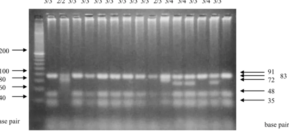
After electrophoresis, the gel was stained with ethid- ium bromide and the size of HhaI digested fragments was estimated by comparison with 20 base pair DNA ladder size marker (FMC) (Fig. 1).

Statistical analysis

We estimated allelic and genotypic frequencies of the APOE for the AD patients and controls by counting alleles and genotypes and calculating sample propor- tions. Comparisons of allele frequencies and genotype frequencies of APOE were made using chi-squared analysis.

RESULTS

The genotype frequencies are shown in Table 1. The most frequent genotype for all subjects was e3/e3 (AD subjects, 36.7%; control subjects, 58.2%). The frequencies of APOE alleles in AD patients were 18.3% ( e2), 58.3% (e3), and 23.3% (e4). The corre- sponding frequencies in controls were 13.3% ( e2), 72.5% ( e3), and 14.2% (e4). The frequency of the APOE e2 allele in AD patients was not significantly different from that in controls. When statistical analy- sis was conducted after the exclusion of the APOE e2 allele, the frequency of the APOE e4 allele in AD patients was significantly higher than that in controls ( c

2

= 4.11, d.f. = 1, P < 0.05).

DISCUSSION

The main object of the present study was to examine

the distribution of the APOE alleles in Korean AD

patients. In contrast with previous reports, our results

represented relative higher frequency of the APOE

e2 allele in AD patients and controls. In particular,

the frequency of the APOE e2 allele in our AD

patients was much higher than previous reports in

other racial groups including Caucasians, Hispanics,

African Americans, and Japanese.

15

The frequency of

the APOE e3 allele in our AD patients was similar to

that in Caucasians and African Americans, but lower

than that in Hispanics and Japanese.

15

The frequency

(3)

of the APOE e4 allele in our AD patients was lower than that in Caucasians, African Americans, and Japanese, but slightly higher than that in Hispanics.

15

Compared with controls, the frequency of the APOE e2 allele in our AD patients was not significantly different from that in controls. The frequency of the APOE e4 allele in our AD patients was 23.3%, which was significantly higher than that in controls.

These results do not support that the APOE e2 allele protects the development of AD, but do support that the APOE e4 allele plays a role as a risk factor for AD.

According to previous studies in Koreans,

23,24

the frequency of the APOE e4 allele in AD patients was significantly higher than that in controls and the asso- ciation between the APOE e4 allele and AD was con- sistently reported. These results, including our data, confirm the importance of the APOE e4 allele as a risk factor for AD in Koreans. In contrast with the APOE e4 allele, the role of the APOE e2 allele in the development of AD in Koreans was controversial.

One study reported the APOE e2 allele frequency (4.1%) in Korean AD group did not differ from that

(4.6%) in the control group.

23

The other study reported the same frequency (10.0%) of the APOE e2 alleles in Korean AD patients and controls which was a relatively high percentage compared with other racial groups’ results.

24

Compared with the present study, the frequency of the APOE e2 allele of controls in that study

24

was not significantly different from that in the present study ( c

2

= 1.30, d.f. = 2, P = 0.523).

However, the frequency of the APOE e2 allele of controls in another Korean study

23

was significantly different from that in the present study ( c

2

= 25.79, d.f. = 2, P = 0.000). The reason for this difference is not determined yet, however, probably due to differences in population characteristics. Therefore, there is a strong need to study large samples for further clarifi- cation. The frequency of the APOE e2 allele in our Korean AD patients was higher than that in any other previous reports, but was not significantly different from that in controls. These results suggest that the APOE e2 allele may not play a protective role in the development of AD in Koreans.

The association between the APOE e4 allele and AD has been reported primarily from Caucasian Figure 1. Example of APOE alleles from 15 different people. The three major alleles of APOE differ by single nucleotide substitutions within the amino acid codons at positions 112 (CysÆArg; e3Æe4) and 158 (ArgÆCys; e3Æe2). These sequence differences can be demonstrated by using polymerase chain reaction to amplify DNA obtained from blood samples. In restric- tion isotyping of APOE, the sizes of HhaI digested fragments in each isoform are visualized by UV illumination. The APOE e2 allele was characterized by the presence of the 83 and 91 base pair bands, the APOE e3 allele by the 35, 48, and 91 base pair bands, and the APOE e4 allele by the 19, 35, 48, and 72 base pair bands. Each of the heterozygous types is shown by mixed bands of homozygotes.

Table 1. APOE genotypes and allele frequencies

By genotype By allele

e2/e2 e2/e3 e2/e4 e3/e3 e3/e4 e4/e4 e2 e3 e4

AD (n = 30) 1 5 4 11 8 1 11 35 14

3.3% 16.7% 13.3% 36.7% 26.7% 3.3% 18.3% 58.3% 23.3%

Controls (n = 158) 2 20 18 92 25 1 42 229 45

1.3% 12.7% 11.4% 58.2% 15.8% 0.6% 13.3% 72.5% 14.2%

91 72 48 35 100 83

80 60 40 200

base pair base pair

3/3 2/2 3/3 3/3 3/3 3/3 3/3 3/3 3/3 2/3 3/4 3/4 3/3 3/4 3/3

(4)

populations in the USA and Europe. However, the APOE has been extensively investigated in popula- tions worldwide because of its role in lipid metabo- lism and ischemic cardiovascular disease. Nowadays, the emergence of the APOE e4 allele as a major risk factor for AD has been confirmed in more than 100 studies worldwide. The association between the APOE e4 allele and AD is strongest in Japanese sub- jects, followed by Caucasians, and is seemingly weaker among African Americans and Hispanics.

15

A meta- analysis of 40 studies representing nearly 30 000 APOE alleles concluded that the APOE e4 allele rep- resented a major risk factor for AD in Caucasians, African Americans, Hispanics, and Japanese, across all ages between 40 and 90 years, but that the effect diminished after age 70.

15

The role of the APOE e2 allele in the development of the AD is less clear. A protective effect of the APOE e2 allele for AD has been reported.

14,16

Although a number of studies confirmed the lowered APOE e2 allele frequency in AD patients versus con- trols, others did not find the association. One study reported a significant increase in the frequency of the APOE e2 allele in Italian patients with sporadic AD as well as with familial early-onset AD.

18

An increased APOE e2 allele frequency was also reported for AD patients of African-American descent, while a decreased APOE e2 allele frequency was found in Caucasian AD patients.

25

Furthermore, a Dutch popu- lation-based study presented that the APOE e2 allele was associated with an increased risk of early-onset AD and a reduced survival.

17

Our study also showed a tendency to increase in the frequency of the APOE e2 allele in AD patients in spite of no significant dif- ference in the frequency of the APOE e2 allele between AD patients and controls. There are at least three possible explanations for these discrepancies between studies of the role of the APOE e2 allele in AD patients.

The first explanation for the different allelic associ- ations of the APOE among Koreans and other racial groups with AD is that the associations are due to linkage disequilibrium, rather than to a direct effect of the APOE alleles. The linkage disequilibrium implies that the risk allele of an unknown causal genomic polymorphism is always accompanied by the APOE allele, because APOE and the causal genomic polymorphism are close together on chromosome 19 and recombination during meiosis is unlikely. An AD susceptibility gene may be in the linkage disequilib- rium with the APOE allele in AD patients. Recently, several new polymorphisms within the transcriptional regulatory region of the APOE gene were re- ported.

26,27

Bullido et al. identified a diallelic polymor-

phism in the promoter region of the APOE gene, –491 base pairs upstream of the APOE transcriptional start site (–491 A/T), and found that homozygosity for the –491 A allele was associated with AD, being inde- pendent of APOE e4 allele status.

26

Additionally, they reported in vitro studies, which suggested that the –491 A/T polymorphism altered the level of APOE expression thereby modulating the risk for AD.

26

Because of the close physical distance between the –491 A/T and known APOE polymorphisms and the consistent association of the latter with AD, the association of the –491 A (or –491T) allele with AD reflects the strong linkage disequilibrium between the –491 A (or –491T) allele with the known APOE poly- morphism. Recent study presented a stronger associa- tion between the APOE e2 allele and –491T allele in the Dutch population (P = 0.002 in controls) than in the Spanish one (P = 0.07 in controls).

28

The second possibility is that genes other than APOE may con- found studies. This explanation for the lack of consis- tency in the protective effect of APOE e2 allele for AD is less likely to be the case in our study. The fre- quency of the APOE e4 allele was consistently increased in our AD patients and the APOE e4 allele played a role as a risk factor in the development of AD. Another explanation may be that mutations may have occurred in the rare APOE e2 allele that leads to an increased risk of AD. The new APOE alleles resulting from such mutations may be important determinants of AD risk in some populations but not in others. Such a mechanism cannot be excluded unless the APOE gene is fully sequenced in all patients.

In interpreting the present results, some limitations should be noted. One limitation is that the controls of the present study had a wide range of age distribution and were not age matched. We did not have sufficient old aged controls to compare with AD patients.

However, when we compared controls aged 60 years

or more (n = 31) with controls aged less than 60 years

(n = 127), the frequencies of three different APOE

alleles were not significantly different between them

( c

2

= 4.18, d.f. = 2, P = 0.123). Recent Japanese study

also showed that there were no significant differences

in the distribution of the APOE phenotypes between

the younger (aged 65 years or under) and the older

(aged more than 65 years) subgroups of the normal

healthy controls.

29

Another limitation is that the

number of AD patients was too small to generalize

the study results. In spite of these limitations, it is

noteworthy that this study shows the relative high fre-

quency of the APOE e2 allele in AD patients and

shows no significant difference in the frequency of the

APOE e2 allele between AD patients and controls.

(5)

CONCLUSIONS

In the present study of Korean AD, we presented an increased frequency of the APOE e4 allele in AD patients versus controls and presented no significant difference in the frequency of the APOE e2 allele between AD patients and controls. These results support that the APOE e4 allele plays a role as a risk factor for AD in Koreans and suggest that the APOE e2 allele may not play a protective role in the devel- opment of AD in Koreans. Our results with Korean subjects warrant further study with large samples.

ACKNOWLEDGMENTS

This work was supported by grants from the Institute for Medical Genetics of Keimyung University School of Medicine, Taegu, Korea. We would like to thank Dr Tae-Wan Kim working at Non-Gong Dementia Center, Taegu, Korea for assisting with case recruit- ment and diagnosis.

REFERENCES

1. Mahley RW. Apolipoprotein E: Cholesterol transport protein with expanding role in cell biology. Science 1988; 240: 622–630.

2. Elshourbagy NA, Liao WS, Mahley RW, Taylor JM.

Apolipoprotein E mRNA is abundant in the brain and adrenals as well as in the liver, and is present in other peripheral tissues of rats and marmosets. Proc. Natl Acad. Sci. USA 1985; 82: 203–207.

3. Poirier J, Hess M, May PC, Finch CE. Apolipoprotein E and GFAP-RNA in hippocampus during reactive synaptogenesis and terminal proliferation. Mol. Brain.

Res. 1991; 11: 97–106.

4. Diedrich JF, Minnigan H, Carp RI et al. Neuropatholo- gical changes in scrapie and Alzheimer’s disease are associated with increased expression of apolipoprotein E and cathepsin D in astrocytes. J. Virol. 1991; 65:

4759–4768.

5. Poirier J, Aubert I, Bertrand P, Quirion R, Gauthier S, Nalbantoglu J. Apolipoprotein E4 and cholinergic dys- function in AD: A role for the amyloid/apoE4 complex?

In: Giacobini E, Becker RE (eds). Alzheimer’s Disease:

Therapeutic Strategies. Birkhauser, Boston, 1994; 72–76.

6. Poirier J, Delisle MC, Quirion R et al. Apolipoprotein E4 allele as a predictor of cholinergic deficits and treat- ment outcome in Alzheimer’s disease. Proc. Natl Acad.

Sci. USA 1995; 92: 12 260–12 264.

7. Soininen H, Kosunen O, Helisalmi S et al. A severe loss of choline acetyltransferase in the frontal cortex of Alzheimer patients carrying apolipoprotein epsilon 4 allele. Neurosci. Lett. 1995; 187: 79–82.

8. Davignon J, Gregg RE, Sing CF. Apolipoprotein E poly- morphism and atherosclerosis. Arteriosclerosis 1988; 8:

1–21.

9. Crews DE, Kamboh MI, Mancilha-Carvalho JJ, Kottke B. Population genetics of apolipoprotein A-4, E, and H polymorphisms in Yanomami Indians of northwestern Brazil: Associations with lipids, lipoproteins, and carbo- hydrate metabolism. Hum. Biol. 1993; 65: 211–224.

10. Mayeux R, Stern Y, Ottman R et al. The apolipoprotein e4 allele in patients with Alzheimer’s disease. Ann.

Neurol. 1993; 34: 752–754.

11. Tsuda T, Lopez R, Rogaeva EA et al. Are the associa- tions between Alzheimer’s disease and polymorphisms in the apolipoprotein E and the apolipoprotein CII genes due to linkage disequilibrium? Ann. Neurol. 1994;

36: 97–100.

12. Ueki A, Kawano M, Namba Y, Kawakami M, Ikeda K.

A high frequency of apolipoprotein E4 isoprotein in Japanese patients with late-onset nonfamilial Alzheimer’s disease. Neurosci. Lett. 1993; 163: 166–

168.

13. Strittmatter WJ, Saunders AM, Schmechel D et al.

Apolipoprotein E: High avidity binding to beta-amyloid and increased frequency of type 4 allele in late onset familial Alzheimer disease. Proc. Natl Acad. Sci. USA 1993; 90: 1977–1981.

14. Chartier-Harlin MC, Parfitt M, Legrain S et al.

Apolipoprotein E, epsilon 4 allele as a major risk factor for sporadic early and late-onset forms of Alzheimer’s disease: Analysis of the 19q13.2 chromosomal region.

Hum. Mol. Genet. 1994; 3: 569–574.

15. Farrer LA, Cupples LA, Haines JL et al. Effects of age, sex, and ethnicity on the association between apolipo- protein E genotype and Alzheimer’s disease: A meta- analysis. JAMA 1997; 278: 1349–1356.

16. Corder EH, Saunders AM, Risch NJ et al. Protective effect of apolipoprotein E type 2 allele for late onset Alzheimer disease. Nat. Genet. 1994; 7: 180–184.

17. van Duijn CM, de Knijff P, Wehnert A et al. The apolipoprotein E e2 allele is associated with an increased risk of early-onset Alzheimer’s disease and a reduced survival. Ann. Neurol. 1995; 37: 605–

610.

18. Sorbi S, Nacmias B, Forleo P et al. ApoE allele frequen- cies in Italian sporadic and familial Alzheimer’s disease.

Neurosci. Lett. 1994; 177: 100 –102.

19. American Psychiatric Association. Diagnostic and Sta- tistical Manual of Mental Disorders, 4th edn. American Psychiatric Association, Washington DC, 1994.

20. McKhann G, Drachman D, Folstein M, Katzman R, Price D, Stadlan EM. Clinical diagnosis of Alzheimer’s disease: Report of the NINCDS-ADRDA Work Group under the auspices of Department of Health and Human Services Task Force on Alzheimer’s Disease.

Neurology 1984; 34: 939–944.

21. Maniatis T, Sambrook J, Fritsch EF. Molecular Cloning:

A Laboratory Manual, 2nd edn. Cold Spring Harbor Laboratory, New York, 1989.

22. Emi M, Wu LL, Robertson MA et al. Genotyping and sequence analysis of apolipoprotein E isoform.

Genomics 1988; 3: 373–379.

23. Kim KW, Jhoo JH, Lee KU et al. Association between

(6)

apolipoprotein E polymorphism and Alzheimer’s disease in Koreans. Neurosci. Lett. 1999; 277: 145–

148.

24. Oh MJ, Chung EK, Shin YM, Lee KO, Park YS. The apolipoprotein E genotyping using the PCR-RFLP was useful to linkage analysis of Alzheimer’s disease fami- lies. Exp. Mol. Med. 1997; 29: 161–164.

25. Maestre G, Ottman R, Stern Y et al. Apolipoprotein E and Alzheimer’s disease: Ethnic variation in genotypic risks. Ann. Neurol. 1995; 37: 254–259.

26. Bullido MJ, Artiga MJ, Recuero M et al. A polymor- phism in the regulatory region of APOE associated with risk for Alzheimer’s dementia. Nat. Genet. 1998; 18:

69–71.

27. Lambert JC, Pasquier F, Cottel D, Frigard B, Amouyel P, Chartier-Harlin MC. A new polymorphism in the APOE promoter associated with increased risk of developing Alzheimer’s disease. Hum. Mol. Genet. 1988;

7: 533–540.

28. Roks G, Cruts M, Bullido MJ et al. The –491 A/T poly- morphism in the regulatory region of the apolipopro- tein E gene and early-onset Alzheimer’s disease.

Neurosci. Lett. 1998; 258: 65–68.

29. Nakayama S, Kuzuhara S. Apolipoprotein E phenotypes in healthy normal controls and demented subjects with Alzheimer’s disease and vascular dementia in Mie Pre- fecture of Japan. Psychiatry Clin. Neurosci. 1999; 53:

643–648.

수치

Table 1. APOE genotypes and allele frequencies

참조

관련 문서

1 John Owen, Justification by Faith Alone, in The Works of John Owen, ed. John Bolt, trans. Scott Clark, &#34;Do This and Live: Christ's Active Obedience as the

Second, to see a similarity factor among the relation between a factor which forms peer group and the attitudes toward school life, in case of the

North Korea is not the country which deserves to be invested, but it could play a crucial role in the process of propelling the South Korea's new

Although Korea has been playing a leading role in the development of fusion power generation and Korea is playing a leading role in the world stage,

These results demonstrated that a positive role of Slug and Twist in tumor invasion in CRA, and inverse correlation between Twist and Slug expression

In conclusion, ellipsis plays an important role in English discourse as a grammatical device for drawing the listener's attention to the focal information

Secondly, in the results of the analysis as to the influence of the Taekowndo leader's leadership on the stress of a Taekowndo player, it appeared that in

The HbA1c level is correlated with cardiovascular risk factor in non diabetic elderly population and we suggest that HbA1c may predict cardiovascular risk as