Treatment of Axillary Osmidrosis Using a Subcutaneous Pulsed Nd-YAG Laser
Daejin Kim, Junhyung Kim, Hyeonjung Yeo, Hyukjun Kwon, Daegu Son, Kihwan Han
Department of Plastic and Reconstructive Surgery, Keimyung University School of Medicine, Daegu, Korea
Correspondence: Junhyung Kim Department of Plastic and Reconstructive Surgery, Keimyung University School of Medicine, 56 Dalseong-ro, Jung-gu, Daegu 700-712, Korea
Tel: +82-53-250-7635 Fax: +82-53-255-0632 E-mail: [email protected]
Background Axillary osmidrosis is characterized by an unpleasant odor, profuse sweating, and in some instances, staining of clothes that may socially and psychologically impair affected individuals. Various types of surgical procedures have been developed for the treatment of axillary osmidrosis. This study was undertaken to evaluate the effectiveness of subcutaneous pulsed neodymium: yttrium-aluminum-garnet (Nd-YAG) laser treatment for the treatment of axillary osmidrosis.
Methods Twenty-nine patients with axillary osmidrosis were included in this study. Patients were categorized according to the results of an axillary malodor grading system, and a subcutaneous pulsed Nd-YAG laser was applied to all patients. The treatment area for the appropriate distribution of laser energy was determined using the iodine starch test (Minor’s test) against a grid pattern composed of 2×2 cm squares. The endpoint of exposure was 300 to 500 J for each grid, depending on the preoperative evaluation results. The results were evaluated by measurement of axillary malodor both pre- and postoperatively using the grading system and iodine starch test.
Results The average follow-up period was 12.8 months. Nineteen patients had a fair-to- good result and ten patients had poor results. The postoperative Minor’s test demonstrated that there were remarkable improvements for patients with mild to moderate symptoms.
Complications including superficial second degree burns (n=3) were treated in a conservative manner. A deep second degree burn (n=1) was treated by a surgical procedure.
Conclusions Subcutaneous pulsed Nd-YAG laser has many advantages and is an effective noninvasive treatment for mild to moderate axillary osmidrosis.
Keywords Lasers / Axilla / Starch
Received: 21 Dec 2011 • Revised: 7 Feb 2012 • Accepted: 22 Feb 2012
pISSN: 2234-6163 • eISSN: 2234-6171 • http://dx.doi.org/10.5999/aps.2012.39.2.143 • Arch Plast Surg 2012;39:143-149
No potential conflict of interest relevant to this article was reported.
Original Article
A B
results of axillary osmidrosis include depression, anxiety and various negative social impacts [2].
This condition causes the individual slight discomfort in daily living and/or problems in their social life. Various proce- dures are under investigation. Treating osmidrosis with surgical resection of the apocrine glands is one effective and irreversible method. New treatment methods with at low relapse rate and that reduce the amount of scarring and temporary debilitation associated with surgery are in demand.
Proper diagnosis is required for identifying the appropriate treatment of axillary osmidrosis. Using the method introduced by Park and Shin [3], the iodine starch test was used in this study to determine the degree of severity [4], and laser irradia- tion was performed based on the results. Park and Shin [3] cre- ated classification tools to objectivize the degree of axillary os- midrosis. Based on the degree of severity, patients were treated with subcutaneous pulsed neodymium: yttrium-aluminum- garnet (Nd-YAG) laser [5], and we report the resulting efficacies and observed complications associated with specific grades of diagnosed axillary osmidrosis. Preoperative and postoperative differences were evaluated to identify relapse.
METHODS
Patients
From January 2010 to February 2011, of the 52 patients who visited a plastic surgery clinic reporting conditions of axillary osmidrosis, 29 total individuals (female, 15; male, 14) were selected to be treated with subcutaneous pulsed Nd-YAG laser.
Thirty-three patients were excluded from the study because they did not want an Nd-YAG laser to be used (n=28), or were lost to follow-up (n=5). The ages of the individuals ranged from 11 to 45 years of age (mean, 23.4 years), and the follow-up period ranged from 6 to 18 months with a mean follow-up period of 12.8 months.
Preoperative and postoperative evaluation
Prior to operation, the severity of axillary osmidrosis was deter- mined by the use of a questionnaire which examined perceived levels of malodor and sweating, and use of the classification system defined by Park and Shin [3] (Table 1, Figs. 1, 2). The questionnaire responses regarding malodor and sweating were based on subjective data from the patient as well as their family and friends. The grade was classified into one of three catego- ries: minimal, moderate, and severe.
Table 1. Osmidrosis grading system
Grade Degree of malodor
0 Gauze rubbed on the armpit does not give off any malodor under any circumstances (normal)
1 G auze rubbed on the armpit emits slight malodor only when patients are under stressful conditions (exercising or walking) and no malodor is detected from the rubbed gauze after performing usual daily activities
2 G auze rubbed on the armpit emits strong malodor after performing daily activities but malodor could not be detected at a distance of 1.5 m from the patient
3 Without the use of gauze, prominent malodor could be detected easily from the body at a distance of 1.5 m during daily activity
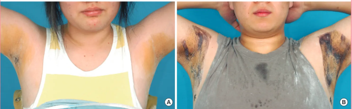
Fig. 1. Preoperative iodine starch test (Minor’s test)
Minor’s solution and starch were applied on both axillae. After 10 to 15 minutes, the color changed from white to blue-black. (A) Preoperative view
of a 13-year-old female with grade 2 osmidrosis. (B) Preoperative view of a 17-year-old male patient with grade 3 osmidrosis showing more deeply
discolored areas than the patient on the left.
Mild osmidrosis was defined as an indistinct malodor and sweating which is unnoticed by others during normal daily ac- tivities, moderate osmidrosis was defined as malodor that could be identified by the patient but could not be detected by others at a distance of 1.5 m from the patient and accompanied by a little perspiration, and severe malodor and sweating could be detected easily from the body by others at a distance of 1.5 m during normal daily activity.
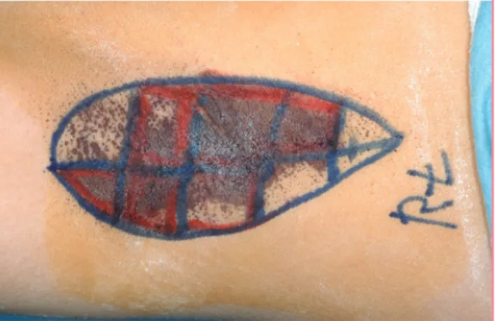
In order to reduce the uneven distribution of laser energy, a grid pattern composed of 2×2 cm squares was drawn on the preoperative bilateral axillae. This was followed by the iodine starch test to identify the preoperative sweating pattern. The iodine starch test (Minor’s test) [4] was used to objectively determine the surface area required for the operation. Areas of deep discoloration resulting from the iodine starch test were
noted and the preoperative bilateral axillae were marked with a different color to make this area distinct (Fig. 2).
Satisfaction was evaluated after the operation using a ques- tionnaire regarding any complications (scarring, burns, hema- toma, loss of hair), degree of malodor, and sweating. The iodine starch test [4] was also used to perform a comparison of the pre- and postoperative area of sweating.
Statistical analysis was performed using SPSS ver. 16.0 (SPSS Inc., Chicago, IL, USA) on MS Windows, with statistical signifi- cance (P<0.05) determined using a paired t-test.
Operative methods
With the patients holding their arms in maximum elevation to expose both axillae, both axillae were draped and 0.005%
chlorhexadine and 70% alcohol were applied. To relieve anxiety, 0.05-0.1 mg/kg of ketamine (Ketamine HCL, Huons, Seoul, Korea, 500 mg, 10 mL) and 2 mg/kg midazolam (Dormicum, Roche, Switzerland, 5 mg, 5 mL) was administered intravenous- ly. Then a 1:100,000 mixture of epinephrine and 1% lidocaine was mixed with normal saline in a 1:1 ratio. The subcutaneous injection was applied to the axilla followed by 5 minutes of compression. After making a hole using an 18-gauge needle, pretunneling was performed using a blunt dissector (Fig. 3A).
Dissection was necessary so that the 1-mm diameter laser can- nula could be inserted close to the subcutaneous apocrine gland targets. The cannula was inserted into the dermal-subdermal junction. The area of transcutaneous illumination indicated the positioning of the tips. The pulsed Nd-YAG laser (BL 3500N, B&B Systems, Seoul, Korea) treatment was performed using a wavelength of 1,064 nm, a pulse duration of 40 Hz and a power of 150 mJ. The endpoints of exposure for each grid were 200 to Fig. 2. Preoperative design
Circle affected area with surgical marker, then mark a 2×2 cm square grid pattern. Deeply discolored areas are marked with additional red lines.
Fig. 3. Operative technique
(A) Multiple puncture points. Typically, three small punctures were used, but, random selection was possible. Adequate multiple punctures aided
the method. Punctures were made with an 18-gauge needle at the anterior, posterior, proximal region, and border of each axilla. (B) Lipolysis was
performed using an neodymium: yttrium-aluminum-garnet laser on the axilla. Laser exposures were 300 to 500 J for each square depending on the
preoperative evaluation results.
300 J in grade 2 cases and 500 J in grade 3 cases, depending on the thickness of the skin, the density of the hair and the degree of severity of axillary osmidrosis (Fig. 3B). Laser assisted lipo- suction was not applied to objectively evaluate the effects of the treatment using only the Nd-YAG laser. No suture or drain was inserted into the cannula insertion site.
Postoperative management
Unlike other operative procedures, subcutaneous pulsed Nd- YAG laser does not require a specific postoperative dressing.
Antibiotic ointment (0.3% ofloxacin ophthalmic ointment, Samil Pharm, Seoul, Korea, 3.5 g) and hydrocellular foam (Al- levyn standard, Smith & Nephew, Largo, FL, USA) was applied to the wounds. This was an outpatient procedure, no restric-
tions in arm movements were required, and patients were able to return immediately to everyday life. The dressing remained in place overnight and was removed the day after the operation.
After removal of the dressing, patients performed a simple exer- cise and then showered.
RESULTS
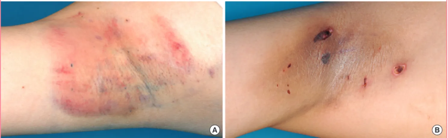
There were no observations of postoperative hematoma, necro- sis, infection, or axillary hair loss in any of the patients (Table 2). The most common complications were contact burns (n=4, 13.8%) caused by the laser cannula. Of these contact burns, most were superficial second-degree burns. However, one case progressed from a superficial to a deep second degree burn, which subsequently necessitated a burn scar operation (Fig. 4).
Every patient involved in the operation was assessed to have grade 2 or 3 osmidrosis (grade 2, n=19; grade 3, n=10) (Table
Fig. 4. Postoperative complications viewed after 10 days
(A) A 36-year-old female showed superficial second degree burns on the right axilla and was treated in a conservative manner. (B) A 40-year-old male showed deep second degree burns on the left axilla and was treated with excision and primary closure.
A B
Fig. 5. Preoperative and postoperative evaluation
0 51.7
65.5
13.8
34.5 34.5
70
60 50 40 30 20 10 0
Grade 0, 1 Grade 2 Grade 3
(%) Preoperatively
Postoperatively
Distribution of axillary osmidrosis by preoperative and postoperative evaluation, according to the osmidrosis grading system.
Table 2. Postoperative evaluation (n = 29)
Postoperative evaluation No. (%)
Subjective assessment Good
Fair Poor
13 (44.8) 6 (20.7) 10 (34.5) Osmidrosis grading system
Grade 0, 1 (preoperatively/postoperatively) Grade 2
Grade 3
0 (0)/15 (51.7) 19 (65.5)/4 (13.8) 10 (34.5)/10 (34.5) Iodine starch test (Minor’s test)
(mean squares of sweating area) (total/grade 2/grade 3) Preoperatively
Postoperatively 21.9/15.3/35.2
12.5
a)/5.3
a)/26.2 Complication
Burn or skin erosion Scar Hematoma or infection Movement limitation Major vessel or nerve injury
4 (13.8) 0 (0) 0 (0) 0 (0) 0 (0)
a)