∙ Received: June 25, 2012. Accepted: October 2, 2012.
∙ Corresponding author : Da-Young Lee
Department of Nuclear Medicine, Severance Hospital, Yonsei University Health System, 250 Seongsanno, Seodaemun-gu, Seoul, 120-752 Tel: +82-2-2228-6051, Fax: +82-2-312-0578
E-mail: [email protected]
Original Article 유방암의 감시림프절 검사에서 유방크기와 체질량지수에 따른 검사시간 변화
연세의료원 세브란스병원 핵의학과, 신구대학교 방사선과1
이다영 ․ 남궁혁 ․ 조석원 ․ 오신현 ․ 임한상 ․ 김재삼 ․ 이창호 ․ 박훈희1
The Variation of Scan Time According to Patientʼs Breast Size and Body Mass Index in Breast Sentinel lymphangiography
Da-Young Lee, Hyuk Nam-Koong, Seok-Won Cho, Shin-Hyun Oh, Jae-Sam Kim, Chang-Ho Lee and Hoon-Hee Park1
Department of Nuclear Medicine in Yonsei University Health System, Korea
1Department of Radiology in Shin-Gu University at Seongnam, Korea
Purpose : At this time, the sentinel lymph node mapping using radioisotope and blue dye is preceded for breast cancer patient’s sentinel lymph node biopsy. But all patients were applied the same protocol without consideration of physical specific character like the breast sizes and body mass indexes. The purpose of this study is search the optimized scan time in breast sentinel lymphangiography by observing how much the body mass index and breast size influence speed of lymphatic flow. Materials and Methods : The Object of this study was 100 breast cancer patients(Female, 100 persons, average age 50.34±10.26 years old)at Severance hospital from October 2011 to December 2011. They were scanned breast sentinel lymphangiography before operation.
This study was performed on Forte dual heads gamma camera (Philips Medical Systems, Nederland B.V.). All patients were intra-dermal injected 99mTc-Phytate 18.5 MBq, 0.5 ml. For 80 patients, we have scanned without limitation of scan time until the lymphatic flow from the lymph node since injection. We measured how long the lymphatic flow time between departures from injects site and arrival to lymph node using stopwatch. After we calculated patient’s Body mass Index and classified as 4 groups. And we measured patient’s breast size and classified 3 groups. The modified breast lymphangiography that changing scan time according to comparison study’s result was performed on 20 patients and was estimated. Results : The mean scan time as breast size was A group 2.48 minutes, B group 7.69 minutes, C group 10.43 minutes. The mean scan time as body mass index was under weight 1.35 minutes, normal weight 2.56 minutes, slightly over 5.62 minutes, over weighted 5.62 minutes. The success rate of modified breast lymphangiography was 85%. Conclusion : As the Body mass index became higher and breast size became bigger, the total scan time is increased. Based on the obtained information, we designed modified breast lymphangiography protocol. At the cases applying that protocol, most of sentinel lymph nodes were visualized as lymphatic pool. In conclusion, we found that the more success rate in modified protocol considering physical individuality than study carrying out in the same protocol. (Korean J Nucl Med Technol 2012;16(2):62-67)
Key Words : Breast cancer, Sentinel Lymphangiography, Breast size, Body Mass Index
서 론
한국의 여성 암 발병률 1위인 유방암은 국립암센터의 국 가암등록 자료에 따르면 5년 생존율이 89%로 조기진단과 치 료가 이루어질 때 비교적 예후가 좋은 암종이라 할 수 있다.
유방암환자의 병기결정 및 예후를 판단하는 중요한 지표로
Fig. 1. The sentinel lymph node mapping.
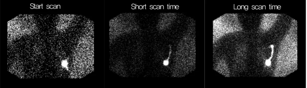
Short scan time
Start scan Long scan time
Fig. 2. Because the sentinel lymphangiography is dynamic study, both injection and scan are started at the same time. The case of short scan time, the sentinel lymph node wasn’t visualized sufficiently within the study. On the other hand, the long scan time study visualized sufficiently the sentinel lymph node.
Fig. 3. Forte dual heads gamma camera (Philips Medical Systems, Nederland B.V.).
유방암의 액와림프절 전이를 꼽을 수 있다.1-3) 침윤성유방암 에서 액와림프절 절제는 유방암수술의 표준술식이 되었지만 최근 조기 유방암환자의 비율이 증가하면서 수술 시 절제한 액와림프절 전이가 발견되지 않는 비율 역시 증가하였다.4) 액와림프절 절제 후 림프부종이나 혈청종 등 여러 합병증이 발생하므로 꼭 필요한 경우에만 액와림프절 절제술을 시행 하는 최소 침습적 수술을 시행하여 불필요한 합병증을 없애 는 것이 필요하다. 유방암은 감시림프절을 타고 액와림프절 로 전이가 이루어진다. 이 감시림프절은 1977년 Canvanas가 암환자의 치료를 위해 종양에서 가장먼저 전이되는 림프절 로써 개념을 도입하였고5) 1933년 Krag은 이 개념을 유방암 환자에게 적용하여 방사성의약품을 주사하여 유방의 감시림 프절을 발견하였다.6) 따라서 감시림프절 생검을 통해 림프절 전이 여부를 판단할 수 있게 되었고 액와림프절 절제술에 비 해 생검을 최소화 할 수 있게 되었다.
유방암 환자의 감시림프절 생검을 위하여 현재 방사성동 위원소와 Blue dye를 이용한 감시림프절 매핑(Mapping)이
선행되고 있다(Fig. 1). 본원의 유방의 림프절 신티그래피는 동적 영상으로 5 Frame, 60sec, 총 5분의 검사시간으로 시행 되었다. 그러나 모든 환자에 대하여 일괄적인 검사방법이 적 용되므로 환자의 유방의 크기나 비만도와 같은 특성이 고려 되지 않아 림프절의 흐름이 느린 환자의 경우, 림프절을 충 분히 형성하지 못한 채 검사가 종료되는 경우가 종종 발생한 다(Fig. 2). 본 연구에서는 환자의 신체적 특성인 체질량지수 (Body Mass Index, 이하 BMI)와 유방의 크기에 따른 림프액 의 속도차이를 관찰하였다. 이를 통해 방사성동위원소를 이 용한 유방의 감시림프절 신티그래피에서 환자의 특성을 고 려한 최적의 검사시간을 도출 내고자 한다.
대상 및 방법
본 연구는 2011년 10월부터 2011년 12월까지 수술직전 유 방의 감시림프절 신티그래피를 시행한 100명(여성 100명, 평 균연령 50.34±10.26)를 대상으로 하였다. 장비는 감마카메라 Forte (Philips Medical Systems, Nederland B.V.)를 사용하였 으며, 방사성의약품은 99mTc-Phytate 18.5 MBq, 0.5 ml를 피 내주사 하였다(Fig. 3).
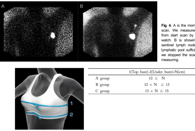
A B
Fig. 4. A is the moment of start scan. We measured the time from start scan by using stop watch. B is showing that the sentinel lymph node made as lymphatic pool sufficiently. Then we stopped the scan and time measuring.
1(Top bust)-2(Under bust)=N(cm)
A group 10 ≦ N
B group 10 < N ≦ 13
C group 13 < N ≦ 15
Fig. 5. We spilt the objects into 3 groups as the global standard of brassiere size. We used cup measurement methods. The cup size can be determined by calculating the difference between top bust size and the under bust size. A, B, C groups mean A, B, C cup sizes of women breasts.
1. 유방크기와 체질량지수에 따른 구분
먼저 80명의 환자를 대상으로 기존의 5분 검사방법대신 충분히 감시림프절에 섭취 될 때까지 시간의 제한 없이 영상 을 획득하였다. 검사시작 직후 주사부위로부터 출발한 림프 액의 흐름이 감시림프절에 섭취 될 때까지의 시간을 측정하 였다. 이때 감마카메라 모니터를 보며 감시림프절의 응집을 이룬 후 더 이상 흐름의 진척이 관찰되지 않을 시 검사를 종 료 하였다(Fig. 4). 30분이 지나도 림프절의 응집을 형성하지 않은 일부 환자의 경우는 검사를 중지하고 30분 후 지연촬영 을 실시하였고, 지연촬영에서도 림프액의 흐름이 관찰되지 않은 경우 자료수집에서 제외하였다. 검사가 완료된 80명의 환자를 두 가지 지표로 나누었다. 첫 번째로 80명의 대상 중 유방의 크기를 측정하여 기준에 따라 A, B, C의 세 군으로 나누었다(Fig. 5). 유방의 크기는 A군 40명, B군 25명 C군 15 명이었다. 두 번째로 80명의 대상 중 체질량지수를 기준하여 저체중, 표준, 과체중, 비만의 네 군으로 나누었다(Fig. 6). 체 질량지수는 저 체중 19명, 표준 33명, 과체중 25명, 비만이 3 명이었다. 총 80명의 대상을 각 군으로 나누어 평균 검사시 간을 구하였다.
2. 환자의 신체조건에 맞춘 변형 유방 림프신티그래피
앞서 구한 평균 검사시간을 적용하여 기존의 검사방법에 서 검사시간만 변화시킨 변형 유방 림프절 신티그래피를 20 명의 환자에게 시행하였다. 검사시행 전 환자의 유방의 크기 와 체질량지수를 구하여 각각 설정해 놓은 검사시간에 적용 하여 검사를 시작하였다. 이들 중 검사시간 내에 감시림프절 응집의 형성이 관찰되면 성공으로 평가 하였고, 검사시간 내 에 형성하지 못한 경우는 실패로 평가하였다(Fig. 7).
결 과
1. 유방크기와 체질량지수에 따른 평균 검사시간
80명의 대상 중 유방의 크기에 따른 평균 검사시간은 A군 2.48분, B군 7.69분, C군 10.43분 이었다(Fig. 8). 80명의 대상 중 체질량지수에 따른 평균 검사시간은 저 체중 1.35분, 표준 2.56분, 과체중 5.62분, 비만 15분이었다(Fig. 9).
Under weight
Normal weight
Slightly overweight
Over weight
~20 20~24 25~29 30~
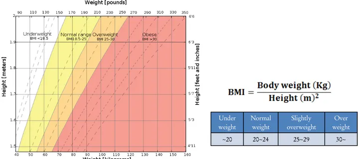
Fig. 6. We spilt the objects into 4groups as the global standard of body mass index. Body mass index is defined as the individual's body weight divided by the square of his or her height. The WHO regard a BMI of less than 20 as underweight, while a BMI greater than 25 is considered slightly overweight and above 30 is considered over weight.
A B
Fig. 7. We estimated a success if the lymph node sufficiently formed lymphatic pool with in the scan time. .A is the case of success. The lymph node sufficiently formed lymphatic pool at left axilla. B is the case of fail. There is lacking uptake in lymph vessel and node.
*Unit: Minutes *Unit: Minutes
Fig. 8. The average scan times as breast sizes. The studies of patients having small breasts needed short scan time. On the other hands, patients having bigger breasts needed longer scan time.
Fig. 9. The average scan times as body mass index. The studies of underweight patients needed short scan time. On the other hands, overweight patients needed longer scan time.
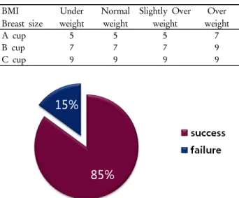
Table 1. The modified protocol was settled by previous study’s average. When the women with Breast size 80B, BMI 25, We subjected the protocol with scan time of 5 frames.
(*Unit: frames, 1Frame=30sec) BMI
Breast size
Under weight
Normal weight
Slightly Over weight
Over weight
A cup 5 5 5 7
B cup 7 7 7 9
C cup 9 9 9 9
Fig. 10. Most of the modified studies was successful. The 17 in 20 studies were estimated as success by sufficiently visualization of sentinel lymph node. And 3 in 20 studies were estimated as failure. The success rate of modified breast lymphangiography was 85%.
2. 변형 유방 림프신티그래피 결과
앞서 얻어진 정보를 바탕으로 각 군별 적절한 검사시간을 적용하여 검사를 시행하였다(Table 1). 결과는 20명 중 성공 평가를 받은 경우는 17회 실패평가를 받은 경우는 3회로 총 85% 성공률을 나타내었다(Fig. 10).
고 찰
본 연구에서는 대상을 구분하는 지표를 체질량지수와 유 방의 크기 두 가지로 삼았다. 그러나 여성에 있어 생리주기, 폐경의 유무, 나이, 임신경험 혹은 가족력과 같이 유방의 상 태에 영향을 주는 인자들이 있다. 좀더 지표들을 확장하고 세분화하여 각 부문별 연구가 이루어져야 할 것이다. 또한 최종결과를 도출하기 위한 두 번째 실험에서 실험대상의 수 가 더 많았다면 더욱 정확한 결론을 얻었을 것이다.
결 론
유방의 크기와 체질량지수에 따른 총 검사 시간은 체질량 지수가 높을수록, 유방의 크기가 클수록 증가하였으며, 얻어 진 정보를 바탕으로 기존 검사방법에서 검사시간만을 변화
시킨 변형 유방 림프절 신티그래피를 적용하였을 때 대부분 의 경우 검사시간 내 림프절을 형성함을 관찰 할 수 있었다.
이를 통해 모든 환자에게 일괄적으로 적용하던 검사방법보 다 개인의 신체적 특성을 고려한 적절한 검사시간을 각각 다 르게 적용하였을 때 검사에서 높은 성공률을 보임을 알 수 있었다, 즉 체질량지수가 높고, 유방의 크기가 큰 환자일수록 감시림프절을 영상화하는데 더 긴 검사시간을 필요로함을 알 수 있었다. 이 연구를 통해 감시림프절 신티그래피에서 보다 효율적인 검사가 진행되고 수술 중 액와림프절 생검 시 에 정확한 위치 정보를 주는데 도움이 될 것이라 사료된다.
요 약
유방암 환자의 감시림프절 생검을 위하여 현재 방사성동 위원소와 blue dye를 이용한 감시림프절 매핑(Mapping)이 선행되고 있다. 현재 모든 환자에 대하여 일괄적인 검사방법 이 적용되므로 환자의 유방의 크기나 비만도와 같은 특성이 고려되지 않아 림프절의 흐름이 느린 환자의 경우, 림프절을 충분히 형성하지 못한 채 검사가 종료되는 경우가 종종 발생 한다. 본 연구에서는 환자의 신체적 특성인 체질량지수와 유 방의 크기에 따른 림프액의 속도차이를 관찰하였다. 이를 통 해 방사성동위원소를 이용한 유방의 감시림프절 신티그래피 에서 환자의 특성을 고려한 최적의 검사시간을 도출하는데 목적을 두었다. 본 연구는 2011년 10월부터 2011년 12월까지 수술직전 유방의 감시림프절 신티그래피를 시행한 100명(여 성 100명, 평균연령 50.34±10.26)를 대상으로 하였다. 장비는 감마카메라 Forte (Philips Medical Systems, Nederland B.V.) 를 사용하였으며, 방사성의약품은 99mTc-Phytate 18.5 MBq, 0.5 ml를 피내주사하였다. 먼저 80명의 환자를 대상으로 기 존의 5분 검사방법대신 충분히 림프절을 형성할 때까지 시간 의 제한 없이 영상을 획득하였다. 이를 통해 환자의 유방크 기와 체질량지수 별 그룹을 나누어 평균 검사시간을 구하였 다. 이 결과를 바탕으로 검사시간을 변화시킨 변형 유방 림 프신티그래피를 20명의 환자에게 시행하여 유용성을 확인하 였다. 80명의 대상 중 유방의 크기에 따른 평균 검사시간은 A그룹 2.48분, B그룹 7.69분, C그룹 10.43분이었다. 80명의 대상 중 체질량지수에 따른 평균 검사시간은 저 체중 1.35분, 표준 2.56분, 과 체중 5.62분, 비만 15분이었다. 앞서 얻어진 정보를 바탕으로 각 그룹별 적절한 검사시간을 적용하여 검 사를 시행하였다. 결과는 20명 중 성공평가를 받은 경우는 17회 실패평가를 받은 경우는 3회로 총 85% 성공률을 나타 내었다. 유방의 크기와 체질량지수에 따른 총 검사 시간은
체질량지수가 높을수록, 유방의 크기가 클수록 증가하였으 며, 얻어진 정보를 바탕으로 기존 검사방법에서 검사시간만 을 변화시킨 변형 유방 림프신티그래피를 적용하였을 때 대 부분의 경우 검사시간 내 림프절을 형성할 수 있었다. 이를 통해 모든 환자에게 일괄적으로 적용하던 검사방법보다 개 인의 신체적 특성을 고려한 적절한 검사시간을 각각 다르게 적용하였을 때 검사에서 높은 성공률을 보임을 알 수 있었다.
REFERENCES
1. Albertini JJ, Lyman GH, Cox C, Yeatman T, et al. Lymphatic mapping and sentinel node biopsy in the patient with breast cancer. JAMA 1996;276:18-22.
2. Giuliano AE, Dale PS, Turner RR, et al. Improved axillary stag- ing of breast cancer with sentinel lymphadenectomy. Ann Surg 1995;222:394-9.
3. Copeland EM III. Is axillary dissection necessary for T1 carci- noma of the breast? J Am Coll Surg 1997;184:397-8.
4. Silverstein MJ, Gierson ED, Waisman JR, et al. Axillary lymph node dissection for T1a breast carcinoma. Is it indicated?
Cancer 1994;73:664-7.
5. Ramon M Cavanas. An Approach For The Treatment Of Penile Carcinoma Cancer 1977;39:456-466.
6. David Krag, Donald Weaver, Akamaru Ashikaga, et al. The Sentinel Node In the Breast Cancer. A multicenter validation study. The New England Journal of Medicine 1998;339.
7. Hyuk Nam-Koong, Hoon-Hee Park, Min-Hye Lee, et al. A study of usefulness of lymphoscintigraphy Day-before-surgery in obese breast cancer's patients. 서울시 KIMES 학술대회 2007.
8. Yeung HW. Lymphoscintigraphy and sentinel node local- ization in breast cancer patients: a comparison between 1-day and 2-day protocol. Journals Nuclear Medicine 2001;42:420-3.
9. Charles E. Cox. Age and body mass index may increase the chance of failure in sentinel lymph node biopsy for women with breast cancer. The Breast Journal 2002;8:88-91.